Digital Poster
Processing & Analysis: Data Processing I
Joint Annual Meeting ISMRM-ESMRMB & ISMRT 31st Annual Meeting • 07-12 May 2022 • London, UK

Digital Poster
Processing & Analysis: Data Processing I
| Computer # | ||||
|---|---|---|---|---|
2840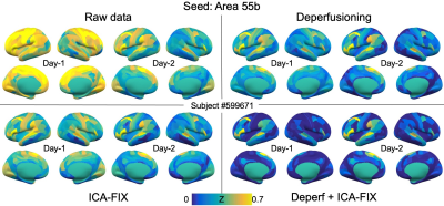 |
89 | Removal of perfusion lag structure before ICA-FIX cleaning boosts both reproducibility and between-subject similarity in fcMRI
Toshihiko Aso1 and Takuya Hayashi1
1Laboratory for Brain Connectomics Imaging, RIKEN Center for Biosystems Dynamics Research, Kobe, Japan
Removal of the perfusion lag structure in the fMRI signal was combined with independent component analysis (ICA)-based cleaning to evaluate its effect on test-retest reproducibility. In HCP test-retest data from 40 subjects, either treatment and the combined method effectively improved the reproducibility of functional connectivity. However, the effect was more evident in between-subject similarity of the connectivity pattern than within-subject similarity. This finding suggests that the perfusion lag structure may be one of the sources of between-subject variability, that may degrade the functional connectivity.
|
||
2841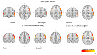 |
90 | Low-grade glioma resting-state fMRI pre-operative maps: a comparison with functional atlas from direct electrode stimulation. Video Permission Withheld
Beatrice Federica Luciani1, Donna Gift Cabalo1, Francesca Saviola1, Luca Zigiotto2,3, Stefano Tambalo1, Domenico Zacà1, Lisa Novello1, Silvio Sarubbo2,3, and Jorge Jovicich1
1CIMeC Center for Mind/Brain Sciences, Trento, Italy, 2Department of Neuroscience, Division of Neurosurgery, S.Chiara Hospital, APSS Trento, Trento, Italy, 3Structural and Functional Connectivity Lab, S.Chiara Hospital, APSS Trento, Trento, Italy A cortical-subcortical functional atlas has been recently proposed from direct-electrode stimulation on low grade glioma patients. The agreement between that atlas and non-invasive pre-operative resting-state functional MRI (rs-fMRI) data has not yet been investigated. In this study, we use pre-operative rs-fMRI data and the RestNeuMap tool to investigate the agreement between language and sensory networks in a group of low grade glioma patients. We find that rs-fMRI networks of language and speech arrest show greater overlap with DES-derived functional atlas than the sensorimotor network. |
||
2842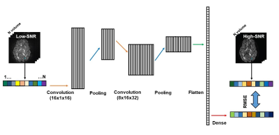 |
91 | Mitigation of noise floor in diffusion MRI using deep learning
Hu Cheng1, Jian Wang2, Sophia Vinci-Booher1, Qiuting Wen3, and Sharlene Newman4
1Psychological and Brain Sciences, Indiana University, Bloomington, IN, United States, 2Shandong Normal University, Jinan, China, 3Indiana University, Indianapolis, IN, United States, 4University of Alabama, Tuscaloosa, AL, United States
Rectified noise floor is a challenging problem for high-resolution diffusion MRI, which cannot be tackled by current denoising methods. We propose a simple deep learning method for correcting noise floor in diffusion MRI. The method is based on 1D CNN model and works on voxel-wise time courses. Therefore, even one dataset of the brain has sufficient number of samples for training, which is a big advantage for practical application. Both simulation and in vivo results show that the method is robust in mitigating the noise floor artifact and restore the true values of diffusion metrics.
|
||
2843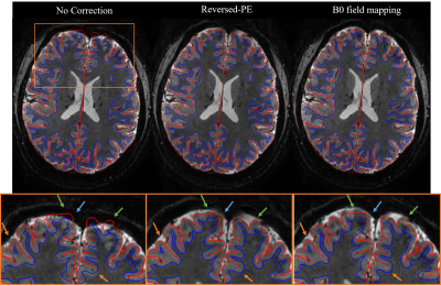 |
92 | Distortion correction of 3D EPI for cortical depth-resolved fMRI at 7T
Vahid Malekian1, Nadine N. Graedel1, Alice L. Hickling1, Ali Aghaeifar1,2, Nadège Corbin1,3, Oliver Josephs1, Eleanor A. Maguire1, and Martina F. Callaghan1
1Wellcome Centre for Human Neuroimaging, UCL Queen Square Institute of Neurology, University College London, London, United Kingdom, 2MR Research Collaborations, Siemens Healthcare Limited, Frimley, United Kingdom, 3Centre de Résonance Magnétique des Systèmes Biologiques, CNRS‐University Bordeaux, Bordeaux, France Geometric distortion is a major concern for cortical depth-dependent fMRI using EPI readouts that suffer from low bandwidth in the phase-encoded direction. Distortions are exacerbated at higher field strengths due to increased B0 field inhomogeneity. In this study, we quantitatively compared two distortion correction methods (B0 field-mapping and reversed-PE) applied to submillimeter (0.8mm isotropic) 3DEPI data at 7T. Cortical alignment was evaluated through comparison with an anatomically-faithful MP2RAGE reference by computing the dice coefficient and normalised mutual information. Both distortion correction methods usefully improve alignment. The reversed-PE approach performed better.
|
||
2844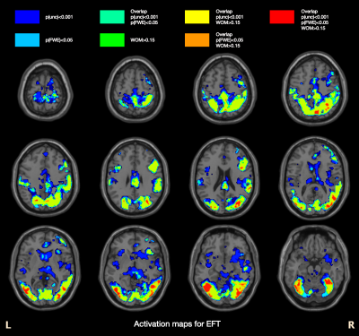 |
93 | Monte Carlo simulations of weighted overlap map thresholds to reduce the risk for type I errors in fMRI
Peter Van Schuerbeek1
1Radiology, UZ Brussel (VUB), Brussels, Belgium
We performed Monte Carlo Simulations to show that the risk for type I errors decreases if a minimum overlap between the individual results is set in addition to the significance of the group results. The activation maps of a real fMRI experiment showed that the combination of a weighted overlap map threshold in combination with a voxel significance threshold seems to lead to less type II errors compared to applying a family wise error (FWE) correction.
|
||
2845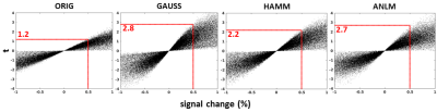 |
94 | Adaptive non-local means filtering as a drop-in preprocessing step to improve statistical sensitivity in task-based fMRI
Ajay Nemani1 and Mark J Lowe2
1Imaging Institute, Cleveland Clinic, Cleveland, OH, United States, 2Cleveland Clinic, Cleveland, OH, United States
Spatial filtering is an important step in the preprocessing of task-based fMRI to improve sensitivity in statistical analyses. This is usually implemented as a pure distance-based filter such as Gaussian filtering or an optimized matched filter. Adaptive non-local means (ANLM) filtering is a patch-based approach that is sensitive to the local noise model, especially at low signal to noise ratio such as fMRI. We show how ANLM filtering is a simple drop-in replacement at the spatial smoothing step of fMRI preprocessing pipeline that compares favorably to other approaches while better preserving local high frequency features.
|
||
2846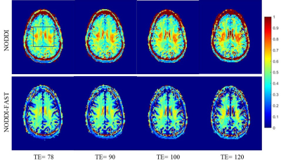 |
95 | NODDI-FAST derived parameters are echo-time independent
Maryam H Alsameen1, Zhaoyuan Gong1, Matthew Kiely1, Curtis Triebswetter1, and Mustapha Bouhrara1
1Magnetic Resonance Physics of Aging and Dementia Unit, National Institute on Aging, NIH, Baltimore, MD, United States In a previous work, we introduced the NODDI-FAST approach to address issues regarding overestimated CSF and neurite density (NDI) fractions in white matter seen with the original NODDI approach. However, in both NODDI-FAST and NODDI signal models, the compartment-specific T2 relaxations are not considered. Therefore, derived parameter estimates, especially NDI, could be dependent on echo time (TE). Here, we show that, as expected, ODI derived values using either NODDI or NODDI-FAST are TE-independent. We also confirm that NDI derived values using NODDI are TE-dependent. More importantly, we show that NDI derived values using NODDI-FAST are TE-independent.
|
||
2847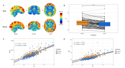 |
96 | Fractal-Based Analysis of fMRI BOLD Signal During Naturalistic Viewing Conditions
Olivia Campbell1,2, Tamara Vanderwal2,3, and Alexander Mark Weber1,2,4,5
1School of Biomedical Engineering, University of British Columbia, Vancouver, BC, Canada, 2BC Children's Hospital Research Institute, Vancouver, BC, Canada, 3Department of Psychiatry, University of British Columbia, Vancouver, BC, Canada, 4Department of Pediatrics, University of British Columbia, Vancouver, BC, Canada, 5Department of Neuroscience, University of British Columbia, Vancouver, BC, Canada
We investigated the difference between the brain’s fractal dynamics during movie-watching and resting-state conditions using 7T fMRI data. During movie-watching, we find that the BOLD signal becomes more scale-invariant and self-similar than during the resting-state. This supports the idea that movie-watching evokes a state of optimal neural functioning that may better reflect the endogenous state of the brain than a fixed cross-hair. We also find that fractal properties differ greatly across functional networks, providing novel information about the temporal dynamics of each network during naturalistic processing. Overall, these findings advance understanding of both fractal dynamics and naturalistic viewing in fMRI.
|
||
2848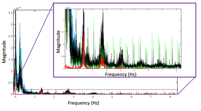 |
97 | Ultra-fast 3D fMRI to explore cardiac-induced fluctuations in BOLD-based functional imaging
Brad Sutton1,2,3, Aaron Anderson1, Benjamin Zimmerman1, Paul Camacho1,3,4, Riwei Jin1,2, Charles Marchini1,2, Olawale Salaudeen1, Natalie Ramsy1,5, Davide Boido6, Serge Charpak7, Andrew Webb8, and Luisa Ciobanu6,9
1Beckman Institute, University of Illinois Urbana Champaign, Urbana, IL, United States, 2Bioengineering, University of Illinois Urbana Champaign, Urbana, IL, United States, 3Neuroscience Program, University of Illinois Urbana Champaign, Urbana, IL, United States, 4Interdisciplinary Health Sciences Institute, University of Illinois Urbana Champaign, Urbana, IL, United States, 5Carle-Illinois College of Medicine, Urbana, IL, United States, 6NeuroSpin CEA, Saclay, France, 7INSERM, Paris, France, 8Department of Radiology, Leiden University Medical Center, Leiden, Netherlands, 9Paris-Saclay University, Saclay, France
Leveraging recent developments in high speed imaging, we use a 3D fMRI acquisition with 60 ms TR to examine the impact of physiological signals on spatiotemporal correlations in BOLD imaging. Given the standard sampling of fMRI with 2D slices, coherent slice-by-slice sampling of physiological signals can lead to many additional components in the fMRI time series. The current approach provides a way to explore the impact of these confounding signals and to test correction methods.
|
||
2849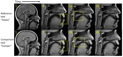 |
98 | Principal Component Characterization of Deformation Variations Using Dynamic Imaging Atlases
Fangxu Xing1, Riwei Jin2, Imani Gilbert3, Georges El Fakhri1, Jamie Perry3, Bradley Sutton2, and Jonghye Woo1
1Radiology, Massachusetts General Hospital, Boston, MA, United States, 2University of Illinois at Urbana-Champaign, Champaign, IL, United States, 3East Carolina University, Greenville, NC, United States
High-speed dynamic magnetic resonance imaging is a highly efficient tool in capturing vocal tract deformation during speech. However, automated quantification of variations in motion patterns during production of different utterances has been a challenging task due to spatial and temporal misalignments between different image datasets. We present a principal component analysis-based deformation characterization technique built on top of established dynamic speech imaging atlases. Two layers of principal components are extracted to represent common motion and utterance-specific motion, respectively. Comparison between two speech tasks with and without nasalization reveals subtle differences on velopharyngeal deformation reflected in the utterance-specific principal components.
|
||
2850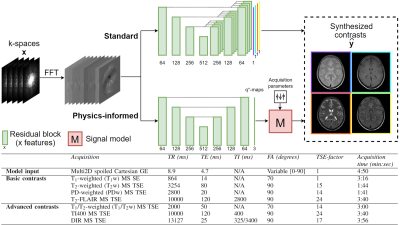 |
99 | Generalizable synthetic multi-contrast MRI generation using physics-informed convolutional networks
Luuk Jacobs1,2, Stefano Mandija1,2, Hongyan Liu1,2, Cornelis AT van den Berg1,2, Alessandro Sbrizzi1,2, and Matteo Maspero1,2
1Department of Radiotherapy, Division of Imaging and Oncology, UMC Utrecht, Utrecht, Netherlands, 2Computational Imaging Group for MR Diagnostics and Therapy, Center for Image Sciences, UMC Utrecht, Utrecht, Netherlands
Synthetic MRI aims to reconstruct multiple MRI contrasts from short measurements of tissue properties. Here, a generalizable physics-informed deep learning-based approach for synthetic MRI was investigated. Acquired data were mapped to effective quantitative parameter maps, here named q*-maps, which are fed to a physical signal model synthesizing four contrasts-weighted images. We demonstrated that from q*-maps, MRI contrasts unseen during training could be synthesized. The proposed method is benchmarked to a standard end-to-end deep learning approach. The two deep learning methods generated similar brain images for healthy subjects and patients with different pathologies.
|
||
 Back to Meeting Home
 Back to the Program-at-a-Glance
The International Society for Magnetic Resonance in Medicine is accredited by the Accreditation Council for Continuing Medical Education to provide continuing medical education for physicians.
 Back to Meeting Home
 Back to the Program-at-a-Glance
 View the Presentation