Digital Poster
Processing & Analysis: Data Processing II
Joint Annual Meeting ISMRM-ESMRMB & ISMRT 31st Annual Meeting • 07-12 May 2022 • London, UK

Digital Poster
Processing & Analysis: Data Processing II
| Computer # | ||||
|---|---|---|---|---|
2899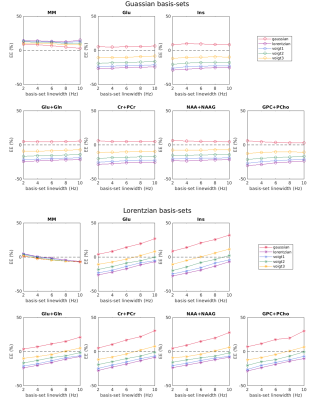 |
105 | Investigating the influences of spectral lineshape and linewidth in LCModel basis-sets on 1H MRS quantification at 7T
Ying Xiao1,2, Ivan Tkáč3, and Lijing Xin1
1Animal imaging and technology (CIBM), EPFL, Lausanne, Switzerland, 2Laboratory for Functional and Metabolic Imaging (LIFMET), EPFL, Lausanne, Switzerland, 3Center for Magnetic Resonance Research, University of Minnesota, Minneapolis, MN, United States In this study, we investigated the 1H MR spectra quantification accuracy using basis sets with different linewidths and lineshapes in LCModel. Simulation results showed that the estimated metabolite concentrations depend on the linewidths and lineshapes of the basis sets as well as the spectral lineshape. This effect was validated with in vivo MR results. We conclude that the spectral lineshape and linewidth of basis sets should be carefully considered for accurate metabolite concentration estimations from short-TE 1H MR spectra using LCModel. |
||
2900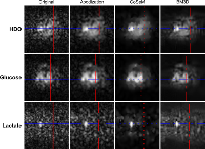 |
106 | Improving SNR in deuterium metabolic imaging: A comparison of methods
Elton Tadeu Montrazi1, Ricardo Martinho1, Qingjia Bao2, Keren Sasson1, Lilach Agemy1, Avigdor Scherz1, and Lucio Frydman1
1Weizmann Institute of Science, Rehovot, Israel, 2Chinese Academy of Sciences, Wuhan, China
Deuterium metabolic imaging (DMI) is a promising approach to study tumor metabolism. Still, DMI’s signal-to-noise ratio (SNR) is limited because of 2H’s low Larmor frequency, coupled to the low concentrations of DMI’s targets. We recently proposed a multi-echo balanced steady-state free precession (ME-bSSFP) method increasing DMI’s SNR, but still find it lacking in some aspects. This work assesses simple apodization, Compressed Sensing Multiplicative (CoSeM) and Block-matching/3D filtering (BM3D) denoising methods for improving DMI’s results. The ability of the latter denoising methods to enhance sensitivity without blurring resolution, is evidenced by pancreatic cancer studies carried at 15.2T.
|
||
2901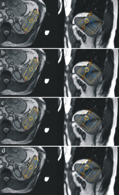 |
107 | Phosphorus MR Spectroscopy of Healthy Human Spleen Video Not Available
Jan Weis1, Maysam Jafar2, and Per Liss3
1Department of Medical Physics, Uppsala University Hospital, Uppsala, Sweden, 2Philips Nordic, Stockholm, Sweden, 3Department of Surgical Sciences, Section of Radiology, Uppsala University Hospital, Uppsala, Sweden
Phosphorous spectra of healthy spleen are useful for studies of splenic malignancies and benign causes of splenomegaly. The main challenge is the relatively small size of the normal spleen and the large distance between human spleen and surface coil. The purpose of this study was to investigate whether it is possible to acquire phosphorous spectra of healthy spleen using single-voxel ISIS sequence on a 3T scanner. We demonstrate that the proposed spectroscopy of human spleen is feasible in a clinically acceptable acquisition time and that transmitter excitation profile and chemical shift displacement errors need to be taken into consideration
|
||
2902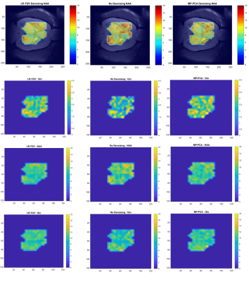 |
108 | MP-PCA and Low-rank noise-reduction in 1H-FID-MRSI data in the rat brain at 14.1T
Brayan Alves1,2, Dunja Simicic1,2,3, Jessie Mosso1,2,3, Ileana Jelescu4, Cristina Cudalbu1,2, and Antoine Klauser1,5
1CIBM Center for Biomedical Imaging, Lausanne, Switzerland, 2Animal Imaging and Technology, EPFL, Lausanne, Switzerland, 3LIFMET, EPFL, Lausanne, Switzerland, 4Service de radiodiagnostic et radiologie interventionnelle, Lausanne University Hospital CHUV, Lausanne, Switzerland, 5Department of Radiology and Medical, Informatics, University of Geneva, Geneva, Switzerland 1H-MRSI is highly challenging and the constant appetite for higher spatial resolution leads to increased search for post-processing methods aiming to reduce the noise variance in 1H-MRSI. The aim of the present study was to implement and test the feasibility of two noise-reduction techniques on preclinical 14.1T fast 1H-FID-MRSI datasets: the MP-PCA based denoising and the low-rank TGV reconstruction. Our results are promising showing an enormous potential of the two noise-reduction techniques towards novel and fast MRSI developments. Further studies will be performed to evaluate if the “apparent” increase in spectral SNR translates in true lower uncertainty in metabolite concentrations. |
||
2903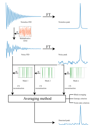 |
109 | A multiplicative denoising method based on compressed sensing for magnetic resonance spectroscopy and imaging
Ricardo P. Martinho1, David Koprivica1, Mihajlo Novakovic1, Michael J. Jaroszewicz1, and Lucio Frydman1
1Department of Chemical and Biological Physics, Weizmann Institute of Science, Rehovot, Israel
2D MRI/S are commonly affected by changes in temperature, field drifts, or motions, leading to multiplicative noise. We introduce here CoSeM (Compressed Sensing Multiplicative) denoising, a method that converts instability-related “t1” noise, into additive noise liable to signal averaging. CoSeM evaluates and discards indirect-domain points that may have been strongly influenced by instabilities, and makes up for this discarding by compressed sensing reconstructions. 2D localized MRS in the brain and 2D abdominal MRI experiments liable to instabilities, evidence 2-3 fold increases in SNR by CoSeM processing. CoSeM is also shown to retain quantitative information –e.g., in T1 mapping experiments.
|
||
2904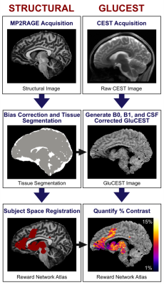 |
110 | Towards a robust and reproducible GluCEST analysis pipeline
Joelle Jee1, Ravi Prakash Reddy Nanga2, Abigail Cember2, Paul Jacobs2, Heather Robinson1, Mark A Elliott2, Ravinder Reddy2, and David R Roalf1
1Psychiatry, University of Pennsylvania, Philadelphia, PA, United States, 2Radiology, University of Pennsylvania, Philadelphia, PA, United States
Robust, reproducible methods are paramount in neuroimaging. This is a challenge for leading-edge and novel techniques at 7T MRI, such as glutamate-weighted chemical exchange saturation transfer (GluCEST). As such, our primary objective is to move towards a robust and reproducible analytical pipeline for GluCEST imaging data. Here we show, using python-based neuroimaging tools, the development of GluCEST-prep. GluCEST-prep incorporates common neuroimaging analysis steps—brain extraction, tissue segmentation, co-registration and normalization—that are optimized for 7T MRI and uses python-based analysis—in place of in-house MATLAB scripts—to generate post-processed 2D or 3D GluCEST images in both native and template space.
|
||
2905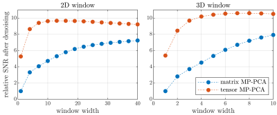 |
111 | Improved MP-PCA denoising through tensor generalization
Jonas Lynge Olesen1,2, Andrada Ianus3, Noam Shemesh3, and Sune Nørhøj Jespersen1,2
1Center of Functionally Integrative Neuroscience (CFIN) and MINDLab, Aarhus University, Aarhus, Denmark, 2Department of Physics and Astronomy, Aarhus University, Aarhus, Denmark, 3Champalimaud Research, Champalimaud Centre for the Unknown, Lisbon, Portugal
A popular SNR-boosting method in MRI is denoising based on principal component analysis with automated rank estimation by exploiting the Marchenko-Pastur distribution of noise singular values (MP-PCA). MP-PCA operates by reshaping data-patches into matrices to discriminate signal from noise using random matrix theory. Here, we generalize MP-PCA to exploit tensor-structured data arising in, e.g., multi-contrast or multicoil acquisitions, without introducing new assumptions. As proof of concept, we demonstrate a substantial increase in denoising performance in a multi-TE DKI dataset, in particular for small sliding windows. This is beneficial especially in cases of rapidly varying contrast or spatially varying noise.
|
||
2906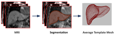 |
112 | Constructing a Mesh-Based Template of the Liver is Consistent as a Function of Cohort Size in the UK Biobank
Marjola Thanaj1, Nicolas Basty1, Madeleine Cule2, Elena P Sorokin2, Jimmy David Bell1, Elizabeth Louise Thomas1, and Brandon Whitcher1
1Research Centre for Optimal Health, School of Life Science, University of Westminster, London, United Kingdom, 2Calico Life Sciences LLC, South San Francisco, CA, United States
Computational atlases provide many advantages in quantifying and modeling differences in the size and shape of internal organs between individuals in population imaging studies. A cohort of UK Biobank participants were analyzed to explore the consistency of templates, which is the average labeled image from a number of subjects constructed across different population sizes, and investigate whether this has an impact on the statistical analysis. We found no differences in our significance areas between templates. Although, for the statistical parametric mapping, a larger sample size that involves multiple hypothesis tests, results in a higher representative power.
|
||
2907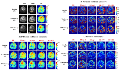 |
113 | Evaluating Reproducibility and Repeatability of Penalty Function Based Methods for Quantitative Intravoxel Incoherent Motion Analysis Video Permission Withheld
Esha Baidya Kayal1, Kedar Khare1, Raju Sharma2, Sameer Bakhshi2, Devasenathipathy Kandasamy2, and Amit Mehndiratta1,2
1Indian Institute of Technology Delhi, Delhi, India, 2All India Institute of Medical Sciences Delhi, Delhi, India Reproducibility and repeatability of penalty-based IVIM analysis methodologies BE+TV and BE+HPF have been evaluated using clinical dataset with osteosarcoma in comparison with existing IVIM analysis methods. IVIM datasets at three time-points during chemotherapy were analyzed and healthy muscle tissue was used as local control for IVIM analysis and evaluation of scan-rescan repeatability and reproducibility. Results showed, parametric maps estimated by BE+TV and BE+HPF methods were observed to have higher reproducibility and lower spatial inhomogeneity than the existing IVIM analysis methods and produced satisfactory inter-scan agreement in parameters estimation proving its repeatability in clinical scenario. |
||
2908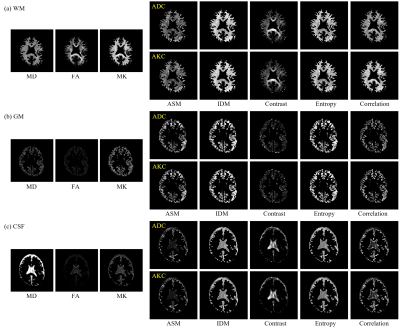 |
114 | Comparison of intra-voxel spatial distributions of diffusion coefficients with intra-voxel spatial distributions of kurtosis coefficients
Suguru Yokosawa1, Toru Shirai1, Yoshitaka Bito2, and Hisaaki Ochi1
1Innovative Technology Laboratory, FUJIFILM Healthcare Corporation, Tokyo, Japan, 2Radiation Diagnostic Systems Division, FUJIFILM Healthcare Corporation, Chiba, Japan
Previously, we have proposed the method for characterizing the intra-voxel spatial distribution of apparent diffusion coefficients (ADC) by using texture analysis and showed that texture features can provide different information from the conventional diffusion tensor imaging. In this study, we extended our method to the apparent kurtosis coefficients (AKC). We compared the features of the intra-voxel spatial distribution of ADC with that of AKC. We found that there is no significant difference in the spatial distribution of AKC from that of ADC, and a short scanning time with a single b-value may be sufficient to obtain information on diffusion distribution.
|
||
2909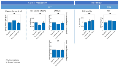 |
115 | Disentangling apparent discordance between ASL-MRI and [18F]-FDG PET following a single dose of the β2-agonist clenbuterol
Courtney A. Bishop1, Gaia Rizzo1, Thomas Lodeweyckx2, Jan de Hoon2, Koen Van Laere3,4, Michel Koole4, Wim Vandenberghe5, Eugenii Rabiner1,6, Renee Martin7, Anthony Ford7, and Gabriel Vargas7
1Invicro, London, United Kingdom, 2Center for Clinical Pharmacology, Department of Pharmaceutical and Pharmacological Sciences, KU Leuven, Leuven, Belgium, 3Division of Nuclear Medicine, University Hospital Leuven, Leuven, Belgium, 4Nuclear Medicine and Molecular Imaging, KU Leuven, Leuven, Belgium, 5Department of Neurology, University Hospital Leuven, Leuven, Belgium, 6Centre for Neuroimaging Sciences, Institute of Psychiatry, Psychology and Neuroscience, King’s College, London, United Kingdom, 7CuraSen Therapeutics, San Carlos, CA, United States
Here we demonstrate that an apparent discrepancy in CBF (from ASL) and CMRglu (from [18F]-FDG PET) following high-dose administration of clenbuterol in healthy volunteers is caused by within-scan rising plasma glucose concentrations. With simulation-based correction, post-clenbuterol CBF changes from ASL agree with blood flow estimates from [18F]-FDG PET, without increase in CMRGlu. ASL-MRI may therefore provide a valuable tool for monitoring the central effects of β2-adrenergic receptor activation in larger, future studies on both healthy volunteers and patients with neurodegenerative disorders.
|
||
 Back to Meeting Home
 Back to the Program-at-a-Glance
The International Society for Magnetic Resonance in Medicine is accredited by the Accreditation Council for Continuing Medical Education to provide continuing medical education for physicians.
 Back to Meeting Home
 Back to the Program-at-a-Glance
 View the Presentation