Digital Poster
Processing & Analysis: Data Analysis I
Joint Annual Meeting ISMRM-ESMRMB & ISMRT 31st Annual Meeting • 07-12 May 2022 • London, UK

Digital Poster
Processing & Analysis: Data Analysis I
| Computer # | ||||
|---|---|---|---|---|
1244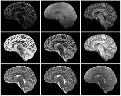 |
40 | Evaluation of quantitative T1 and PD mapping at 7T from an MP2RAGE Sequence optimised to obtain UNI and FLAWS contrast images in a single scan
Ayse Sila Dokumaci1,2, Katy Vecchiato2,3,4, Raphael Tomi-Tricot1,2,5, Pip Bridgen1,2, Michael Eyre1,2, Tobias C. Wood6, Chiara Casella2,4, Jan Sedlacik2,7, Tom Wilkinson1,2, Sharon Giles1,2, Joseph V. Hajnal1,2, Shaihan Malik1,2, Jonathan O’Muircheartaigh2,3,4,8, and David W. Carmichael1,2
1Biomedical Engineering Department, School of Biomedical Engineering and Imaging Sciences, King's College London, London, United Kingdom, 2London Collaborative Ultra high field System (LoCUS), London, United Kingdom, 3Department of Forensic and Neurodevelopmental Sciences, Institute of Psychiatry, Psychology and Neuroscience, King’s College London, London, United Kingdom, 4Centre for the Developing Brain, School of Biomedical Engineering and Imaging Sciences, King’s College London, London, United Kingdom, 5MR Research Collaborations, Siemens Healthcare Limited, Frimley, United Kingdom, 6Department of Neuroimaging, Institute of Psychiatry, Psychology and Neuroscience, King’s College London, London, United Kingdom, 7Radiology Department, Great Ormond Street Hospital for Children, London, United Kingdom, 8MRC Centre for Neurodevelopmental Disorders, King’s College London, London, United Kingdom
The Magnetization Prepared 2 Rapid Acquisition Gradient Echoes (MP2RAGE) sequence is commonly used for 3D structural T1-weighted imaging of the brain at 7T and can be optimised to obtain UNI and clinically relevant FLuid and White Matter Suppression (FLAWS) images within one acquisition. In this study, such a protocol was used together with newly derived analytical equations accounting for partial Fourier acquisitions, and B1+ maps, in a dedicated fitting algorithm to produce quantitative T1 and arbitrarily scaled PD-maps. These maps were evaluated in children and adults at 7T demonstrating a significant T1 reduction with age.
|
||
1245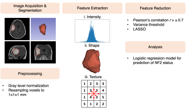 |
41 | Meningiomas with NF2-Loss Exhibit Strong Radiomics Correlations on Contrast Enhanced T1-Weighted MRI at 3T
Esra Sümer1, Kübra Tan2, Ayça Ersen Danyeli3, Ozge Can4, M. Necmettin Pamir5, Alp Dinçer6, Koray Özduman5, and Esin Ozturk-Isik1
1Institute of Biomedical Engineering, Bogazici University, İstabul, Turkey, 2Health Institutes of Turkey, İstanbul, Turkey, 3Department of Medical Pathology, Acibadem Mehmet Ali Aydinlar University, İstanbul, Turkey, 4Department of Medical Engineering, Acibadem Mehmet Ali Aydinlar University, İstanbul, Turkey, 5Department of Neurosurgery, Acibadem Mehmet Ali Aydinlar University, İstanbul, Turkey, 6Department of Radiology, Acibadem Mehmet Ali Aydinlar University, İstanbul, Turkey Meningioma is the most common primary intracranial tumor in adults, and loss of NF2 function in meningiomas has been associated with a more aggressive biology, shorter time to recurrence and shorter overall survival. In this work, radiomics based biomarkers of NF2 copy number loss (NF2-L) have been assessed. Lower grey-level co-occurrence matrix cluster shade with wavelet high-high-low pass filter, lower original image first order minimum, and higher first order skewness with wavelet low-low-high pass filter were observed in tumors with NF2-L compared to tumors with no copy number loss. The classification accuracy of NF2 molecular subsets was 0.80±0.03 (precision=0.85±0.04, recall=0.73±0.05). |
||
1246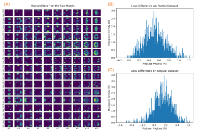 |
42 | Mapping Transient Co-Activity Patterns of Brain with High- and Low- Intensity Frames of fMRI Data
Kaiming Li1 and Xiaoping Hu1
1Department of Bioengineering, UC Riverside, Riverside, CA, United States
Framewise co-activity patterns of functional MRI data may reflect the transient synchronization and coordination across brain regions. High-intensity frames of resting state fMRI have revealed granulated co-activation patterns that resembled the resting state brain networks. However, whether the low-intensity frames carry such information remains unclear. The present study trained variational autoencoder models with both positive values and negative values of normalized fMRI time series respectively and evaluated the two models on a separate dataset. We found the two models were very similar, suggesting that the negative-value frames can reflect transient brain co-activity patterns as the positive ones do.
|
||
1247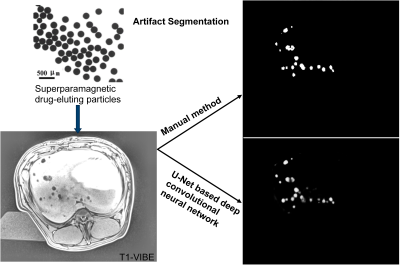 |
43 | U-Net-based deep convolutional neural network for detection of superparamagnetic drug-eluting particles used for liver chemoembolization
NING LI1,2, Cyril Tous1,2, Phillip Fei1,2, Ivan P Dimov1,2, Simon Lessard1,2, Urs O. Häfeli3, Sylvain Martel4, An Tang1,2, and Gilles Soulez1,2
1Centre de recherche du Centre hospitalier de l’Université de Montréal (CRCHUM), Montreal, QC, Canada, 2Université de Montréal, Montreal, QC, Canada, 3University of British Columbia, Vancouver, BC, Canada, 4Polytechnique Montréal, Montreal, QC, Canada Magnetic steering of superparamagnetic drug-eluting particles (SMDEPs) loaded with anti-tumor drugs across hepatic arteries is a promising technique to perform segmental liver embolization for patients with hepatocellular carcinomas (HCCs). These aggregates (20 ± 6 SMDEPs) are sequentially released with a specially designed injector through a catheter in the proper hepatic artery of a swine placed in an MRI. Manual segmentation was previously done to localize and count the particles (volume of the artifact) in each lobe. We propose to train a U-net over this pre-existing database. Dice similarity coefficient, accuracy, and precision were respectively 99.1%, 98.3%, and 84.6%. |
||
1248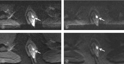 |
44 | Assessment of anal fistula using advanced diffusion-weighted imaging techniques: value of ZOOMit and RESOLVE Video Not Available
Yue Qin1, Dayong Jin1, Xin Li1, Liyao Liu1, Yinhu Zhu1, Juan Tian1, Yifan Qian1, Shaoyu Wang2, and Boyuan Jiang1
1Xi'an Daxing Hospital, Xi'an, China, 2Siemens Healthineers, Ltd., Xi'an, China
This study investigated ZOOMit and RESOLVE DWI sequences to compare of the image quality of anal canal and the visibility of the fistula. The results showed that both ZOOMit DWI and RESOLVE DWI obtained high quality images, ZOOMit DWI sequence had higher fistula visibility and SNR of the fistula than those of RESOLVE DWI sequence. Our findings also suggest that ZOOMit DWI can be used as the preferred sequence for anal MR diffusion imaging.
|
||
1249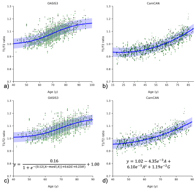 |
45 | Investigation of myelin using T1/T2 ratio imaging across multiple datasets: comparable or not?
Riona Fumi1, Hanna Maksimuk2,3, James H Cole4,5, and Sjoerd B Vos2,4,6
1Queen Square Institute of Neurology, University College London, London, United Kingdom, 2Neuroradiological Academic Unit, Queen Square Institute of Neurology, University College London, London, United Kingdom, 3Department of Radiology, Brest Regional Clinical Hospital, Brest, Belarus, 4Centre for Medical Image Computing, Department of Computer Science, University College London, London, United Kingdom, 5Dementia Research Centre, Queen Square Institute of Neurology, University College London, London, United Kingdom, 6Centre for Microscopy, Characterisation, and Analysis, The University of Western Australia, Perth, Australia
Cortical T1/T2 ratios (T1/T2r) have been used to assess myelination along the lifespan and investigate demyelination in neurological diseases. Creating a comprehensive normative population from open-access datasets could facilitate widespread adoption of this technique. Here we investigate comparability between two such publicly available datasets, OASIS3 and CamCAN. We find that they vary in terms of offset, lifespan trajectory, and presence (OASIS3) or absence (CamCAN) of hemispheric asymmetry. This variability exists despite both studies use the same scanner model, and is likely increased across different scanner models and vendors. These findings indicate that combining data from different studies requires advanced normalisation.
|
||
1250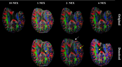 |
46 | Noise reduction in fractional anisotropy maps using deep learning based denoising Video Not Available
Seema S Bhat1, Pavan Poojar2,3, Chennagiri Rajarao Padma2, and Hanumantharaju MC4
1Department of Information Science and Engineering, Dayananda Sagar College of Engineering, Bengaluru, India, 2Department of Medical Electronics, Dayananda Sagar College of Engineering, Bengaluru, India, 3Columbia University in the City of New York, Newyork, NY, NY, United States, 4Department of Electronics and Communications, BMS Institute of Technology and Management, Bangalore, India
Denoising is an alternative for enhancing signal-to-noise ratio in high b-value diffusion imaging instead of prolonged acquisition time. We experimented a deep learning based denoising method on prospective high b-value DWI and visualized the impact of denoising using fractional anisotropy(FA) maps. Experiment was repeated for three different signal averages:1,2 4-NEX and two different slice thickness 1mm and 5mm with gold standard reference of 10-NEX images. The current work obtained average peak signal-to-noise ratio >34dB and SSIM >0.94 after denoising for FA maps. The PSNR and SSIM values in FA maps were modestly increased after denoising.
|
||
1251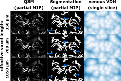 |
47 | Resolution-dependency of arterial and venous density estimates and vessel distance maps in deep gray matter
Hendrik Mattern1 and Oliver Speck1,2,3,4
1Biomedical Magnetic Resonance, Otto von Guericke University, Magdeburg, Germany, 2German Center for Neurodegenerative Disease, Magdeburg, Germany, 3Center for Behavioral Brain Sciences, Magdeburg, Germany, 4Leibniz Institute for Neurobiology, Magdeburg, Germany
The detection of vessel in MRI depends on the imaging resolution used. Hence, subsequent quantification with vessel densities and vessel distance mapping (VDM) is resolution dependent. In this study, the resolution dependency of both metrics was evaluated for arteries and veins in deep gray matter regions by retrospective downsampling of high-resolution vessel images. Quantitative comparison shows that with decreasing resolution the estimated vessel distances increase, while vessel densities decline. The resolution dependency of vessel densities and distances can be modeled linearly. The here found scaling factors may improve inter-study comparability of vessel densities and VDM.
|
||
1252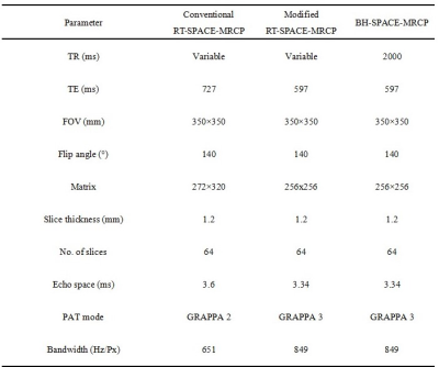 |
48 | MRCP at 1.5 T: comparing image quality and acquisition time between respiratory-triggered and breath-hold SPACE sequences Video Not Available
Xin Li1, Yue Qin1, Dayong Jin1, Yinhu Zhu1, Liyao Liu1, Yifan Qian1, Juan Tian1, and Shaoyu Wang2
1Xi'an Daxing Hospital, Xi'an, China, 2Siemens Healthineers, Ltd., Xi'an, China
This study used magnetic resonance cholangiopancreatography (MRCP) to compare the image quality and acquisition time of conventional respiratory-triggered (RT), modified RT, and breath-hold (BH) sampling perfection with application-optimized contrasts using different flip evolutions (SPACE) sequences in patients with biliary and pancreatic disorders. Our results suggested that modified RT-SPACE-MRCP sequence can significantly reduced acquisition time and had better image quality. The MRCP sequence can be selected flexibly according to the patient's situation to obtain good diagnostic images.
|
||
1253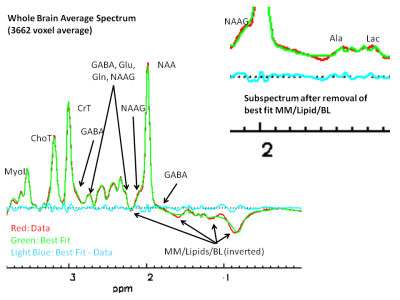 |
49 | Quantifying metabolites in large brain areas by 3D Echo Planar Spectroscopic Imaging spectral averaging for a study of cigarette smoking
Jeffry R Alger1,2,3,4, Maylen Perez Diaz5, Joseph O'Neill5, Dara Ghahremani5, Andy C Dean5, Sulaiman Sheriff6, Andrew A Maudsley6, and Edythe London5
1NeuroSpectroScopics LLC, Sherman Oaks, CA, United States, 2University of California, Los Angeles, Los Angeles, CA, United States, 3Advanced Imaging Research Center, University of Texas Southwestern Medical Center, Dallas, TX, United States, 4Hura Imaging Inc, Calabasas, CA, United States, 5Department of Psychiatry and Biobehavioral Sciences, University of California, Los Angeles, Los Angeles, CA, United States, 6Radiology, University of Miami, Miami, FL, United States
Spectral averaging of thousands of magnetic resonance spectroscopic imaging voxel spectra over large brain volumes provides a means of assessing low-level and/or hard-to-quantify metabolite differences over large brain volumes between groups of individuals.
|
||
1254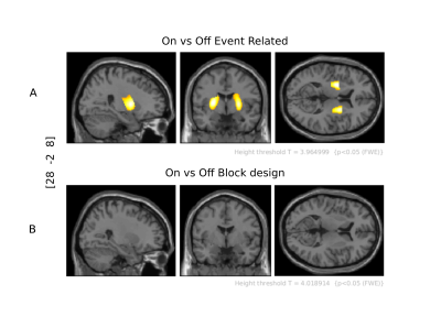 |
50 | Different Brain Areas Require Different Analysis Models—fMRI Observations in Parkinson’s Disease.
Renzo Torrecuso1, Karsten Mueller1, Stefan Holiga1,2, Dusan Urgosik3, Josef Vymazal4, Tomas Sieger5, Filip Růžička6, Evzen Růžička7, Matthias Schroeter8, Robert Jech3, and Harald E. Möller1
1NMR, Max Planck Institute for Human Cognitive and Brain Sciences, Leipzig, Germany, 2Roche Pharma Research and Early Development, Roche Innovation Center Basel, Basel, Switzerland, 3Department of Neurology, Charles University in Prague | CUNI, Prague, Czech Republic, 4| CULS · Faculty of Environmental Science, Czech University of Life Sciences Prague, Prague, Czech Republic, 5Department of Neurology and Center of Clinical Neuroscience, Charles University in Prague, Prague, Czech Republic, 6Department of Environmental Engineering, Faculty of Technology Tomas Bata University in Zlín, Zlin, Czech Republic, 7Neurology, General University Hospital in Prague, Prague, Czech Republic, 8Clinic for Cognitive Neurology, University Hospital Leipzig, Leipzig, Germany
Foreseeing how specific brain areas respond in time to a stimulus can be a prerequisite for a successfully conceived fMRI experiment. We demonstrate that in medicated Parkinson’s disease patients, putamen's activation peaks around the onset of tapping but does not persist throughout the tapping block, whereas sustained activation is observed in the motor cortex. Consequently, in the widely used tapping paradigm “On vs. Off L-DOPA”, the drug effect remains undetected if statistical analysis apply a block design instead of an event-related one. Ignoring this information can lead to fallacious conclusions which suggests using different models to investigate different brain regions.
|
||
1255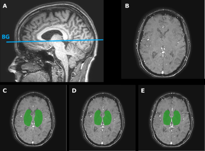 |
51 | Test-retest reliability of velocity pulsatility in perforating arteries in the basal ganglia at 3T MRI
Rick J. van Tuijl1, Stanley D.T. Pham1, Birgitta K Velthuis1, Ynte M. Ruigrok2, Geert Jan Biessels2, and Jaco J.M. Zwanenburg1
1Radiology, UMC Utrecht, Utrecht, Netherlands, 2Neurology, UMC Utrecht, Utrecht, Netherlands
We studied the test-retest reliability of assessing perforating arteries of the BG with 2D-PC velocity measurements at 3T-MRI, with and without repositioning of the patient. We showed reproducible assessment of Ndetected, Vmean and vPI in the perforating arteries, independent of slice planning. The relative imprecision was smallest for Vmean (12%) and highest for vPI (32%). The interrater-reliability assessment showed high consistency between two raters (mean variability <5% for all parameters). We found very similar Limits of Agreement with and without repositioning, which indicated that uncertainty in measuring these parameters was dominated by scanner and physiological noise, rather than by planning.
|
||
1256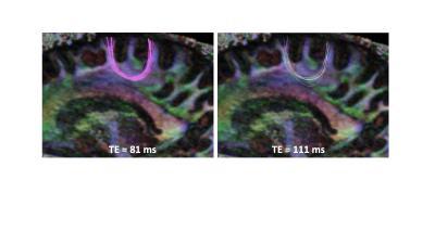 |
52 | T2-dependence of deep and superficial white matter tractography
Yogesh Rathi1, Lipeng Ning1, Congyu Liao2, Yang Ji3, Carl-Fredrik Westin1, Fan Zhang1, Nikolaos Makris1, Lauren J O'Donnell1, and Kawin Setsompop2
1Harvard Medical School, Boston, MA, United States, 2Stanford University, Stanford, CA, United States, 3Oxford University, Oxford, United Kingdom
Tractography is typically obtained from diffusion MRI data acquired at a particular echo time (TE). Recent works have shown differential T2-relaxation times of tissue microstructure and fiber tracts. However, estimation of T2 in fiber bundles have typically been done at the last stage (after tractography). In this work, we show that tractography itself is affected by the T2 (and hence TE) of the fibers. Specifically, we show that the ability to trace accurately depends on the T2 of the fiber bundle. This is specifically true for the superficial white matter (u-fibers) which has higher myelination and iron content.
|
||
1257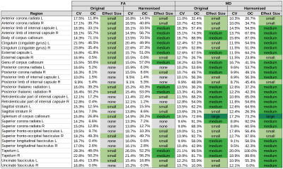 |
53 | Impact of Diffusion Signal Harmonisation on Voxel-Wise Analysis of Mild Traumatic Brain Injury
Stefan Winzeck1,2, Sophie Richter2, Maira Siqueira Pinto3, Evgenios N. Kornaropoulos4, Virginia F.J. Newcombe2, Ben Glocker1, David K. Menon2, and Marta M. Correia5
1BioMedIA, Department of Computing, Imperial College London, London, United Kingdom, 2University Division of Anaesthesia, Department of Medicine, University of Cambridge, Cambridge, United Kingdom, 3Universitair Ziekenhuis Antwerpen, Antwerp, Belgium, 4Clinical Sciences, Lund, Diagnostic Radiology, Lund University, Lund, Sweden, 5MRC Cognition and Brain Sciences Unit, University of Cambridge, Cambridge, United Kingdom
The comparison of voxel-wise regression analysis of a multi-centre diffusion MRI study showed that similar results could be achieved regardless of whether prior data harmonisation was applied or not. However, harmonisation had a positive impact on increasing the effect size found between mild traumatic brain injury patients and controls.
|
||
1258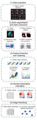 |
54 | Combining Shape Descriptor and Image Ranking to the Identification of Alzheimer’s Disease in Anatomical Regions using T1-weighted MR Images
Kaue Tartarotti Nepomuceno Duarte1, Richard Frayne1, David Gobbi2, Paulo Sergio Martins3, and Marco Antonio Garcia de Carvalho3
1Radiology, University of Calgary, Calgary, AB, Canada, 2CIPAC, University of Calgary, CALGARY, AB, Canada, 3University of Campinas, Limeira, Brazil
Alzheimer's Disease is one of the most prevalent types of dementia that affects human behaviour and cognitive skills. The progression of this dementia occurs at different rates in distinct parts of the brain, thus also affecting their brain shape. We proposed an approach based on shape feature extraction and image ranking to identify which regions are more predictive for each level of the AD progression. For each stage, we highlighted the sub-cortical regions that suggested a strong correlation in the AD levels. In fact, the results showed a predictive pattern, which aligns to the state-of-the-art.
|
||
 Back to Meeting Home
 Back to the Program-at-a-Glance
The International Society for Magnetic Resonance in Medicine is accredited by the Accreditation Council for Continuing Medical Education to provide continuing medical education for physicians.
 Back to Meeting Home
 Back to the Program-at-a-Glance
 View the Presentation