Digital Poster
Processing & Analysis: Data Analysis II
Joint Annual Meeting ISMRM-ESMRMB & ISMRT 31st Annual Meeting • 07-12 May 2022 • London, UK

Digital Poster
Processing & Analysis: Data Analysis II
| Computer # | ||||
|---|---|---|---|---|
1329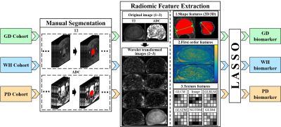 |
35 | Radiomic biomarker extracted from PI-RADS 3 patients support more efficient and robust prostate cancer diagnosis: a multi-center study Video Not Available
Longfei Li1,2, Rui Yang3, Xin Chen4, Cheng Li2, Hairong Zheng2, Yusong Lin1, Zaiyi Liu4, and Shanshan Wang2
1the Collaborative Innovation Center for Internet Healthcare , School of Information Engineering, Zhengzhou University, Zhengzhou, China, 2Paul C. Lauterbur Research Center for Biomedical Imaging, Shenzhen Institute of Advanced Technology, Chinese Academy of Sciences, Shenzhen, China, 3Department of Urology, Renmin Hospital of Wuhan University, Wuhan, China, 4Department of Radiology, Guangdong Provincial People’s Hospital, Guangzhou, China
Prostate Imaging Reporting and Data System (PI-RADS) based on multi-parametric MRI classifies patients into 5 categories (PI-RADS 1-5) for routine clinical diagnosis guidance. However, there is no consensus on whether PI-RADS 3 patients should go through biopsies. Mining features from these hard samples (HS) is meaningful for physicians to achieve accurate diagnoses. Currently, the mining of HS biomarkers is insufficient, and the effectiveness and robustness of HS biomarkers for prostate cancer diagnosis have not been explored. In this study, biomarkers from different data distributions are constructed. Results show that HS biomarkers can achieve better performances in different data distributions.
|
||
1330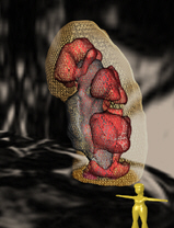 |
36 | Workflow development for kidney segmentation using a U-net model using 11000 MRI data sets from the German National Cohort (NAKO/GNC) study
Martin Buechert1, Jan Lipovsek2, Marco Reisert2, Harald Horbach2, Wilfried Reichardt2, Christopher Schlett2, Fabian Bamberg2, Peggy Sekula 2, Anna Köttgen2, and Elias Kellner2
1Magnetic Resonance Development and Applicatione Center, Medical Center, University of Freiburg, Faculty of Medicine, Freiburg, Germany, 2Medical Center, University of Freiburg, Faculty of Medicine, Freiburg, Germany
A processing pipeline for kidney segmentation using a hierarchical patch-based stack of U-nets was implemented and applied to abdominal MRI images of the German National Cohort study. The training data set included 300 cases and the final net was applied to the dataset of 11,207 MRIs. The compartments cortex, medulla and hilus could be segmented very robustly with the network. The relation of first parameters based on the segmentation withsex, age, weight, subject size and BMI are presented. This is an optimal starting point to identify more advanced biomarkers and their correlations, especially with kidney functional parameters.
|
||
1331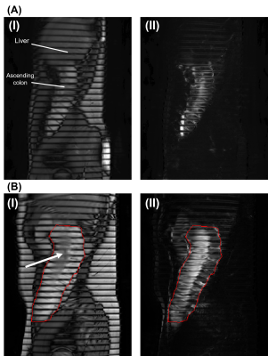 |
37 | Assessing Inter-Observer Variability of MRI Tagging of the Colon Contents in Healthy Human Subjects
Meshari T Alshammari1,2, Ali S Alyami3, Victoria Wilkinson-Smith3, Luca Marciani1, Gordon W. Moran1, and Caroline L. Hoad3,4
1Translational Medical Sciences and National Institute for Health Research (NIHR) Nottingham Biomedical Research Centre, Nottingham University Hospitals NHS Trust and University of Nottingham, Nottingham, United Kingdom, 2Department of Diagnostic Radiology, College of Applied Medical Sciences, University of Hail, Hail, Saudi Arabia, 3NIHR Nottingham Biomedical Research Centre (BRC), Nottingham University Hospitals NHS Trust and University of Nottingham, Nottingham, United Kingdom, 4Sir Peter Mansfield Imaging Centre, School of Physics and Astronomy, University of Nottingham, Nottingham, United Kingdom MRI Tagging techniques have been applied to the GI tract to assess bowel contractions and colon content mixing. Two independent datasets of healthy adults were used to evaluate the dependence of the percentage Coefficient of variation (%CoV) measurement on inter-observer variability in the ascending colon and descending colon. There was little difference between both AC and DC measurements of the %CoV with high inter-rater agreement (intra-class correlation coefficient > 0.95). This technique could be used to provide objective measures for the motility assessment of the colon in inflammatory and functional bowel diseases.
|
||
1332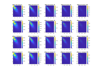 |
38 | Optimization of GLCM Texture Analysis settings in Liver Radial T2 Maps: Fibrotic vs Healthy Liver
Diana Bencikova1,2, Veronika Janacova1, Marcus Raudner1, Ahmed Ba-Ssalamah1, Siegfried Trattnig1,2, and Martin Krssak3
1Department of Biomedical Imaging and Image-Guided Therapy, Medical University Vienna, Vienna, Austria, 2Christian Doppler Laboratory for Clinical Molecular Imaging, MOLIMA, Vienna, Austria, 3Division of Endocrinology and Metabolism, Department of Medicine III, Medical University Vienna, Vienna, Austria
GLCM texture analysis is a promising technique for characterizing and classifying tissue pathologies. In liver applications it has been mostly performed on T2-weighted images. Fast radial T2 mapping techniques enable acquiring T2 maps in clinically feasible measurement time (during breath-hold). Here, we explored the performance of the different settings of GLCM texture analysis in liver T2 maps, which might be more advantageous compared to T2-weighted images. We identified the grey-level quantization of 8bits and direction of 90° as the best setting for discrimination between fibrotic and healthy livers.
|
||
1333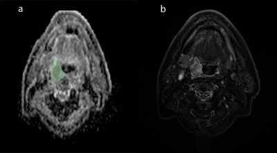 |
39 | Analyzing Radiomic Features in Magnetic Resonance Images of Head and Neck Cancer during Radiation Therapy: Preliminary Results
Victor Fritz1, Martin Schwartz1,2, Jens Kübler3, Simon Böke4, Jonas Habrich5, Daniel Zips4, Daniela Thorwarth5, Konstantin Nikolaou3, and Fritz Schick1
1Department of Diagnostic and Interventional Radiology, University of Tuebingen, Section on Experimental Radiology, Tuebingen, Germany, 2University of Stuttgart, Institute of Signal Processing and System Theory, Stuttgart, Germany, 3Department of Diagnostic and Interventional Radiology, University of Tuebingen, University Hospital Tuebingen, Tuebingen, Germany, 4Department of Radiation Oncology, University of Tuebingen, University Hospital and Medical Faculty, Tuebingen, Germany, 5Department of Radiation Oncology, University of Tuebingen, Section for Biomedical Physics, Tuebingen, Germany
The aim of this work was to identify potential texture features as imaging biomarkers for monitoring treatment response in head and neck cancer (HNC). For this purpose, a total number of 93 texture features were extracted on segmented calculated ADC maps and compared at baseline and in the early treatment phase of radiation therapy (RT). Fifteen texture features showed a statistically difference in the course of RT. In particular, features suggesting that ADC-based HNC texture became finer but more heterogeneous changed significantly. Presented preliminary results offer initial findings that will be systematically investigated in upcoming studies.
|
||
1334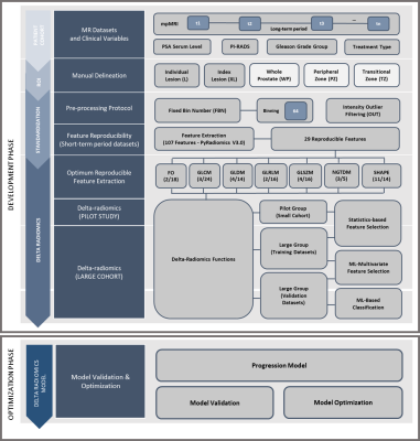 |
40 | Developing a Delta Radiomics Framework for Prostate Cancer Progression Biomarkers in Patients under Active Surveillance: Pilot Study
Dyah Ekashanti Octorina Dewi1, Mohammed R. S. Sunoqrot1, Gabriel Addio Nketiah1, Elise Sandsmark2, Sverre Langørgen2, Helena Bertilsson3, Mattijs Elschot1,2, and Tone Frost Bathen1,2
1Department of Circulation and Medical Imaging, Norwegian University of Science and Technology, Trondheim, Norway, 2Department of Radiology and Nuclear Medicine, St. Olavs Hospital, Trondheim University Hospital, Trondheim, Norway, 3Department of Urology, St. Olavs Hospital, Trondheim University Hospital, Trondheim, Norway
Identification of progressive lesions over time is important to monitor the prostate cancer progression in active surveillance. Delta Radiomics, computed as the relative changes of features among scans, provides temporal evolution of features that may indicate clinical changes along time periods. This pilot study aims to develop a framework to identify progressive lesions on two long-term scans in prostate T2WI based on Delta-radiomics. The current results show that four Delta-radiomics features from GLDM and Shape feature groups have potentials to identify progressive lesions compared to other selected reproducible features.
|
||
| 1335 | 41 | Quantitative analysis of dynamic-contrast enhanced MRI by dispersion analysis for improved prostate cancer diagnosis
Simona Turco1, Catarina Dinis Fernandes2, Razvan Miclea3, Ivo Schoots4, Peet Nooijen5, Hans van der Linden5, Jelle Barentsz6, Stijn Heijmink 7, Hessel Wijkstra2, and Massimo Mischi2
1Electrical Engineering, Eindhoven Univeristy of Technology, Eindhoven, Netherlands, 2Electrical Engineering, Eindhoven University of Technology, Eindhoven, Netherlands, 3Abdominal radiology, Maastricht University Medical Center, Maastricht, Netherlands, 4Abdominal radiology, Erasmus Medical Center, Rotterdam, Netherlands, 5Uropathology, Jeroen Bosch Ziekenhuis, 's-Hertogenbosch, Netherlands, 6Radiology, Radboud university medical center, Nijmegen, Netherlands, 7Radiology, Netherlands Cancer Institute, Amsterdam, Netherlands
Multiparametric MRI, including T2-weighted, diffusion-weighted, and dynamic contrast enhanced (DCE) imaging, is the recommended imaging modality for prostate cancer (PCa). Although the role of DCE-MRI has been greatly limited in recent years, only qualitative analysis of DCE is currently performed. Here we propose magnetic resonance dispersion imaging (MRDI) to obtain quantitative parametric maps from DCE-MRI. Comparing the performance of two radiologists, our results show that some clinically-significant PCa are only found by interpretation of either mpMRI alone or MRDI maps alone, suggesting that combined interpretation of MRDI and mpMRI may improve PCa diagnosis.
|
||
 Back to Meeting Home
 Back to the Program-at-a-Glance
The International Society for Magnetic Resonance in Medicine is accredited by the Accreditation Council for Continuing Medical Education to provide continuing medical education for physicians.
 Back to Meeting Home
 Back to the Program-at-a-Glance
 View the Presentation