Digital Poster
Managing Motion in the Brain & Body II
Joint Annual Meeting ISMRM-ESMRMB & ISMRT 31st Annual Meeting • 07-12 May 2022 • London, UK

Digital Poster
Managing Motion in the Brain & Body II
| Computer # | ||||
|---|---|---|---|---|
2053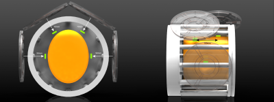 |
43 | On the concept of long-range short-wave motion tracking
Christoph Michael Schildknecht1 and Klaas Paul Prüssmann1
1Institute for Biomedical Engineering, ETH Zurich and University of Zurich, Zürich, Switzerland
Current motion tracking modalities operate either in frequency bands that are also occupied by the MRI scanner or interact heavily with the environment. As an alternative, the frequency band of 1-5 MHz can be utilized for motion tracking and has been demonstrated to deliver high performance. Due to the limited sensitivity at a great distance of current broadband systems, we show in this work how short-wave motion tracking can be done at long ranges, enabling independent and robust motion tracking with fewer geometric constraints and greater ease of use.
|
||
2054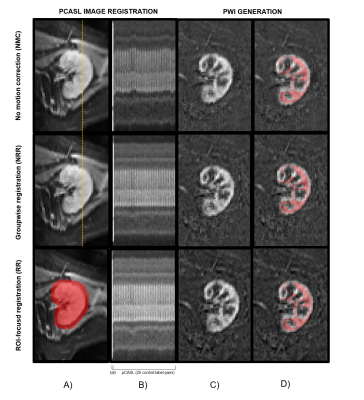 |
44 | ROI-focused Non-Rigid Groupwise Registration approach for Motion Correction in ASL Renal Blood Flow Imaging
Anne Oyarzun1, Rebeca Echeverria-Chasco2,3, Paloma L. Martin Moreno4, Nuria Garcia-Fernandez4, Gorka Bastarrika2,3, María A. Fernández-Seara1,3, and Arantxa Villanueva1,3,5
1Electrical, Electronics and Communications Engineering Department, Universidad Pública de Navarra, Pamplona, Spain, 2Radiology, Clínica Universidad de Navarra, Pamplona, Spain, 3IdiSNA, Instituto de Investigación Sanitaria de Navarra, Pamplona, Spain, 4Nephrology, Clínica Universidad de Navarra, Pamplona, Spain, 5ISC, Instituto de Smart Cities, Pamplona, Spain
Motion correction methods are a prerequisite in multiple-image registration tasks. We implemented a non-rigid groupwise registration method and ROI-focused non-rigid groupwise registration method for renal pCASL images. We evaluated the temporal signal variation in the renal cortex after motion correction using both methods. Motion correction technique shows statistically significant improvement on the tSNR and ROI-focused method performs statistically better.
|
||
2055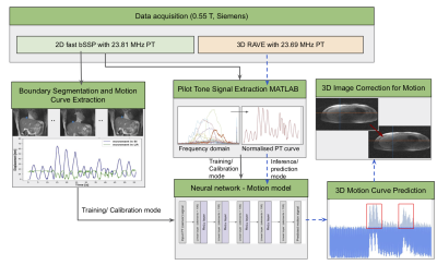 |
45 | A deep learning image based calibration model to predict motion using auxiliary sensors
Radhika Tibrewala1, Mahesh B Keerthivasan2, Kai Tobias Block1, Jan Paska1, and Ryan Brown1
1CAI2R, NYU School of Medicine, New York, NY, United States, 2Siemens Medical Solutions USA, New York, NY, United States
For MRI hindered by motion artifacts sensors are able to provide a surrogate for bulk motion, but may not be tissue specific. In this work, we use deep learning to build a motion model with an auxiliary pilot tone sensor during a fast-image calibration step. A neural network is used to learn the correlation between the pilot tone signal and the pixel-wise liver displacement which is predicted using an automatic segmentation model. The motion model is used to predict displacement on low frame rate images, thus offering the opportunity to perform motion resolved reconstruction.
|
||
2056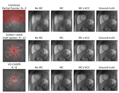 |
46 | Virtual coil concept applied to motion corrected and low rank undersampled reconstructions
Gastao Cruz1, Camila Munoz1, René M. Botnar1, and Claudia Prieto1
1King's College London, London, United Kingdom
Partial Fourier strategies leverage the Hermitian symmetric properties of k-space into higher acceleration factors. Motion corrected (MC) reconstructions incorporate generalized motion into the process. Contrast resolved reconstructions, e.g. low rank inversion (LRI), exploit temporal redundancies in highly accelerated acquisitions. Both the MC and LRI reconstructions are usually ill-posed and can benefit from additional sources of encoding. The Virtual Coil Concept (VCC) has been recently introduced, combining Parallel Imaging with Partial Fourier. Here we investigate the combination of VCC with MC and VCC with LRI to enable further acceleration of both approaches.
|
||
2057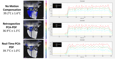 |
47 | Magnetic Resonance Thermometry Motion Compensation during Focused Ultrasound Controlled Hyperthermia in a Small Animal Model
Suzanne Wong1,2, Claire Wunker3,4, Ben Keunen2, Maryam Siddiqui5, Karolina Piorkowska2, Yael Babichev4, Warren Foltz6, Rebecca Gladdy3,4, Samuel Pichardo5, Adam Waspe2, and James Drake1,2
1Biomedical Engineering, University of Toronto, Toronto, ON, Canada, 2Neuroscience and Mental Health, The Hospital for Sick Children, Toronto, ON, Canada, 3Institute of Medical Sciences, University of Toronto, Toronto, ON, Canada, 4Lunenfeld-Tanenbaum Research Institute, Mount Sinai Hospital, Toronto, ON, Canada, 5Hotchkiss Brain Institute, University of Calgary, Calgary, AB, Canada, 6Radiation Physics, University Health Network, Toronto, ON, Canada
Magnetic resonance guided high intensity focused ultrasound (MRgHIFU) has gained interest over the past decade due to its ability to administer controlled hyperthermia for localized drug release. One of the main challenges is that MR thermometry is highly susceptible to motion artifacts. A hybrid principal component analysis and projection onto dipole fields motion artifact removal method was applied in real-time during controlled hyperthermia in a murine model using a small-animal MRgHIFU system (Bruker 7T MRI and IGT HIFU). For a target temperature of 40.5°C to be maintained, a significant increase in ultrasound power was required when tissue motion was observed.
|
||
2058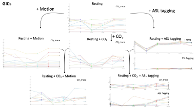 |
48 | Novel methods for correcting motion regression errors caused by global intensity changes in scans of cerebrovascular function
Ryan Beckerleg1, Joseph Whittaker1,2, Daniel Gallichan3, and Kevin Murphy1
1Cardiff University Brain Research Imaging Centre (CUBRIC), School of Physics and Astronomy, Cardiff University, Cardiff, United Kingdom, 2Max Planck Institute for Human Cognitive and Brain Sciences, Leipzig, Germany, 3Cardiff University Brain Research Imaging Centre (CUBRIC), School of Engineering, Cardiff University, Cardiff, United Kingdom
Previously we showed that when global intensity changes (GIC’s) are present in data (e.g., CO2 stimuli during measurement of CVR, ASL tagging), volume registration algorithms misrepresent such signal change as motion. Here we look at motion estimates derived from VRA’s, an external marker-less camera, and novel data-derived techniques, to evaluate if this problem can be overcome. We determined that in most cases where GIC’s are present, the use of an ICA in correction can improve erroneous results from VRA estimates. This isn’t the case in all scans however and the GIC causing this error should be removed prior to correction.
|
||
2059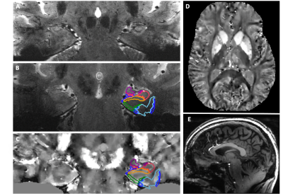 |
49 | Prospective Motion Correction for a Full Neuroimaging Protocol in Elderly Subjects at 7T
Mackenzie Carlson1, Phillip DiGiacomo1, Brian Burns2, Pascal Spincemaille3, Yi Wang3, Julian Maclaren1,4, Murat Aksoy4, Brian Rutt1, and Michael Zeineh1
1Stanford University, Stanford, CA, United States, 2GE Healthcare, Stanford, CA, United States, 3Weill Cornell Medicine, New York City, NY, United States, 4Hobbitview Inc., San Jose, CA, United States
Correction of head motion during MRI scanning is critical to achieving high-resolution images to study and quantify small regions such as the hippocampus in Alzheimer’s disease. Our optical prospective motion correction prototype system has previously demonstrated improved image quality in volunteers at 7T, but clinical translatability has been lacking. In this work, we implemented an integrated power supply and applied prospective correction to a broad set of sequences, including mGRE and FSE, for a clinically relevant research protocol with emphasis on hippocampal iron imaging. Metrics achievable using our protocol include high-resolution cortical QSM, hippocampal subfield QSM and volume.
|
||
2060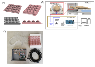 |
50 | MoCoPad: A new soft sensor system for fast head motion detection and tracking in MRI
Saikat Sengupta1,2, Mishek Musa3, and Yue Chen4
1Vanderbilt University Institute of Imaging Science, Nashville, TN, United States, 2Department of Radiology, Vanderbilt University Medical Center, Nashville, TN, United States, 3Department of Mechanical Engineering, University of Arkansas, Fayetteville, AR, United States, 4Department of Biomedical Engineering, Georgia Institute of Technology, Atlanta, GA, United States
In this abstract we present initial feasibility results of a novel pneumatic sensor-based head motion detection and tracking system for MRI. The system comprises of head pad with a built in matrix of air pressure sensors, which replaces the standard head pad in an MRI scan and allows fast sequence agnostic tracking of head motions without the need for navigators, head markers or camera systems. Here, we show initial results for motion detection and head pose estimation in phantom studies using a motion model trained outside the scanner.
|
||
2061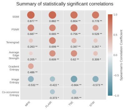 |
51 | Evaluating the match of image quality metrics with radiological assessment in a dataset with and without motion artifacts
Hannah Eichhorn1, Simon Chemnitz-Thomsen1,2, Evangelos Vouros1,3, Nitesh Shekhrajka4, Robert Frost5,6, André van der Kouwe5,7, and Melanie Ganz1,8
1Neurobiology Research Unit, Copenhagen University Hospital, Rigshospitalet, Copenhagen, Denmark, 2Department of Mathematical Sciences, University of Copenhagen, Copenhagen, Denmark, 3Niels Bohr Institute, University of Copenhagen, Copenhagen, Denmark, 4Department of Radiology, Copenhagen University Hospital, Rigshospitalet, Copenhagen, Denmark, 5Athinoula A. Martinos Center for Biomedical Imaging, Massachusetts General Hospital, Charlestown, MA, United States, 6Department of Radiology, Harvard Medial School, Boston, MA, United States, 7Department of Radiology, Harvard Medical School, Boston, MA, United States, 8Department of Computer Science, University of Copenhagen, Copenhagen, Denmark Currently no image quality metric, used for evaluating the performance of artifact correction or image reconstruction methods, is sensitive to all possible image artifacts. This complicates the choice of a proper quantitative quality measure. To provide assistance with this choice, we investigated the correlation of commonly used metrics with radiological evaluation on a dataset acquired with and without motion. The full-reference metrics SSIM and PSNR correlated most strongly with observer scores. Among the reference-free metrics Image Entropy, Average Edge Strength and Tenengrad measure showed a consistent correlation for all investigated sequences. |
||
2062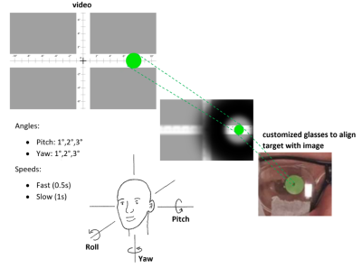 |
52 | Choreography Controlled (ChoCo) by synchronized video for motion artifact reproducibility and dataset generation
Oscar Albert Dabrowski1, Sébastien Courvoisier2, Jean-Luc Falcone3, Antoine Klauser2, Julien Songeon3, Michel Kocher2, Bastien Chopard3, and Francois Lazeyras2
1Computer science, University of Geneva, Geneva, Switzerland, 2CIBM, Geneva, Switzerland, 3University of Geneva, Geneva, Switzerland
In the age of artificial intelligence, there is a need for simple and inexpensive frameworks aimed at performing standardized reproducible motion-controlled experiments allowing the creation of datasets of motion corrupted in vivo MRI scans. Focused on human brain imaging, we propose ChoCo: a choreography controlled protocol for reproducible head motion and an ad-hoc hardware device for synchronization between an MRI scanner and a movie displaying motion to be performed. A proof of concept demonstrated that 6 participants were able to reproduce head choreographies accurately. The resulting motion corrupted brain images show qualitatively similar artifacts, confirming consistent motion reproduction among subjects.
|
||
2063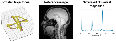 |
53 | Rotation estimation for cloverleaf navigators using a k-space map simulation
Tamsin Edwards Lambourne1,2, André J. W. van der Kouwe1,3, and Robert Frost1,3
1Athinoula A. Martinos Center for Biomedical Imaging, Department of Radiology, Massachusetts General Hospital, Charlestown, MA, United States, 2Northeastern University, Boston, MA, United States, 3Department of Radiology, Harvard Medical School, Boston, MA, United States
Short <5 ms cloverleaf navigators have been demonstrated for prospective motion correction at a rate of 71 Hz in steady-state FLASH. The original cloverleaf approach required a 12 second k-space mapping pre-scan to reliably deal with out-of-plane rotations. Here we investigate simulation of a k-space map based on a low spatial resolution pre-scan. The approach shows potential for real-time detection of rigid-body rotation in the context of real-time self-correction of the cloverleaf orientation.
|
||
 Back to Meeting Home
 Back to the Program-at-a-Glance
The International Society for Magnetic Resonance in Medicine is accredited by the Accreditation Council for Continuing Medical Education to provide continuing medical education for physicians.
 Back to Meeting Home
 Back to the Program-at-a-Glance
 View the Presentation