Digital Poster
Pancreaticobiliary & Bowel
Joint Annual Meeting ISMRM-ESMRMB & ISMRT 31st Annual Meeting • 07-12 May 2022 • London, UK

| Computer # | ||||
|---|---|---|---|---|
2032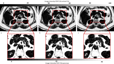 |
22 | Acceleration of chemical shift encoding-based water-fat imaging for pancreatic proton density fat fraction mapping in a single breath-hold
Selina Rupp1, Mingming Wu1, Jessie Han1, Kilian Weiss2, Johannes M Peeters3, Christina Holzapfel4, Marcus M Makowski1, Daniela Junker1, and Dimitrios C Karampinos1
1Technical University of Munich, München, Germany, 2Philips GmbH Market Dach, Hamburg, Germany, 3Philips Healthcare, Best, Netherlands, 4Institute for Nutritional Medicine, School of Medicine, Technical University of Munich, München, Germany With rising prevalence of obesity and metabolic syndrome comes an increasing demand of noninvasive detection of nonalcoholic fatty pancreas disease. Chemical shift encoding-based water-fat separation based on a multi-echo gradient echo acquisition enables pancreatic fat fraction (PDFF) mapping. The pancreas is a small organ and requires high spatial resolution which results in prolonged breath-hold duration for PDFF mapping. The present work aims to accelerate high-resolution single-breath-hold pancreas PDFF mapping using a methodology combining sparse sampled acquisitions with compressed sensing and deep learning reconstructions. |
||
2033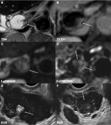 |
23 | Value of high b-value computed diffusion-weighted imaging in intraductal papillary mucinous neoplasia of the pancreas
Felix N. Harder1, Kilian Weiss2, Eva Jung1, Omar Kamal1, Sean McTavish1, Anh T. Van1, Markus M. Graf1, Ihsan E. Demir3, Veit Phillip4, Helmut Friess3, Roland M. Schmid4, Fabian K. Lohöfer1, Marcus R. Makowski1, Georgios A. Kaissis1, Dimitrios C. Karampinos1, and Rickmer F. Braren1
1Institute of Diagnostic and Interventional Radiology, Technical University of Munich, School of Medicine, Munich, Germany, 2Philips GmbH Market DACH, Hamburg, Germany, 3Department of Surgery, Technical University of Munich, School of Medicine, Munich, Germany, 4Department of Medicine II, Technical University of Munich, School of Medicine, Munich, Germany
The incidence of intraductal papillary mucinous neoplasms (IPMN), which represent a potential precursor of pancreatic cancer, is increasing. We herein investigate the potential of computed high b-value diffusion-weighted imaging (cDWI) in detection and stratification of IPMN. cDWI at b = 1000 s/mm2 increases lesion detection rates as well as diagnostic sensitivity and specificity regarding the presence of malignant imaging features. Combining cDWI and high-resolution reduced field-of-view imaging further increases diagnostic accuracy. cDWI seems particularly promising in view of the need for non-invasive lesion detection and characterization due to the recent paradigm shift from surgery to active surveillance of IPMN.
|
||
2034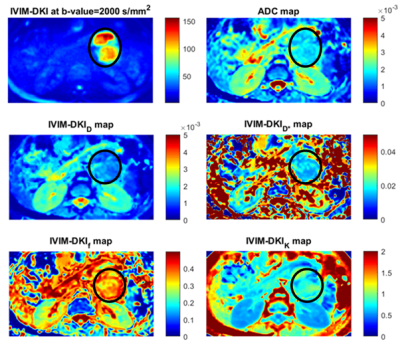 |
24 | Machine learning-based texture analysis of IVIM-DKI for classification of benign and malignant pancreatic masses
Archana Vadiraj Malagi1, Sivachander Shivaji2, Devasenathipathy Kandasamy2, Pramod Garg3, Siddhartha Datta Gupta4, Shivanand Gamanagatti 2, Raju Sharma2, and Amit Mehndiratta1,5
1Centre for Biomedical Engineering, Indian Institute of Technology Delhi, New Delhi, India, 2Department of Radiodiagnosis, All India Institute of Medical Sciences Delhi, New Delhi, India, 3Department of Gastroenterology, All India Institute of Medical Sciences Delhi, New Delhi, India, 4Department of Pathology, All India Institute of Medical Sciences Delhi, New Delhi, India, 5Department of Biomedical Engineering, All India Institute of Medical Sciences Delhi, New Delhi, India
Tumor heterogeneity could be detected non-invasively utilizing textural indicators from IVIM-DKI, which have a high potential for early prognosis of pancreatic lesions. A novel technique was investigated for tumor prediction model utilizing IVIM-DKI with total variation penalty function, in which we employed combinations of texture characteristics from IVIM-DKI parameters. In this study, texture characteristics of the kurtosis(k) parameter had the high accuracy:93% and AUC:1, and combinations of all IVIM-DKI parameters' textural features after feature reduction had accuracy:84% and AUC:0.91 for classifying benign and malignant pancreatic lesions. Whole-volumetric texture analysis of IVIM-DKI can be employed for characterization of pancreatic lesions.
|
||
2035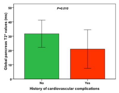 |
25 | Pancreatic iron is a marker of cardiovascular complications in sickle cell disease
Antonella Meloni1, Laura Pistoia1, Massimiliano Missere2, Riccardo Righi3, Antonino Vallone4, Ada Riva5, Vincenzo Positano1, Luciana Rigoli6, Francesco Massei7, Valentina Carrai8, Gianna Alberini1, Filippo Cademartiri1, and Alessia Pepe1
1Fondazione G. Monasterio CNR-Regione Toscana, Pisa, Italy, 2Gemelli Molise SpA, Fondazione di Ricerca e Cura "Giovanni Paolo II", Campobasso, Italy, 3Ospedale del Delta, Lagosanto (FE), Italy, 4Azienda Ospedaliera "Garibaldi" Presidio Ospedaliero Nesima, Catania, Italy, 5Ospedale “SS. Annunziata” ASL Taranto, Taranto, Italy, 6Policlinico "G. Martino", Messina, Italy, 7Azienda Ospedaliero Universitaria Pisana, Pisa, Italy, 8Azienda Ospedaliero - Universitaria Careggi, Firenze, Italy
We systematically explored the link between pancreatic iron detected by the T2* Magnetic Resonance Imaging technique and cardiovascular complications in a cohort of 63 patients with sickle cell disease (SCD). Patients with cardiovascular complications (supraventricular arrhythmias, heart failure, pulmonary hypertension, myocardial infarction, deep vein thrombosis, leg ulcers) showed significantly lower global pancreas T2* values than patients free of cardiovascular complications but comparable global heart T2* values. Pancreatic iron overload (T2*<26 ms) was associated with 7.5 times higher odds of cardiovascular complications.
|
||
2036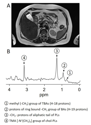 |
26 | 1H-MRS of bile in the gallbladder in subjects without and with hepatic steatosis
Jürgen Machann1,2,3, Norbert Stefan2,3,4, Andreas L Birkenfeld2,3,4, and Fritz Schick1
1Section on Experimental Radiology, Department of Diagnostic and Interventional Radiology, University Hospital Tübingen, Tübingen, Germany, 22. Institute for Diabetes Research and Metabolic Diseases of the Helmholtz Centre Munich at the University of Tübingen, University of Tübingen, Tübingen, Germany, 33. German Center for Diabetes Research (DZD), Tübingen, Germany, 44. Department of Internal Medicine, Division of Endocrinology, Diabetology and Nephrology, University Hospital Tübingen, Tübingen, Germany
1H-MRS of bile was performed in 54 healthy subjects at increased risk for metabolic diseases. Additionally, intrahepatic lipids (IHL) were quantified. Concentration of main peaks of bile was calculated in relation to non-suppressed water signal. Large interindividual differences in concentration of the bile-peaks with significantly lower concentrations in subjects with increased IHL were detected. Males show a tendency towards higher concentrations in all bile-peaks as well as in IHL (n.s.). It remains speculative, whether or not these MRS-derived results might play a role in the pathogenesis of insulin resistance – further analyses with assignable metabolic and laboratory data are recommended.
|
||
2037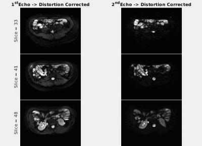 |
27 | Distortion Correction in Abdominal DW-MRI using Dual-Echo EPI for Improved Evaluation of Crohn’s Disease
Cemre Ariyurek1, Onur Afacan1, and Sila Kurugol1
1Radiology, Boston Children's Hospital and Harvard Medical School, Boston, MA, United States
In MR enterography, diffusion-weighted MRI (DW-MRI) is commonly used to evaluate Crohn’s disease (CD). Echo-planar imaging (EPI) used in DW-MRI suffers from geometric distortion due to time-varying susceptibility field changes in the bowel. Hence, we implemented a DW-MRI sequence using a dual-echo EPI with opposing encoding polarities. Thus, the distortion field can be estimated dynamically and corrected retrospectively. The proposed method was evaluated in DW-MRI data acquired on suspected CD patients by visually and quantitatively assessing distortion corrected volumes based on anatomical similarity with T2-HASTE and uncertainty of estimated diffusion parameters of the intravoxel incoherent motion model.
|
||
2038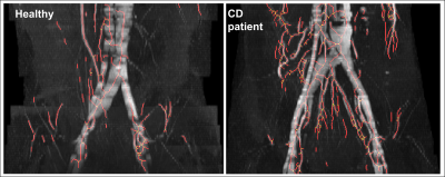 |
28 | Quantification of The Comb Sign in Crohn’s Disease Using Time of Flight Magnetic Resonance Angiography
Iyad Naim1,2, Caroline Hoad2,3, Gordon Moran1,2, and Penny Gowland2,3
1School of Medicine, University of Nottingham, Nottingham, United Kingdom, 2National Institute of Health Research, Nottingham Biomedical Research Centre (BRC) at Nottingham University Hospitals NHS Trust and University of Nottingham, Nottingham, United Kingdom, 3School of Physics and Astronomy, University of Nottingham, Nottingham, United Kingdom
Crohn’s disease (CD) is an inflammatory bowel disease affecting 115k people in the UK. The ‘Comb Sign’ describes increased vascularity and arborization of vessels in the mesentery, due to intestinal inflammation. Radiologists observe this sign on cross-sectional imaging as an indication for CD, but it has never been quantified. We used time of flight MR angiography to visualize the vessels in the abdomen without using contrast agents. Images were analyzed using in-house developed software that traces vessels and counts branching points. Scans from 15 healthy volunteers(HV) and 6 CD patients showed patients had more vessel branching points compared to HV.
|
||
2039 |
29 | The feasibility of DCE-MRI in presurgical evaluation of tumor budding grade in rectal cancer Video Not Available
Wenjun Hu1, Anliang Chen2, Wan JUN Dong2, Xiwei JUN Li2, Yue Wang2, Yuhui Liu2, and Ailian Liu1
1Department of Radiology, the First Affiliated Hospital of Dalian Medical University, Dalian, China, 2The First Affiliated Hospital of Dalian Medical University, Dalian, China
Tumor budding (TB) is a histopathologic characteristic which has led to a growing interest in the prognosis prediction of cancers of various sites. Preoperative prediction of TB status may help personalize treatment in rectal cancer cancer patients. Previous studies showed MRI-based and PET/CT based radiomics can predict TB status, however, the high cost and complex post-processing limit their clinical applications. Dynamic contrast-enhanced (DCE) MRI has been extensively evaluated aggressiveness of rectal cancer. Results of this study indicate Kep values can effectively differentiate high from intermediate-low grade TB.
|
||
2040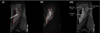 |
30 | MRI Tagging of Colonic Contents in Healthy Subjects: Reliability of the Measurement with Time
Meshari T Alshammari1,2, Luca Marciani1, Gordon W. Moran1, and Caroline L. Hoad3,4
1Translational Medical Sciences and National Institute for Health Research (NIHR) Nottingham Biomedical Research Centre, Nottingham University Hospitals NHS Trust and University of Nottingham, Nottingham, United Kingdom, 2Department of Diagnostic Radiology, College of Applied Medical Sciences, University of Hail, Hail, Saudi Arabia, 3NIHR Nottingham Biomedical Research Centre (BRC), Nottingham University Hospitals NHS Trust and University of Nottingham, Nottingham, United Kingdom, 4Sir Peter Mansfield Imaging Centre, School of Physics and Astronomy, University of Nottingham, Nottingham, United Kingdom We aimed to investigate the temporal variation of the MR colonic tagging technique to measure mixing of colonic contents following a water based laxative drink by acquiring multiple scans on healthy participants. The study results indicate that time did not influence the measurement of colonic content mixing, however the scans were variable over the 10 minute acquisition window which suggests multiple scans are needed to accurately assess this parameter. Multiple measurements of this non-invasive technique can be used in future studies that aim to look at bowel motility and treatment response of IBD patients, using the same water-based laxative. |
||
2041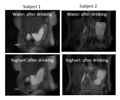 |
31 | Gastric emptying whilst upright
Hannah Askill 1, Emily Gaudoin1, Dipendra Jayantilal Mistry1, Olivier Mougin1, Caroline Hoad1,2, Hayfa Sharif1,2, Luca Marciani1,2, and Penny A Gowland1,2
1Sir Peter Mansfield Imaging Centre,, University of Nottingham, Nottingham, United Kingdom, 2Nottingham Biomedical Research Centre, Nottingham, United Kingdom
Gastric accommodation and gastric tone are key measure in functional diseases such as gastroparesis, dyspepsia, and even reflux. Whilst gastric accommodation has been assessed using conventional MRI such measures will have been be perturbed by the subject lying supine. Gastric tone is either assessed using a barostat or pressure sensors. This work investigate the gastric response to a meal in the upright position with the aim of analysing the data to study accommodation and markers of tone in future.
|
||
2042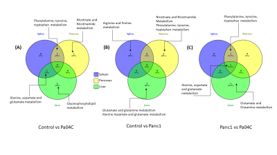 |
32 | Systemic metabolic dysregulation common across multiple organs caused by cancer and cancer-induced cachexia
Raj Kumar Sharma1, Santosh K. Bharti1, Paul T Winnard1, Marie-France Penet1, and Zaver M. Bhujwalla1,2,3
1Division of Cancer Imaging Research, The Russell H. Morgan Department of Radiology and Radiological Science, The Johns Hopkins University School of Medicine, Baltimore, MD, United States, 2Sidney Kimmel Comprehensive Cancer Center, The Johns Hopkins University School of Medicine, Baltimore, MD, United States, 3Radiation Oncology and Molecular Radiation Sciences, The Johns Hopkins University School of Medicine, Baltimore, MD, United States Analysis of organ metabolism with cancer growth can unravel systemic changes that occur with cancer and with cancer-induced cachexia. Cancer-induced cachexia, commonly observed with pancreatic cancer, causes tissue wasting, intolerance to chemotherapy, and poor quality of life. Here, we identified alterations of metabolic pathways in the spleen, liver, pancreas, lung, heart, brain and kidney following growth of cachexia-inducing and non-cachexia inducing pancreatic cancer xenografts. These results highlight the systemic metabolic changes that occur with cancer and with cancer induced cachexia that may lead to the development of organ-based early biomarkers as well to organ-based metabolic treatment strategies. |
||
 Back to Meeting Home
 Back to the Program-at-a-Glance
The International Society for Magnetic Resonance in Medicine is accredited by the Accreditation Council for Continuing Medical Education to provide continuing medical education for physicians.
 Back to Meeting Home
 Back to the Program-at-a-Glance
 View the Presentation