Digital Poster
Prostate, Techniques & Detection of Cancer
Joint Annual Meeting ISMRM-ESMRMB & ISMRT 31st Annual Meeting • 07-12 May 2022 • London, UK

Digital Poster
Prostate, Techniques & Detection of Cancer
| Computer # | ||||
|---|---|---|---|---|
0823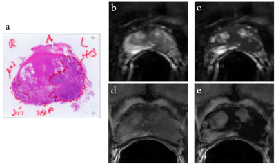 |
48 | Enhanced T2-weighted images using Luminal Water Imaging and U-Net based segmentation for prostate cancer diagnosis.
Candice JX Ip1,2, Silvia D Chang3,4,5, Shirin Sabouri1, Andrew Yung1, Stefan Reinsberg2, and Piotr Kozlowski1,2,3,4,5
1UBC MRI Research Center, University of British Columbia, Vancouver, BC, Canada, 2Department of Physics and Astronomy, University of British Columbia, Vancouver, BC, Canada, 3Department of Urologic Sciences, University of British Columbia, Vancouver, BC, Canada, 4Vancouver Prostate Centre, Vancouver, BC, Canada, 5Department of Radiology, The University of British Columbia, Vancouver, BC, Canada
Clinical prostate carcinoma (PCa) detection with MRI has focused on qualitative assessment. However, novel Luminal Water Imaging (LWI) provides quantitative information with promising results for detecting PCa. To overcome shortcomings of clinical prostate MRI protocol, we propose using LWI to augment T2-weighted (T2W) images to improve image contrast for PCa detection while maintaining anatomical details needed for radiological diagnosis. Here, we investigate automatic segmentation models and various weighting functions of LWI parameter maps to generate semi-quantitative T2W images that also preserves anatomical detail. Our results show that a combined T2W and LWI parameter image provides enhanced detection of PCa.
|
||
0824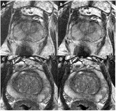 |
49 | Radiological Evaluation of Model-Based Reconstruction for Through-Plane Resolution Enhancement in T2-Weighted Spin-Echo Prostate MRI
Eric A. Borisch1, Adam T. Froemming1, Roger C. Grimm1, Akira Kawashima2, Joshua D. Trzasko1, and Stephen J. Riederer1
1Radiology, Mayo Clinic, Rochester, MN, United States, 2Radiology, Mayo Clinic, Phoenix, AZ, United States
Results of a radiological review of a previously described Model Based Iterative Reconstruction (MBIR) compensating for slice profile arising in a thick and overlapping-slice acquisition of the prostate are detailed. Blinded review by two radiologists indicated improvements in SNR, contrast fidelity, and diagnostic quality.
|
||
0825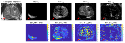 |
50 | Incorporating compartmental T2 measurements into Restriction Spectrum Imaging of the prostate
Christopher C Conlin1, Roshan A Karunamuni2, Christine H Feng2, Ana E Rodriguez-Soto1, Joshua M Kuperman1, Dominic Holland3, Rebecca Rakow-Penner1, Michael E Hahn1, Anders M Dale1,3,4, and Tyler M Seibert2,5
1Department of Radiology, University of California San Diego, La Jolla, CA, United States, 2Department of Radiation Medicine and Applied Sciences, University of California San Diego, La Jolla, CA, United States, 3Department of Neurosciences, University of California San Diego, La Jolla, CA, United States, 4Halıcıoğlu Data Science Institute, University of California San Diego, La Jolla, CA, United States, 5Department of Bioengineering, University of California San Diego, La Jolla, CA, United States
Restriction Spectrum Imaging (RSI) examines diffusion in discrete tissue compartments to better detect and characterize prostate cancer. T2 information from these compartments may further improve prostate cancer evaluation. In this study, RSI data was acquired using multiple echo times to measure both compartmental T2 and diffusion in patients with suspected prostate cancer. A multivariable model was then developed to identify cancer from compartmental T2 and diffusion measurements. Significant differences in compartmental T2 were observed between normal and cancerous prostatic tissue. However, the multivariable model did not significantly improve cancer detection performance over diffusion measurements alone.
|
||
0826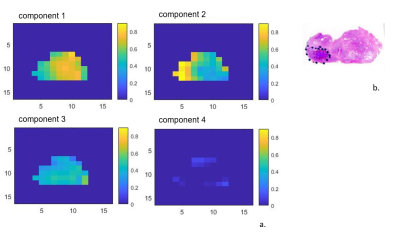 |
51 | MCR-ALS application for prostate cancer localization
Angeliki Stamatelatou1, Carlo Giuseppe Bertinetto2, Jeroen Jansen2, Geert Postma2, Arend Heerschap1, and Tom Scheenen1
1Radiology and Nuclear Medicine, Radboud University Medical Center, Nijmegen, Netherlands, 2Analytical Chemistry & Chemometrics, Institute for Molecules and Materials, Nijmegen, Netherlands Three-dimensional MRSI data of the prostate was analyzed with a Multivariate Curve Resolution-Alternating (MCR) approach for rapid automated localization and classification of cancer and healthy tissue. This data-driven method was used to extract common spectroscopic components without a need of prior knowledge, and compared to fitting a linear combination of prior knowledge models (LCModel). The MCR method identified components with known prostate metabolites and residual lipid and water signals Altogether, our approach can be considered as a step towards the development of an automated tool for classification of prostate MRSI spectra, avoiding subjective human intervention. |
||
0827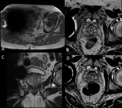 |
52 | 1.5T Prostate MRI Utilizing Deep Learning Reconstruction in Patients with Hip Prosthesis
Melany B Atkins1, Arnaud Guidon2, Michael Vinski3, Thomas Schrack4, Heidi Harris5, and Ersin Bayram6
1Radiology, Fairfax Radiological Consultants, Arlington, VA, United States, 2GE Healthcare, Boston, MA, United States, 3GE Healthcare, Lynchburg, VA, United States, 4Fairfax Radiological Consultants, Fairfax, VA, United States, 5GE Healthcare, Waukesha, WI, United States, 6GE Healthcare, Houston, TX, United States
While 3T offers SNR benefits for prostate imaging, patients with hip prostheses are problematic due to susceptibility artifacts and implant heating concerns. These issues are better managed at 1.5T but 1.5T lacks the SNR versus 3T. Deep learning reconstruction (DLR) may provide the SNR boost to clear this hurdle. Feasibility of 1.5T prostate MRI with DLR in patients with hip prosthesis is explored in up to 20 subjects focusing on two key contrasts in the PI-RADS guideline: T2Wand DWI. Preliminary results suggest that 1.5T with DLR might be the preferred option for patients with hip implants.
|
||
0828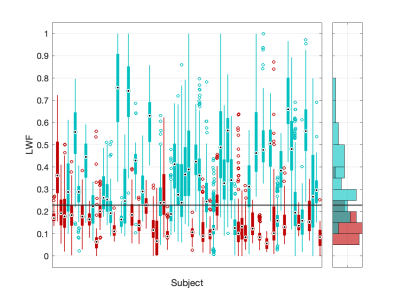 |
53 | Fast Fitting of Luminal Water Fraction in Prostate Cancer
David Atkinson1, Giorgio Brembilla1, Fiona Gong1, and Shonit Punwani1
1Centre for Medical Imaging, University College London, London, United Kingdom The use of a fast, direct, Luminal Water Fraction fitting technique is compared with a more commonly used iterative method on data from 76 prostate cancer patients with biopsy Gleason grade available. The fast method shows a similar Receiver Operating Characteristic Area Under the Curve (ROC-AUC) when used to classify an image region as clinically significant or non-significant cancer. |
||
0829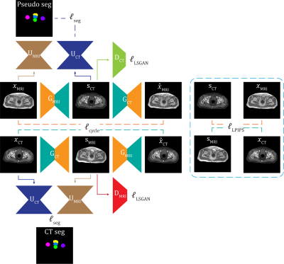 |
54 | CycleSeg-v2: Improving unpaired MR-to-CT Synthesis and Segmentation with pseudo label and LPIPS loss
Huan Minh Luu1, Gyu-sang Yoo2, Won Park2, and Sung-Hong Park1
1Bio and Brain Engineering, Korea Advanced Institute of Science and Technology, Daejeon, Korea, Republic of, 2Department of Radiation Oncology, Samsung Medical Center, Seoul, Korea, Republic of
Radiotherapy treatment typically requires both CT and MRI as well as labor intensive contouring for effective planning and treatment. Deep learning can enable an MR-only workflow by generating synthetic CT (sCT) and performing automatic segmentation on the MR data. However, MR and CT data are usually unpaired and limited contours are available for MR data. In this study, we proposed CycleSeg-v2 that extends the previously proposed CycleSeg to work with unpaired data. To ensure robust training, we employed LPIPS loss in addition to pseudo label. Experiments with data from prostate cancer patients showed that CycleSeg-v2 improved upon previous approaches.
|
||
0830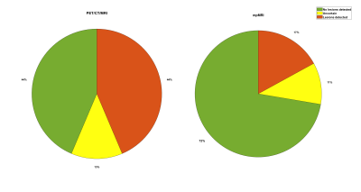 |
55 | PSMA-PET vs mpMRI in prostate cancer patients with biochemical recurrence
Ingerid Skjei Knudtsen1, Bendik Skarre Abrahamsen1, Kirsten Margrete Selnæs2, Mattijs Elschot1, Sverre Langorgen3, Thomas Morten Keil3, Håkon Johansen3, Helena Bertilsson4,5, Dag Linthoe Halvorsen4, Torgrim Tandstad5,6, and Tone Frost Bathen1
1Department of Circulation and Medical Imaging, Norwegian University of Science and Technology, Trondheim, Norway, 2Department of Nuclear Medicine and Medical Physics, St. Olavs Hospital, Trondheim University Hospital, Trondheim, Norway, 3Department of Radiology and Nuclear Medicine, St. Olavs Hospital, Trondheim University Hospital, Trondheim, Norway, 4Department of Urology, St. Olavs Hospital, Trondheim University Hospital, Trondheim, Norway, 5Department of Clinical and Molecular Medicine, Norwegian University of Science and Technology, Trondheim, Norway, 6Cancer Clinic, St. Olavs Hospital, Trondheim University Hospital, Trondheim, Norway
The PSMA prostate recurrence study is a multisenter prospective trial where the overall aim is to evaluate the diagnostic accuracy of 18F/68Ga-PSMA PET for detection of disease in patients with biochemical recurrence. In this preliminary analysis, we have compared the clinical imaging report of PET/CT/mpMRI to a separate evaluation of mpMRI where the radiologist was blinded to the PET results. Of 94 patients included so far, recurrent disease and/or distant metastasis was detected in 44 % (median PSA 0.4 ng/ml), according to the imaging protocol while the detection rate was 17 % for the mpMRI study scoring.
|
||
 Back to Meeting Home
 Back to the Program-at-a-Glance
The International Society for Magnetic Resonance in Medicine is accredited by the Accreditation Council for Continuing Medical Education to provide continuing medical education for physicians.
 Back to Meeting Home
 Back to the Program-at-a-Glance
 View the Presentation