Digital Poster
Prostate & Whole-Body Imaging
Joint Annual Meeting ISMRM-ESMRMB & ISMRT 31st Annual Meeting • 07-12 May 2022 • London, UK

| Computer # | ||||
|---|---|---|---|---|
0925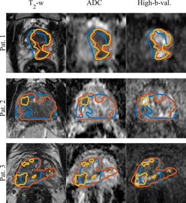 |
45 | Automatic Prostate Tumor Segmentation: Does the Convolutional Neural Network Learn How the Tumor Looks, or What the Radiologist Sees?
Deepa Darshini Gunashekar1, Lars Bielak1,2, Benedict Oerther 3, Matthias Benndorf 3, Anca Grosu 2,3, Constantinos Zamboglou2,3, and Michael Bock 1,2
1Dept.of Radiology, Medical Physics, Medical Center University of Freiburg, Faculty of Medicine, University of Freiburg, Freiburg, Germany, 2German Cancer Consortium (DKTK), Partner Site Freiburg, Freiburg, Germany, Freiburg im Breisgau, Germany, 3Dept.of Radiology, Medical Center University of Freiburg, Faculty of Medicine, University of Freiburg, Freiburg, Germany
A convolutional neural network was implemented to automatically segment tumors in multi-parametric MRI data. The influence of the variability in the ground truth data was evaluated for automated prostate tumor segmentation. Therefore, the agreement between the predictions of the CNN was measured with co-registered whole mount histopathology images and the tumor contours drawn by an expert radio-oncologist. The results indicate that the network can discriminate tumor from healthy tissue rather than mimicking the radiologist.
|
||
0926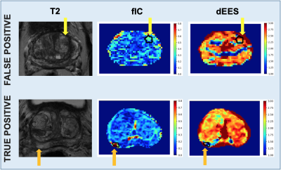 |
46 | VERDICT-MRI Analysis of False Positives in Prostate Mp-MRI
Snigdha Sen1, Vanya Valindria1, Hayley Pye2, Alistair Grey3, Aiman Haider4, Alex Freeman4, Caroline Moore3, Hayley Whitaker2, Shonit Punwani5, Saurabh Singh5, and Eleftheria Panagiotaki1
1Centre for Medical Image Computing, University College London, London, United Kingdom, 2Molecular Diagnostics and Therapeutics Group, University College London, London, United Kingdom, 3Department of Urology, University College London Hospitals NHS Foundation Trust, London, United Kingdom, 4Department of Pathology, University College London Hospitals NHS Foundation Trust, London, United Kingdom, 5Centre for Medical Imaging, University College London, London, United Kingdom
Mp-MRI has moderate specificity for the diagnosis of prostate cancer, resulting in many false positive results and unnecessary biopsies for patients with other prostatic diseases. This work presents an approach using VERDICT-MRI to investigate differences in microstructural parameter estimates between normal prostate tissue, false positive lesions and clinically-significant cancer. The VERDICT intracellular volume fraction and intrinsic diffusivity in the extracellular-extravascular space can discriminate false positive lesions with greater accuracy than the clinical ADC from mp-MRI. These results show VERDICT has the potential to identify false positives and reduce unnecessary biopsies in prostate patients.
|
||
0927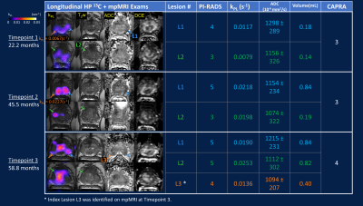 |
47 | Hyperpolarized 13C Pyruvate MR-Assisted Longitudinal Risk Stratification in Prostate Cancer Patient – From Surveillance to Surgery Video Not Available
Hsin-Yu Chen1, Hao G. Nguyen2, Zhen J. Wang1, Michael A. Ohliger1, Jeffry P. Simko2, Matthew R. Cooperberg2, Daniel Gebrezgiabhier1, Lucas Carvajal1, Jeremy W. Gordon1, John Kurhanewicz1, Rahul Aggarwal2, Daniel B. Vigneron1, and Robert A. Bok1
1Radiology and Biomedical Imaging, University of California, San Francisco, San Francisco, CA, United States, 2Helen Diller Family Comprehensive Cancer Center, University of California, San Francisco, San Francisco, CA, United States
Serial hyperpolarized 13C+mpMRI and paired metabolic image-guided biopsy were employed in a new integrative active surveillance (AS) paradigm, exemplified by a patient who initially enrolled in AS for 5 years, but later underwent radical prostatectomy upon progression. Metabolic biomarker kPL images detected early progression preceding radiologic and clinical parameters. Final whole-gland histopathology also linked kPL and PI-RADS targets to multifocal tumors, where kPL spatially reflected intratumoral heterogeneity, and high kPL regions correlated with adverse pathologic features such as G4 cribriform pattern, extracapsular extension, and lymphovascular invasion. Results indicated HP 13C may help optimize AS by improving clinical risk calculation.
|
||
0928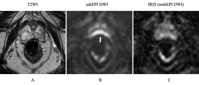 |
48 | Comparison of single-shot EPI DWI and multi-shot EPI DWI in prostate MRI
Ayumu Kido1, Akira Yamamoto1,2, Mitsuru Takeuchi3, Yu Ueda4, Jaladhar Neelavalli5, and Tsutomu Tamada1
1Radiology, Kawasaki Medical School, Kurashiki, Japan, 2Radiology, Kawasaki Medical School, Kurashiki-shi, Japan, 3Radiology, Radiolonet Tokai, Nagoya, Japan, 4Philips Japan, Tokyo, Japan, 5Philips India, Bangalore, India
Single-shot EPI (sshEPI) DWI still suffers from distortion and blurring. Multi-shot EPI (mshEPI) DWI called IRIS enables to reduce image distortion and blurring. A total of 142 patients with suspected prostate cancer (PC) underwent mpMRI including sshEPI DWI and IRIS under the same scan time. The image quality and diagnostic performance of the lesions were compared between sshEPI DWI and IRIS. IRIS improved image distortion and blurring compared to sshEPI DWI, and also suggested a similar or higher diagnostic performance compared to sshEPI DWI.
|
||
0929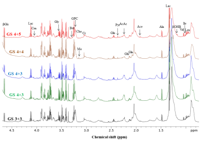 |
49 | Targeted Metabolomic Profiling of Blood Plasma in Aggressive Prostate Cancer Patients using NMR Spectroscopy
Virendra Kumar1, Pradeep Kumar1, Rajeev Kumar2, S. Datta Gupta3, Sanjay Thulkar4, N. R. Jagannathan5, and Rama Jayasundar1
1Department of NMR & MRI Facility, All India Institute of Medical Sciences, New Delhi, India, 2Department of Urology, All India Institute of Medical Sciences, New Delhi, India, 3Department of Pathology, All India Institute of Medical Sciences, New Delhi, India, 4Department of Radio-diagnosis, IRCH, All India Institute of Medical Sciences, New Delhi, India, 5Dept. of Radiology, Chettinad Academy of Research, Kelambakkam, Tamil Nadu, India
Gleason score (GS) is currently clinical predictor of prostate cancer (PCa) mortality. The present study investigates the metabolic profile of blood plasma for distinguishing aggressive PCa patients from non aggressive using targeted 1H-NMR metabolomics. A significantly higher concentration of lactate, pyruvate, choline, dimethylamine, glucose, betaine and lower concentration of leucine, isoleucine, valine, histidine, phenylalanine, and tyrosine were observed in patients with GS 8-10 as compared to patients with GS 7 and 6. Our results suggested that pathway alterations, amino acids, phospholipids and glucose related to PCa progression. Identifying biomarker/s associated with GS can be improved diagnosis and treatment.
|
||
0930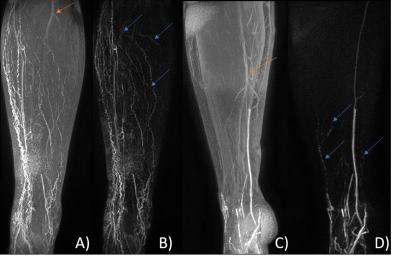 |
50 | Semi-Quantitative Analysis of Dynamic Contrast-Enhanced Magnetic Resonance Lymphangiography in the Legs of Primary Lymphoedema Patients
Michael Mills1, Malou Van Zanten1, Julian Pearce2, Bernard Ho2, Kristiana Gordon1,2, Peter S Mortimer1,2, Pia Ostergaard1, and Franklyn A Howe1
1St George's University of London, London, United Kingdom, 2St George's University Hospitals NHS Foundation Trust, London, United Kingdom
Despite its prevalence, methods for investigating lymphoedema (chronic tissue swelling due to abnormal lymphatic functioning) remain limited. Dynamic Contrast-Enhanced Magnetic Resonance Lymphangiography (DCE-MRL) allows identification of lymph vessels in both healthy and lymphoedematous limbs, however quantitative analyses are lacking. DCE-MRL data was acquired for 30 minutes in lymphoedematous (n=7) and healthy limbs (n=4), with estimates of lymphatic vessel signal characteristics compared between groups. Maximum enhancement compared to baseline was significantly increased in healthy volunteers, while other metrics were similar. Larger groups and longer imaging sessions are likely required to detect additional differences.
|
||
0931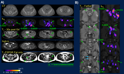 |
51 | Integrative Hyperpolarized 13C Metabolic and Standard-of-Care Abdominopelvic Staging/Restaging MRI for Patients with Advanced Solid Tumors Video Not Available
Hsin-Yu Chen1, Michael A. Ohliger1, Zhen J. Wang1, Mary E. Frost1, Lucas Carvajal1, Philip M. Lee1, Jeremy W. Gordon1, John Kurhanewicz1, Peder E.Z. Larson1, Robert A. Bok1, Rahul Aggarwal2, and Daniel B. Vigneron1
1Radiology and Biomedical Imaging, University of California, San Francisco, San Francisco, CA, United States, 2Helen Diller Family Comprehensive Cancer Center, University of California, San Francisco, San Francisco, CA, United States
Harnessing unique metabolic insight from hyperpolarized (HP) 13C MRI, this study describes an integrated approach toward routine cancer staging and restaging using combined HP 13C plus standard of care (SOC) MRI with commercially-available 13C and 1H array coils and a time-efficient adaptive protocol. A series of patients with osseous, liver, nodal metastases, and locally advanced primary tumors suggested HP 13C not only detected high metabolic activity in progressive tumors, but it also revealed inter- and intratumoral metabolic heterogeneity. Results indicated HP 13C-MRI can greatly complement SOC-MRI in assessing therapeutic response and guiding clinical decisions.
|
||
0932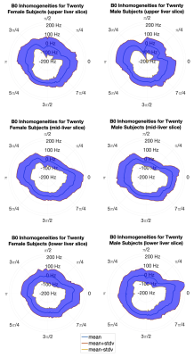 |
52 | B0 inhomogeneity in the abdominal subcutaneous fat at 3T
Aidan Tollefson1, Nate Roberts1, Ali Pirasteh1,2, and Diego Hernando1,2
1Medical Physics, University of Wisconsin, Madison, WI, United States, 2Radiology, University of Wisconsin, Madison, WI, United States
B0 inhomogeneities often lead to failures in chemical shift-based fat suppression, particularly in complex susceptibility environments such as the abdomen. To optimize the application of fat suppression, characterization of B0 inhomogeneities in fat regions is needed. However, such characterization has not been reported for abdominal fat. Therefore, the purpose of this work was to measure the magnitude and variability of B0 inhomogeneity at 3T in abdominal subcutaneous fat. This study shows that several subcutaneous fat regions exhibit B0 inhomogeneities large enough to induce failures in common fat suppression methods. These measurements may guide future optimization of fat suppression methods.
|
||
 Back to Meeting Home
 Back to the Program-at-a-Glance
The International Society for Magnetic Resonance in Medicine is accredited by the Accreditation Council for Continuing Medical Education to provide continuing medical education for physicians.
 Back to Meeting Home
 Back to the Program-at-a-Glance
 View the Presentation