Digital Poster
Applications of Perfusion Techniques I
Joint Annual Meeting ISMRM-ESMRMB & ISMRT 31st Annual Meeting • 07-12 May 2022 • London, UK

Digital Poster
Applications of Perfusion Techniques I
| Computer # | ||||
|---|---|---|---|---|
1215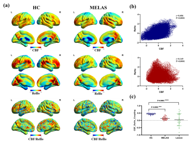 |
11 | Altered neurovascular coupling in patients with MELAS evaluated by combining cerebral blood flow and regional homogeneity Video Permission Withheld
Rong Wang1, Yuxin Li1, Jie Lin1, and Yong Zhang2
1Huashan Hospital, Shanghai, China, 2GE Healthcare, Shanghai, China, Shanghai, China
This is the first study that used a combined arterial spin labeling (ASL) and resting-state fMRI approach to assess the neurovascular coupling in patients with mitochondrial myopathy, encephalopathy, lactic acidosis and stroke-like episodes (MELAS), which may provide a new mechanistic perspective into understanding numerous brain diseases.
|
||
1216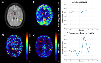 |
12 | Non-invasive assessment of blood-brain barrier permeability in multiple sclerosis patients by arterial spin labeling.
Andre Monteiro Paschoal1, Mateus Boaventura1, Leonie Petitclerc2, Matthias J.P. van Osch2, Maria Concepcion Garcia Otaduy1, Samira Luisa Apostolos-Pereira3, Dagoberto Callegaro3, and Carolina de Medeiros Rimkus1
1Institute of Radiology and Department of Radiology and Oncology, University of Sao Paulo, Sao Paulo, Brazil, 2C.J. Gorter Center for high field MRI, Department of Radiology, Leiden University Medical Center, Leiden, Netherlands, 3Departamento de Neurologia, Hospital das Clinicas da Faculdade de Medicina, University of Sao Paulo, Sao Paulo, Brazil
Multiple sclerosis is a demyelinating disease affecting the central nervous system. Although MS lesions can be identified by conventional MRI, the evaluation of active lesions in normal appearing gray and white matter requires measures of blood-brain barrier permeability (BBB), which is commonly assessed through the dynamic contrast enhanced (DCE) acquisition. When repeated scans are needed in the MS patients follow-up, repeated gadolinium injection is not recommended. This study successfully showed the feasibility of advanced arterial spin labeling sequences as an alternative to evaluate BBB permeability, whose results of water-exchange times across the BBB were in agreement with DCE measurements.
|
||
1217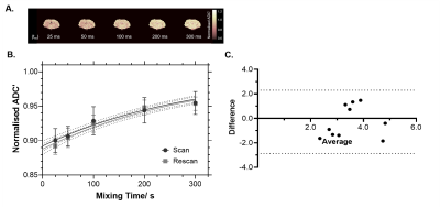 |
13 | Implementation of non-invasive Filter-Exchange Imaging (FEXI) to measure blood-brain barrier water exchange in the rat brain
Yolanda Ohene1,2, Elizabeth Powell3,4, Samo Lasič5,6, Geoff J. M Parker3,7,8, Laura M Parkes 1,2, and Ben R Dickie 2,9
1Division of Neuroscience and Experimental Psychology, University of Manchester, Manchester, United Kingdom, 2Geoffrey Jefferson Brain Research Centre, University of Manchester, Manchester, United Kingdom, 3Centre for Medical Image Computing, UCL, London, United Kingdom, 4Department of Computer Science, UCL, London, United Kingdom, 5Danish Research Centre for Magnetic Resonance, Copenhagen University Hospital Amager and Hvidovre, Copenhagen, Denmark, 6Random Walk Imaging, Lund, Sweden, 7Department of Medical Physics and Biomedical Engineering, UCL, London, United Kingdom, 8Bioxydyn Limited, Manchester, United Kingdom, 9Division of Informatics, Imaging and Data Sciences, University of Manchester, Manchester, United Kingdom
We have developed a preclinical FEXI sequence optimised to measure water exchange across the blood-brain barrier in the rat brain. Simulated synthetic data estimated an apparent exchange rate (AXR) of 1.22 s-1, using the in-vivo acquisition parameters and exchange rate constant = 2.5 s-1, reflecting in-vivo measurements. The experimental normalised AXR in the rat brain was measured at 3.34 ± 1.14 s-1 (scan) and 3.62 ± 0.96 s-1 (rescan), with CoV = 26%, n = 10. This technique is a promising non-invasive tool which can be applied to in a wide range of disease models, including neurodegeneration, stroke and neuroinflammation.
|
||
1218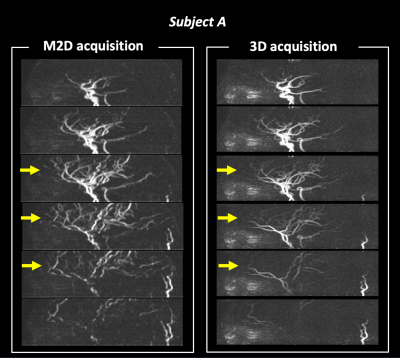 |
14 | Improved visualization of intracranial peripheral arteries with multiple 2D slice ASL-MRA and super-resolution
Yuriko Suzuki1, Peter Jezzard1, Ioannis Koktzoglou2,3, and Thomas Okell1
1Wellcome Centre for Integrative Neuroimaging, FMRIB, Nuffield Department of Clinical Neurosciences, University of Oxford, Oxford, United Kingdom, 2Department of Radiology, NorthShore University HealthSystem, Evanston, IL, United States, 3Pritzker School of Medicine, University of Chicago, Chicago, IL, United States
In recent years, magnetic resonance angiography (MRA) using arterial spin labeling (ASL) technique has been developed aiming to be a non-invasive potential alternative to X-ray angiography. When blood flow is slow (e.g. in elderly patients, especially with steno-occlusive diseases), however, ASL-MRA suffers from poor vessel visualization, which hinders its clinical utility in those patients. In this study, instead of using a 3D volume acquisition commonly used in brain MRI, we propose to separately scan multiple 2D slices to reduce the saturation of arterial blood signal and improve the visualization of small peripheral arteries, in combination with a super-resolution reconstruction.
|
||
1219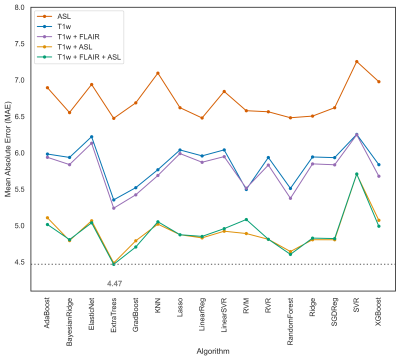 |
15 | Cerebrovascular brain-age
M.B.J. Dijsselhof1, M. Barboure1, M. Stritt2, W. Nordhøy3, A.M. Wink1, L.T. Westlye4,5,6, J.H. Cole7, F. Barkhof1,8, J. Petr9, and H.J.M.M. Mutsaerts1
1Department of Radiology and Nuclear Medicine, Amsterdam Neuroscience, Amsterdam University Medical Center, Amsterdam, Netherlands, 2Mediri GmbH, Heidelberg, Germany, 3Department of Diagnostic Physics, Division of Radiology and Nuclear Medicine, Oslo University Hospital, Oslo, Norway, 4Norwegian Centre for Mental Disorders Research (NORMENT), Institute of Clinical Medicine, University of Oslo & Division of Mental Health and Addiction, Oslo University Hospital, Oslo, Norway, 5Department of Psychology, University of Oslo, Oslo, Norway, 6KG Jebsen Centre for Neurodevelopmental Disorders, University of Oslo, Oslo, Norway, 7Dementia Research Centre, Queen Square Institute of Neurology; Centre for Medical Image Computing, University College London, London, United Kingdom, 8Queen Square Institute of Neurology and Centre for Medical Image Computing, University College London, London, United Kingdom, 9Helmholtz-Zentrum Dresden-Rossendorf, Dresden, Germany The structural brain-age makes predictions based on changes in tissue integrity. Adding cerebrovascular MRI biomarkers may add sensitivity to physiological and metabolic changes, hence complementing structural brain-age, and possibly improving its early pathology sensitivity. Baseline and follow-up T1w, FLAIR, and ASL data of 233 healthy participants and combinations of features and algorithms were used to predict ‘Cerebrovascular brain-age’. The ExtraTrees algorithm utilising T1w, ASL, and FLAIR features performed best and showed good longitudinal reproducibility. |
||
1220 |
16 | Novel arterial spin labelling (ASL) brain injury symmetry assessment in retired professional athletes: a preliminary study
Ethan Danielli1,2, Beatriz Padrela3, Mitchell Doughty1,2, Jan Petr4, Henk Mutsaerts3,5, and Michael D Noseworthy1,2,6
1School of Biomedical Engineering, McMaster University, Hamilton, ON, Canada, 2Imaging Research Centre, St. Joseph's Healthcare Hamilton, Hamilton, ON, Canada, 3Radiology and Nuclear Medicine, Amsterdam University, Amsterdam, Netherlands, 4Institute of Radiopharmaceutical Cancer Research, Helmholtz-Zentrum Dresden-Rossendorf, Dresden, Germany, 5Ghent Institute for Functional and Metabolic Imaging, Ghent University, Ghent, Belgium, 6Department of Electrical and Computer Engineering, McMaster University, Hamilton, ON, Canada
3D PCASL scans were acquired for seventeen aging, retired professional football players with a history of head traumas. Left, right and bilateral CBF and ASL spatial coefficient of variation (sCoV) values were examined for twelve concussion-related ROIs. A Z-scoring approach was applied, with outliers defined as mild, moderate, or severe injury burden (IB). An IB symmetry index was also calculated. Outliers were detected in all 12 ROIs, and the anterior parahippocampal gyrus and inferior frontal gyrus pars opercularis had the highest CBF and ASL sCoV IB, respectively. IB was not biased towards the left or right hemisphere.
|
||
1221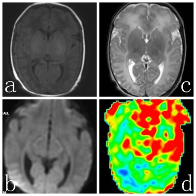 |
17 | The Value of 3D arterial spin labeling in early diagnosis and prognostic grouping of Full-Term neonatal hypoxic-ischemic encephalopathy
Chang Liu1 and Zhang Yong2
1Department of Radiology, Southern District, The First Affiliated Hospital of USTC, Division of Life, HEFEI, China, 2MR Research,GE Healthcare,Shanghai,China, Shanghai, China
3D-ASL technology can reflect cerebral blood flow perfusion status,which is helpful for early diagnosis of HIE.The predictive value of ASL for the prognosis of full-term HIE patients is higher than that of DWI.
|
||
1222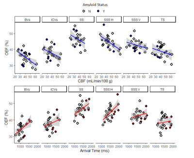 |
18 | Oxygen Extraction Fraction is higher in elderly participants with a higher cardiovascular disease risk; a combined QSM and ASL study
John McFadden1, Julian Matthews1, Lauren Scott1, Karl Herholz1, Ben Dickie2,3, Hamied Haroon1, Oliver Sparasci1,4, Saadat Ahmed1, Natalia Kyrtata1,5, Geoffrey JM Parker6,7, Hedley C A Emsley1,8,9, Maélène Lohezic10, and Laura M Parkes1,3
1Division of Neuroscience and Experimental Psychology, University of Manchester, Manchester, United Kingdom, 2Division of Informatics, Imaging and Data Science, University of Manchester, Manchester, United Kingdom, 3Geoffrey Jefferson Brain Research Centre, Manchester Academic Health Science Centre, Manchester, United Kingdom, 4Greater Manchester Mental Health NHS Foundation Trust, Manchester, United Kingdom, 5University Hospital of Morecambe Bay NHS Foundation Trust, Lancaster, United Kingdom, 6Centre for Medical Image Computing, University College London, London, United Kingdom, 7Bioxydyn Limited, Manchester, United Kingdom, 8Lancaster Medical School, Lancaster University, Lancaster, United Kingdom, 9Department of Neurology, Lancashire Teaching Hospitals NHS Foundation Trust, Preston, United Kingdom, 10GE Healthcare, Manchester, United Kingdom
We investigated quantitative susceptibility mapping (QSM) and arterial spin labelling (ASL) to measure cerebrovascular parameters in elderly people with a range of vascular disease risk (QRisk) and cognitive impairment. Cerebral blood flow (CBF) and arterial transit time (ATT) were derived from ASL and oxygen extraction fraction (OEF) from QSM. Regional estimates were obtained by matching six large veins to their draining territories. QRisk had a significant positive relationship with ATT. OEF had a significant compensatory increase with altered ATT and CBF. Both OEF and ATT were found to be elevated with impaired cognition.
|
||
1223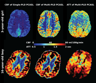 |
19 | Comparing Single and Multiple Delays methods for ASL Perfusion Quantification on Pediatric Neuroimaging
Moss Y Zhao1, Kristen Yeom1, Elizabeth Tong1, Rui Miguel Duarte1,2, Michael Moseley1, and Greg Zaharchuk1
1Department of Radiology, Stanford University, Stanford, CA, United States, 2Department of Neuroradiology, Hospital Beatriz Ângelo, Lisbon, Portugal
This study compared the CBF measurements using single and multiple delay ASL on 28 children without neurological or cerebrovascular disorders. Results showed that CBF of multiple delay ASL was significantly higher than the values of single delay ASL by a mean of 18%. CBF of both ASL techniques increased with age.
|
||
1224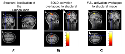 |
20 | Quantitative functional brain mapping imaging using Arterial Spin Labelling for safe neurosurgery
Giannina Rita Iannotti1,2, Isaure Nadin1, Quentin Tourdot3, Shahan Momjian1, Karl Schaller1, Karl Lovblad2, and Frédéric Grouiller4,5
1Department of Neurosurgery, University Hospital of Geneva, Geneva, Switzerland, 2Department of Radiology and Medical Informatics, University Hospital of Geneva, Geneva, Switzerland, 3Faculty of Pharmacy, University of Montpellier, France, Montpellier, France, 4Laboratory of Behavioral Neurology and Imaging of Cognition, Department of Fundamental Neuroscience, University of Geneva, Geneva, Switzerland, 5Swiss Center for Affective Sciences, University of Geneva, Geneva, Switzerland
Brain functional mapping is fundamental in pre-surgical workflow to target areas to be preserved. This work aims at validating ASL as alternative to BOLD to map eloquent cortex, by offering a quantitative measure of the perfusion associated to neuronal activity. Thirty healthy subjects underwent TMS motor mapping and executed a clenching-motor-task in a 3T-MR-scanner, while ASL and BOLD volumes were acquired simultaneously. The comparison between the global maxima of BOLD and ASL activations revealed that ASL localizes significantly deeper and more anteriorly than BOLD. ASL global maximum was found closer than BOLD to the most representative point of TMS mapping.
|
||
 Back to Meeting Home
 Back to the Program-at-a-Glance
The International Society for Magnetic Resonance in Medicine is accredited by the Accreditation Council for Continuing Medical Education to provide continuing medical education for physicians.
 Back to Meeting Home
 Back to the Program-at-a-Glance
 View the Presentation