Digital Poster
Applications of Perfusion Techniques II
Joint Annual Meeting ISMRM-ESMRMB & ISMRT 31st Annual Meeting • 07-12 May 2022 • London, UK

Digital Poster
Applications of Perfusion Techniques II
| Computer # | ||||
|---|---|---|---|---|
1302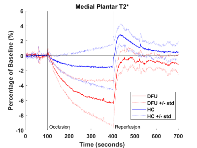 |
8 | Dynamic BOLD MRI Shows Greater Foot Ischemia and Blunted Reperfusion Following Cuff-Occlusion Challenge in Diabetic Patients with Foot Ulcers
Scott J. Edwards1, Jingting Yao2, Marcos Coutinho Schechter1, Maya Fayfman1, Gabriel Santamarina1, Paula Nesbeth1, Vincent Giacalone1, Gerado Blanco1, Rabindra Tirouvanziam1, Jessica Alvarez1, Benjamin Risk1, Ravi Rajani1, and David A. Reiter1
1Emory University, Atlanta, GA, United States, 2Department of Radiology, Massachusetts General Hospital, Boston, MA, United States
Diabetic foot ulcer (DFU) patients often face incomplete wound healing which leads to a long-term state of inflammation and risk of infection. Prolonged wound healing is thought to result in part from impaired tissue perfusion due to microvascular disease. This study applies dynamic blood-oxygenation-level-dependent (BOLD) imaging to quantify reperfusion of tissue after induced occlusion and ischemia in the foot. When compared to a healthy population, DFU patients show an increased ischemic state during cuff occlusion and decreased reperfusion after release. Quantification of cuff-occlusion parameters may assist in the classification of DFUs and be used to predict the prognosis and enhance treatment plan.
|
||
1303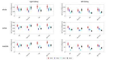 |
9 | Investigating the influence of ECG-triggering and respiration strategy on renal perfusion quantification using arterial spin labelling
Irène Brumer1, Simon Jonscher1, Indre Gineitaite1, Rebeca Echeverria-Chasco2, Lothar R. Schad1, María Asunción Fernández-Seara2, and Frank G. Zöllner1
1Computer Assisted Clinical Medicine, Mannheim Institute for Intelligent Systems in Medicine, Medical Faculty Mannheim, Heidelberg University, Mannheim, Germany, 2Department of Radiology, Clínica Universidad de Navarra, Pamplona, Spain
In this study, we investigated the influence of ECG-triggering and respiration strategy choice on renal perfusion. To this end, pCASL data of healthy volunteers was acquired at 3T. Acquisitions and processing followed the PARENCHIMA consensus. Processing consisted of groupwise registration, manual whole kidney segmentation and automated cortex/medulla segmentation. All calculated perfusion values were close to the expected range for healthy individuals. Preliminary results from four subjects suggest the influence of cardiac cycle is negligible and the choice of respiration strategy has little effect on renal perfusion values calculated from pCASL data, however additional data is necessary for more complete evaluation.
|
||
1304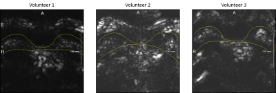 |
10 | Exploration of Velocity-Selective Inversion Arterial Spin Labeling for Breast Imaging
Mareike Alicja Buck1,2, Simon Konstandin1,3, Nora-Josefin Breutigam1, and Matthias Günther1,2,3
1Fraunhofer MEVIS, Bremen, Germany, 2University Bremen, Bremen, Germany, 3mediri GmbH, Heidelberg, Germany
In this work, the application of velocity-selective inversion ASL in healthy breast tissue is demonstrated for the first time. The measurements show a low measured signal in breast tissue. However, the prominent fat artifacts that appear in the images will need to be addressed in future work. The technique presented here has nonetheless been shown to hold promise in the field of non-invasive breast cancer diagnostics with further development, particularly the inclusion of background suppression to get clear perfusion signal.
|
||
1305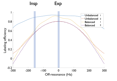 |
11 | Impact of breathing induced off-resonance on labeling efficiency in renal Arterial Spin Labeling
Manuel Taso1 and David C Alsop1
1Division of MRI Research, department of Radiology, Beth Israel Deaconess Medical Center, Harvard Medical School, Boston, MA, United States
Renal Arterial Spin Labeling (ASL) is becoming more widely used as a non-invasive technique for measuring renal perfusion and hence renal function. Free-breathing acquisitions with retrospective motion-correction are commonly used as it facilitates workflow and is robust against uncooperative patients. However, PCASL, especially the balanced implementation in which the same net gradient is used for both label and control, has been shown to be sensitive to off-resonance leading to a loss of labeling efficiency. We propose here a pilot study measuring B0 shifts during free-breathing to simulate its impact on free-breathing renal PCASL perfusion imaging.
|
||
1306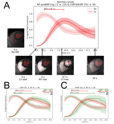 |
12 | Pulmonary transit time derived from routine perfusion cardiac magnetic resonance scans for non-invasive assessment of hemodynamics
Martin Segeroth1, David Winkel1, Shan Yang1, Alexander Sauter1, Jens Bremerich1, Michael Zellweger2, Christian Müller2, and Philip Haaf2
1Clinic for radiology and nuclear medicine, Universitätsspital Basel, Basel, Switzerland, 2Clinic for cardiology, Universitätsspital Basel, Basel, Switzerland
Pulmonary transit time (PTT) derived from routine cardiac magnetic resonance perfusion scans is a robust and easily obtainable noninvasive parameter of hemodynamics. PTT correlates with both LVEF, RVEF and NT-proBNP and could serve as a new global non-invasive parameter with a discriminative character for detecting cardiopulmonary dysfunction. We demonstrate that PTT has a high diagnostic accuracy in terms of exclusion and inclusion of heart failure as assessed by NT-proBNP, even in patients with preserved LVEF and RVEF.
|
||
1307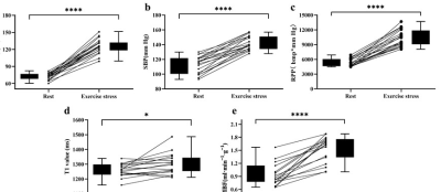 |
13 | Assessment of myocardium native T1 and perfusion using exercise CMR with a novel MRI-compatible supine ergometer Video Not Available
Bo He1 and Fabao Gao1
1Department of Radiology, West China Hospital, Sichuan University, Chengdu, China
This study investigated the feasibility of a novel compact MRI-compatible ergometer to evaluate myocardial tissue characteristics and blood flow. The results showed a significant increase in heart rate, RPP, native T1 and MBF by using this ergometer in healthy controls. Our study has shown an excellent reproducibility in measuring free-breathing native myocardial T1, MBF and MPR during exercise. The pilot testing demonstrated that the novel compact MRI-compatible ergometer was successful at inducing a cardiac stress state and can able to characterise exercise physiology at every stage allowing high quality MR imaging during the stress.
|
||
1308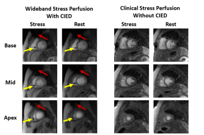 |
14 | Clinical evaluation of accelerated wideband cardiac perfusion pulse sequence in stress testing in patients with a CIED
Sarah M Schwartz1, Lexiaozi Fan1, Bradley D Allen1, Jeremy D Collins2, Kyungpyo Hong1, James C Carr1, Brandon Benefield1,3, Amit R Patel4, Daniel Kim1, and Daniel C Lee1,3
1Department of Radiology, Northwestern University Feinberg School of Medicine, Chicago, IL, United States, 2Department of Radiology, Mayo Clinic, Rochester, MN, United States, 3Department of Medicine, Division of Cardiology, Northwestern University Feinberg School of Medicine, Chicago, IL, United States, 4Department of Radiology, University of Chicago, Chicago, IL, United States
This study evaluates visual scores of image quality (conspicuity, artifact, noise; Likert scale 1[worst]-5[best], 3 acceptable) produced by a wideband cardiac perfusion sequence in patients with CIED and a standard sequence in matching non-device patients in the setting of vasodilated stress perfusion imaging. The median conspicuity scores were not significantly different between wideband (4) and standard (4). While the median artifact score was significantly worse for wideband (4) than standard (5), it was above the acceptable cutoff. The median noise score was significantly better for wideband than standard. Our wideband perfusion sequence produces diagnostically acceptable image quality in CIED patients.
|
||
1309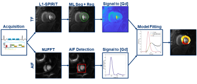 |
15 | Evaluation of Automatic Processing of Quantitative Perfusion Images in Patients with Suspected Coronary Artery Disease
Matthew Van Houten1, Xue Feng1, Yang Yang2, Patricia Rodriguez Lozano1, Christopher Kramer1, and Michael Salerno3
1University of Virginia, Charlottesville, VA, United States, 2Icahn School of Medicine at Mount Sinai, New York, NY, United States, 3Stanford University, Stanford, CA, United States
We developed high resolution quantitative perfusion sequence, along with DCNN network to automatically segment the image sets for motion correction. We deployed the acquisition and post-processing on patients with suspected coronary artery disease and compared the results to their coronary angiography findings. Our sequence and processing successfully match the angiography results.
|
||
1310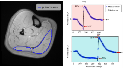 |
16 | BOLD MRI Perfusion in Peripheral Skeletal Muscle: Revealing the cuffing duration dependency Video Permission Withheld
Stefanie Eriksson1,2, Jonathan Arvidsson1,2, Suhela Abubakar Mohammad1, Edvin Johansson3, and Kerstin Lagerstrand1,2
1Department of Medical Radiation Sciences, Institute of Clinical Sciences, Sahlgrenska Academy, University of Gothenburg, Gothenburg, Sweden, 2Department of Medical Physics and Biomedical Engineering, Sahlgrenska University Hospital, Gothenburg, Sweden, 3Antaros Medical, Mölndal, Sweden
This study demonstrates the cuffing duration dependence of BOLD T2*-time curve parameters in peripheral skeletal muscle in the calf. This was made possible by the repeated measurements of healthy individuals using two different cuffing paradigms. The cuffing paradigms consisted of an ischemic period induced by cuffing of the thigh for 1.5 or 5 minutes, as well a 5 minute period of reactive hyperaemia. Systematic comparison of derived parameters was done from a descriptive representation of the T2* time curve.
|
||
1311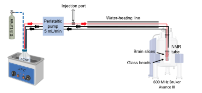 |
17 | Perfusion system for studying dynamic metabolomics in rat brain slices exposed to oxygen and glucose deprivation using 1H and 31P NMR
Deborah Hill1, Alicja Molska1, Trygve Andreassen1, and Marius Widerøe1
1Department of Circulation and Medical Imaging, Norwegian University of Science and Technology (NTNU), Trondheim, Norway
We designed an NMR-compatible bioreactor that allows real-time metabolic measurements of viable rat brain slices under normal conditions and under conditions mimicking hypoxic-ischemic (HI) brain injury. Special emphasis was put on providing physiological temperature in the system, a significant factor in the HI model. 1H NMR spectra were acquired to assess changes in brain metabolites. Peaks of lactic acid, NAA, glutamate, aspartate, and creatine were detected. By acquiring a time series of proton spectra, we observed significantly increased lactate levels over time when switching from normoxia to hypoxia while other metabolites remained stable over the whole experiment.
|
||
 Back to Meeting Home
 Back to the Program-at-a-Glance
The International Society for Magnetic Resonance in Medicine is accredited by the Accreditation Council for Continuing Medical Education to provide continuing medical education for physicians.
 Back to Meeting Home
 Back to the Program-at-a-Glance
 View the Presentation