Digital Poster
Flow Head to Toe I
Joint Annual Meeting ISMRM-ESMRMB & ISMRT 31st Annual Meeting • 07-12 May 2022 • London, UK

| Computer # | ||||
|---|---|---|---|---|
0791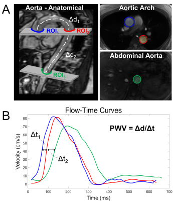 |
16 | Effects of Respiration on Aortic Pulse Wave Velocity using Retrospective Respiratory Gated Radial 2D Phase Contrast MRI
Grant S Roberts1, Zach S Bernhardt2, Sarah Lose2, Alyssa Pandos2, Kevin M Johnson1,3, Laura B Eisenmenger3, Ozioma Okonkwo2, and Oliver Wieben1,3
1Dept of Medical Physics, University of Wisconsin - Madison, Madison, WI, United States, 2Dept of Medicine, University of Wisconsin - Madison, Madison, WI, United States, 3Dept of Radiology, University of Wisconsin - Madison, Madison, WI, United States
Pulse wave velocity (PWV) is a non-invasive biomarker related to vascular stiffness and cardiovascular risk. Cine 2D phase contrast (2DPC) MRI can be used to assess aortic PWV. However, the effects of respiration on PWV measures remain unexplored. Here, we assessed the effects of respiration by retrospectively gating free breathing radially sampled 2DPC MRI data in both inspiration and expiration. Two axial slices were obtained in the aortic arch and abdominal aorta and time shift methods were used to calculate PWV between the ascending and abdominal aorta. PWV measurements from inspiration (7.58m/s) were significantly decreased relative to expiration (8.42m/s).
|
||
0792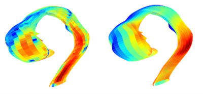 |
17 | Deep-Learning based Wall Shear Stress Assessments from 3D Aortic Shape
Bharath Ambale Venkatesh1, Nadjia Kachenoura2, Kevin Bouaou2, Thomas Dietenbeck2, Rithvik Rithvik Swamynathan3, Alban Redheuil2, Elie Mousseaux4, and Joao A C Lima3
1Radiology, Johns Hopkins University, Baltimore, MD, United States, 2Sorbonne Université, Paris, France, 3Johns Hopkins University, Baltimore, MD, United States, 4Université de Paris, Hôpital Européen Georges Pompidou, Paris, France
We develop deep learning for full aortic wall shear stress assessment using 3D aortic shapes and ascending aortic waveforms as input flow. Technically, this would reduce the acquisition time to less than a minute and the post-processing time to a few seconds.
|
||
0793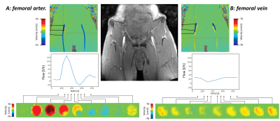 |
18 | Reproducibility of 4D flow MR angiography in peripheral vessels: A combined phantom and in vivo study
Martin Sieben1, Andreas Deistung1, Maik Rothe1, Richard Brill1, Walter Alexander Wohlgemuth1, and Alexander Gussew1
1University clinic and policlinic for Radiology, University Hospital Halle (Saale), Halle (Saale), Germany
Time-resolved 3D phase contrast MRA (4D flow MRA) is a technique providing morphological and quantitative 3D hemodynamic information of the blood vessel network without contrast agents and with a high temporal resolution. This study was performed to systematically validate flow measurements in peripheral vessels with high resolution, based on examinations in 4D flow phantom and in femoral vessels of healthy subjects. For this purpose, exams were run with varying spatial resolution and velocity encoding factors. It was shown, that the applied approach enables accurate and reproducible flow measurements by using venc factors of 120cm/s and isotrop spatial resolution of 1mm.
|
||
0794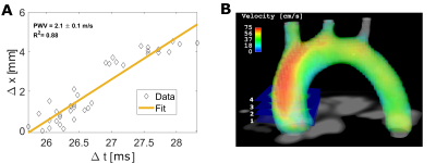 |
19 | Local and Global aortic pulse wave velocity in the murine aortic arch assessed by 4D flow magnetic resonance imaging
Patrick Winter1,2, Kristina Andelovic2,3, Thomas Kampf2,4, Susanne Schnell1, Alma Zernecke3, Wolfgang Rudolf Bauer5, Peter Michael Jakob2, and Volker Herold2
1Department of MR Physics, University of Greifswald, Greifswald, Germany, 2Department of Experimental Physics V, University of Wuerzburg, Wuerzburg, Germany, 3Institute of Experimental Biomedicine, University Clinics Wuerzburg, Wuerzburg, Germany, 4Department of Diagnostic and Interventional Neuroradiology, University Clinics Wuerzburg, Wuerzburg, Germany, 5Department of Medical Clinic and Policlinic I, University Clinics Wuerzburg, Wuerzburg, Germany As atherosclerosis is one of the main causes of death in industrial nations, noninvasive imaging modalities for studying its underlying mechanisms are in great demand. The quantification of hemodynamic parameters such as pulse wave velocity (PWV) assessed by flow sensitive magnetic resonance imaging (MRI) is a promising tool to observe plaque progression in preclinical models. Mostly, a global PWV value is assessed, however, previous studies already pointed to heterogeneous elasticity profiles in the presence of atherosclerotic plaques. Here, we present the measurement of local PWV values in the murine aortic arch assessed by 4D-flow MRI for spatially resolved elasticity measurements. |
||
0795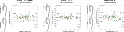 |
20 | Fully automated aortic pulse wave velocity calculation from 4D flow MRI with AI-based segmentation
Ethan M I Johnson1, Haben Berhane1, Michael Scott1, Kelly Jarvis1, Alex Barker2, and Michael Markl1
1Northwestern University, Chicago, IL, United States, 2University of Colorado Anschutz Medical Campus, Aurora, CO, United States
Pulse wave velocity (PWV) is an important measure of vessel stiffness and can be quantified from 4D flow MRI. Accurate assessment of PWV requires placement of multiple analysis planes in an imaged volume, which typically is performed manually to some degree, such as through use of a vessel segmentation made by an experienced human observer. Here we present evaluation of a fully-automated method for calculating PWV in the thoracic aorta, incorporating an artificial intelligence (AI) convolutional neural network to segment the aorta. Estimates of PWV using AI segmentations were compared those from human-observer segmentations and found to be similarly reliable.
|
||
0796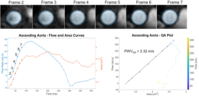 |
21 | Effect of Temporal Resolution on Flow-Area Aortic Pulse Wave Velocity Measures Using Phase Contrast and Balanced SSFP MRI: A Preliminary Study
Tarun Naren1, Grant Steven Roberts1, and Oliver Wieben1
1Medical Physics, University of Wisconsin - Madison, Madison, WI, United States
The flow-area (QA) method is a promising phase contrast (PC) technique to assess local aortic pulse wave velocity (PWV). However, validation for reduced temporal resolutions is lacking. Data analysis and automated post-processing are compromised because of varying PC image quality throughout the cardiac cycle. In this study, we acquire PC and bSSFP data at three temporal resolutions and measure QAPWV using PC data alone and by combining PC flow measures with bSSFP areas. We found that the highest temporal resolution datasets (10ms) provide the most accurate PWV measures and the method combining bSSFP and PC was less accurate.
|
||
0797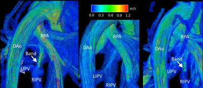 |
22 | 4D Flow MRI Analysis of Flow, Velocity, and Cardiac Flow Compartments in a Swine Model of Pulmonary Hypertension
Daniel P Seiter1, Betty Allen2,3, Diana M Tabima3, Timothy A Hacker4, Phil Corrado1, Thekla H Oechtering5, Scott B Reeder1,5, Naomi C Chesler6,7, and Oliver Wieben1,5
1Medical Physics, University of Wisconsin-Madison, Madison, WI, United States, 2Surgery, University of Wisconsin-Madison, Madison, WI, United States, 3Biomedical Engineering, University of Wisconsin-Madison, Madison, WI, United States, 4Medicine, University of Wisconsin-Madison, Madison, WI, United States, 5Radiology, University of Wisconsin-Madison, Madison, WI, United States, 6Edwards Lifesciences Foundation Cardiovascular Innovation and Research Center, University of California, Irvine, Irvine, CA, United States, 7Biomedical Engineering, University of California, Irvine, Irvine, CA, United States
Pulmonary hypertension is a known consequence of left heart failure. However, little is known about the pulmonary vascular and right ventricular changes caused by increased pulmonary venous pressure independent of left heart failure. By surgically banding the inferior pulmonary vein confluence in swine, we created pulmonary venous hypertension without damage to the left heart. Here, we report results from 4D flow MRI analysis of vessel flow, vessel velocity, cardiac flow compartments, and pressures measured via right heart catheterization in this novel model.
|
||
0798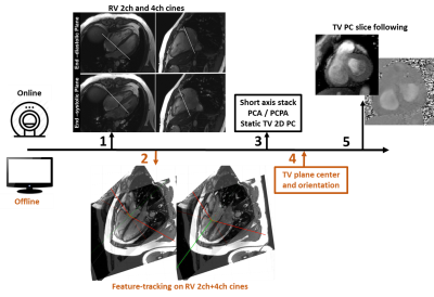 |
23 | Towards 2D phase-contrast of tricuspid valve flow
Jerome Lamy1, Felicia Seemann2, Ricardo Gonzales Vera3, Einar Heiberg4, and Dana Peters1
1Yale University, New Haven, CT, United States, 2National Institutes of Health, Bethesda, MD, United States, 3University of Oxford, Oxford, United Kingdom, 4Lund University, Lund, Sweden
Tricuspid valve flow evaluation is important for evaluation of regurgitation, used as a surrogate of pressures in the RV and as a criterion for evaluation of diastolic dysfunction. While 4D flow methods have interesting possibilities for retrospective valve flow evaluation its practical use is still limited. Here we present a method for direct tricuspid valve flow evaluation in a single breath-hold using 2D valve-following phase contrast sequence with a dynamic slice plane.
|
||
0799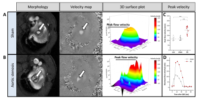 |
24 | Identification of Subtle Alterations for Demarcation of Disease Transition in Murine Aortic Valve Stenosis by Multiparametric MRI
Christine Quast1, Christoph Jacoby1, Malte Kelm1, and Ulrich Flögel1
1Heinrich Heine University, Düsseldorf, Germany
Aortic valve stenosis (AS) is one of the most frequent valve diseases in the elderly with relevant prognostic impact. Becausesufficient experimental models were lacking, we recently refined a murine model of gradable experimental AS closely mimicking disease progression in humans. Here, we aimed at developing a comprehensive MRI approach for simultaneous assessment of changes in valvular, myocardial as well as aortic function in mice. We demonstrate that in this murine model high resolution MRI is capable to reliably display transvalvular aortic flow profiles with concomitant quantification of structural and functional changes inaortic valve, left ventricle, and ascending aorta.
|
||
0800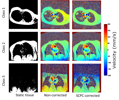 |
25 | Clinical impact of automatic failure mode evaluation using self-calibrated phase contrast correction in phase contrast cardiac MRI
Ana Beatriz Solana1, Savine C.S. Minderhoud2,3, Piotr A. Wielopolski3, Juan A. Hernandez-Tamames3,4, Ricardo P.J. Budde2,3, Willem A. Helbing5, Martin A. Janich1, and Alexander Hirsch2,3
1GE Healthcare, Munich, Germany, 2Department of Cardiology, Erasmus MC, University Medical Center Rotterdam, Rotterdam, Netherlands, 3Department of Radiology and Nuclear Medicine, Erasmus MC, University Medical Center Rotterdam, Rotterdam, Netherlands, 4Department of Imaging Physics, TU Delft, Delft, Netherlands, 5Department of Pediatric Cardiology, Erasmus MC, University Medical Center Rotterdam, Rotterdam, Netherlands
Phase contrast (PC) cardiac MRI is clinically used to quantify flow. The quantification accuracy is diminished by background phase errors. Stationary tissue-based background phase correction algorithms are commercially available, but their accuracy is still under evaluation. Here, we investigate if a self-calibrated phase contrast (SCPC) algorithm which includes automatic failure mode classification, can improve accuracy in a large single-vendor multi-scanner study including 346 PC scans. Our results show that SCPC improves flow quantification accuracy and can identify most PC scans which are unreliable regardless of stationary tissue correction or not.
|
||
 Back to Meeting Home
 Back to the Program-at-a-Glance
The International Society for Magnetic Resonance in Medicine is accredited by the Accreditation Council for Continuing Medical Education to provide continuing medical education for physicians.
 Back to Meeting Home
 Back to the Program-at-a-Glance
 View the Presentation