Digital Poster
New CMR Methods for Anatomy & Function (Micro- to Macro-) I
Joint Annual Meeting ISMRM-ESMRMB & ISMRT 31st Annual Meeting • 07-12 May 2022 • London, UK

Digital Poster
New CMR Methods for Anatomy & Function (Micro- to Macro-) I
| Computer # | ||||
|---|---|---|---|---|
1554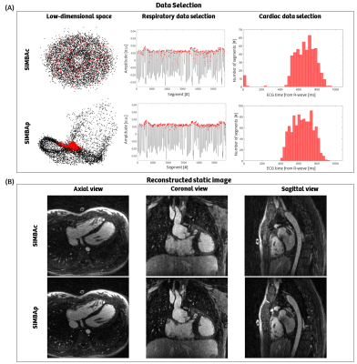 |
9 | Automated subject-specific pixels selection for improved image reconstruction in free-running coronary MRA using SIMBA
Ludovica Romanin1,2, Christopher W. Roy2, Milan Prsa3, Tobias Rutz4, Estelle Tenisch2, Matthias Stuber2,5, and Davide Piccini1,2
1Advanced Clinical Imaging Technology, Siemens Healthcare AG, Lausanne, Switzerland, 2Department of Diagnostic and Interventional Radiology, Lausanne University Hospital and University of Lausanne, Lausanne, Switzerland, 3Division of Pediatric Cardiology, Woman-Mother-Child Department, Lausanne University Hospital and University of Lausanne, Lausanne, Switzerland, 4Service of Cardiology, Heart and Vessel Department, Lausanne University Hospital and University of Lausanne, Lausanne, Switzerland, 5Center for Biomedical Imaging (CIBM), Lausanne, Switzerland
This work proposes an automated subject-specific individual pixels selection as an alternative to a fixed coil selection for the initialization of the input data to a similarity-driven multi-dimensional binning algorithm (SIMBA) for free-running motion-suppressed whole-heart acquisitions. By selecting timeseries with a high low-frequency energy content, we include only pixels with respiratory and cardiac information. Compared to the original method, this leads to a more accurate choice of end-expiration and diastolic phases for the reconstruction of sharp whole-heart and coronary images. Moving forward, the method needs to be refined, optimized and tested to further improve the image quality.
|
||
1555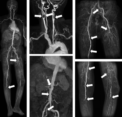 |
10 | One-stop subtractionless first-pass CS-whole-body MRA with once contrast injection of 0.15mmol/kg dose: first clinical experience Video Permission Withheld
Qing FU1, Jia-wei Wu1, Jing-yang Li1, Peng Sun2, and Xiang-chuang Kong1
1Department of Radiology, Union Hospital, Tongji Medical College, Huazhong University of Science and Technology, Wuhan, China, 2Philips Healthcare, Beijing, China
Conventional subtracted whole-body CE-MRA is time-consuming and requires experiences for operators. Instead, one-stop subtractionless CS-MRA in our study could depict the whole-body arterial vasculatures with only once contrast injection and only 0.15 mmol/kg dose, which assisted by multiple techniques including reverse k-space filling mode, total imaging matrix system, automatic table moving, automatic coil selection and especially the CS technique. The comparative results supported it feasible with accepted image qualities and a faster acquisition time in clinical use.
|
||
1556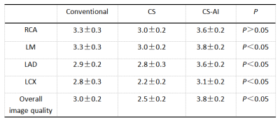 |
11 | Accelerated whole-heart mDixon coronary MRA with a deep learning constrained CompressedSENSE: a feasibility study
Xi Wu1,2, Jiayu Sun1, Xiaoyong Zhang3, and Zhigang Wu4
1Department of Radiology, West China Hospital of Sichuan University, Chengdu, China, 2North Sichuan Medical College, Nanchong, China, 3Philips Healthcare, Chengdu, China, 4Philips Healthcare, Shenzhen, China Currently, modified dixon (mDixon) gradient echo sequence is widely used for respiratory navigated whole-heart coronary magnetic resonance angiography (MRA) for the evaluation of coronary anatomy and abnormalities. However, the main drawback of this approach is that the scan time is longer and prone to interference with motion artifacts. In this study, we investigated the utility of whole-heart coronaryMRA using accelerated mDixonwith compressed sensing (CS) and artificial intelligence(AI) technologies at 3Tesla. The initial results showedtheCS-AI mDixontechnique has potential to be the most viable alternative to enhance the clinical workflow of coronary MRA. |
||
1557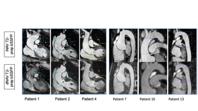 |
12 | Contrast-free 3D Cartesian MRA of the thoracic aorta in 3 min: preliminary clinical evaluation
Anastasia Fotaki1, Camila Munoz1, Alina Hua1, Yaso Emmanuel2, Karl Philip Kunze1,3, Radhouene Neji1,4, Pier Giorgio Masci1, René Botnar1,5, and Claudia Prieto1,5
11School of Biomedical Engineering and Imaging Sciences, King's College London, London, United Kingdom, 2Guy's and St Thomas' Hospital, London, United Kingdom, 3MR Research Collaborations, Siemens Healthcare Limited, Frimley, United Kingdom, 42MR Research Collaborations, Siemens Healthcare Limited, Frimley, United Kingdom, 5Escuela de Ingeniería, Pontificia Universidad Católica de Chile, Santiago, Chile
Cardiovascular Magnetic Resonance Angiography is established for serial monitoring of thoracic aortic disease. Limitations of the current approach include the diaphragmatic navigation, leading to long acquisition times and the correction of the aortic motion, solely in one direction. We propose a T2-prep bSSFP sequence that incorporates image-based navigation and inline non-rigid motion-correction for efficient, free-breathing and isotropic aortic imaging. Comparison between the conventional and the research sequence shows similar diagnostic confidence with both techniques in much shorter acquisition time [3.11.1min (iNAV) versus 11.5 min3.4 (dNAV),P<0.0001] and good agreement of aortic measurements between both techniques, holding promise for forthcoming clinical adoption.
|
||
1558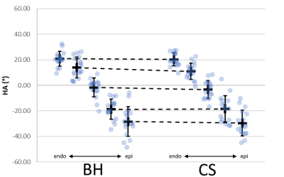 |
13 | Investigating the clinical viability of a control system-based slice-following sequence for free-breathing cardiovascular DTI
Stephen G Jermy1,2, Elizabeth M Tunnicliffe3, Ntobeko A B Ntusi2,4, and Ernesta M Meintjes1,2
1Division of Biomedical Engineering, Department of Human Biology, University of Cape Town, Cape Town, South Africa, 2Cape Universities Body Imaging Centre (CUBIC), University of Cape Town, Cape Town, South Africa, 3Oxford Centre for Magnetic Resonance Research (OCMR), Radcliffe Department of Medicine, University of Oxford, Oxford, United Kingdom, 4Department of Medicine, University of Cape Town, Cape Town, South Africa
A prospective respiratory motion correction control system, capable of slice-following, was tested on a 3 T clinical MRI scanner. The performance of the control system was compared against the most common respiratory motion compensation technique, namely multiple breath-holds. Both techniques produced similarly consistent results. The control system technique was able to reduce the acquisition time by acquiring data with 100% respiratory efficiency
|
||
1559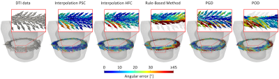 |
14 | Comparison of Interpolation Methods for Ex-vivo and In-vivo Cardiac Diffusion Tensor Imaging
Johanna Stimm1, Sebastian Kozerke1, and Christian Stoeck1,2
1Institute for Biomedical Engineering, University and ETH Zurich, Zuerich, Switzerland, 2Division of Surgical Research, University Hospital Zurich, University Zurich, Zurich, Switzerland
To address the challenge of low resolution and limited number of slices in in-vivo cDTI, and the need for 3D structural information in image-based modeling, we compare five interpolation techniques of predominant cardiomyocyte orientation: two low-rank models, one rule-based method and two tensor interpolation approaches. The direct tensor interpolation approaches result in the smallest errors, followed by the low-rank models and the rule-based method. In view of an optimal experimental design in-vivo, the ex-vivo experiments suggest a larger benefit of increasing in-plane resolution rather than SNR, and a non-linear increase in error for less than five short-axis slices.
|
||
1560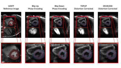 |
15 | Comparison of Blip-Up and Blip-Down EPI Distortion Correction Methods for Cardiac Diffusion Tensor Imaging
Tyler E Cork1,2,3, Matthew J Middione1,2, Michael Loecher1,2, Congyu Liao1, Kévin Moulin4,5, Kawin Setsompop1,6, and Daniel B Ennis1,2,7
1Department of Radiology, Stanford University, Stanford, CA, United States, 2Division of Radiology, Veterans Administration Health Care System, Palo Alto, CA, United States, 3Department of Bioengineering, Stanford University, Stanford, CA, United States, 4CREATIS Laboratory, University of Lyon, Lyon, France, 5Department of Radiology, University Hospital Saint-Etienne, Saint-Etienne, France, 6Department of Electrical Engineering, Stanford University, Stanford, CA, United States, 7Cardiovascular Institute, Stanford University, Stanford, CA, United States To maintain minimal scan times, cardiac Diffusion Tensor Imaging (cDTI) uses an echo-planar imaging (EPI) readout. Off-resonance, that causes geometric distortion in EPI, remains an obstacle that degrades image quality and can affect the underlying quantitative information. In cDTI, the lung/liver/heart interface amplifies the effect of geometric distortion. Distortion correction algorithms, such as TOPUP and DR-BUDDI, have proved to adequately correct distortion in neuroimaging, but limited work has been done for the heart. A first look at comparing these two correction strategies head-to-head was evaluated and resulting in TOPUP as a slightly better tool addressing distortion correction in the heart. |
||
1561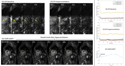 |
16 | A comparison of oxygenation-sensitive cardiac MRI methods after hyperventilation
Qi Huang1,2, Jason Mendes1, Ganesh Adluru1, and Edward DiBella1
1Utah Center for Advanced Imaging Research (UCAIR), University of Utah, Salt Lake City, UT, United States, 2Biomedical Engineering, University of Utah, Salt Lake City, UT, United States Here we study SSFP-based OS-CMR and EPI-based T2*/T2 methods for detection of myocardial oxygenation changes. These techniques have been used previously, and OS-CMR has been investigated in many studies. However, quantifying T2* and T2 every heartbeat may add information to the semi-quantitative OS-CMR method. Here a limited study of subjects on a 3T scanner is performed. Preliminary quantitative results including comparisons between the methods are shown. |
||
1562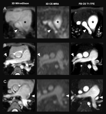 |
17 | Evaluation of non-contrast free-breathing compressed SENSE T1-TFE cardiac MRI at 3T in young children with congenital heart diseases
Inka Ristow1, Shuo Zhang2, Caroline-Viktoria Hancken3, Maria Stark4, Carsten Rickers5, Christoph Katemann2, Peter Bannas1, Gerhard Adam1, Jochen Herrmann3, Lennart Well1, and Julius Matthias Weinrich3
1Department of Diagnostic and Interventional Radiology and Nuclear Medicine, University Medical Center Hamburg-Eppendorf, Hamburg, Germany, 2Philips GmbH Market DACH, Hamburg, Germany, 3Department of Diagnostic and Interventional Radiology and Nuclear Medicine, Section of Pediatric Radiology, University Medical Center Hamburg-Eppendorf, Hamburg, Germany, 4Institute of Medical Biometry and Epidemiology, University Medical Center Hamburg-Eppendorf, Hamburg, Germany, 5University Heart Center, Adult Congenital Heart Disease Unit, University Medical Center Hamburg-Eppendorf, Hamburg, Germany High-quality cardiac MRI at 3T in sedated children with congenital heart disease (CHD) is challenging, because of field inhomogeneities and free breathing motions. Therefore, we aimed to employ the non-contrast free-breathing generic T1-weighted turbo-field echo (TFE) sequence with compressed sensing reconstruction for assessment of vessel diameters and evaluation of the intra- and extracardiac structures in children with CHD aged <5 years. Our results show a highly reliable clinical applicability for intra- and extracardiac structures with significantly higher accuracy in comparison to contrast-enhanced MR-angiography and 3D-Dixon sequences. |
||
1563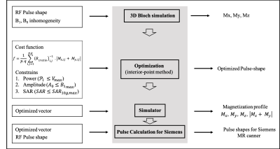 |
18 | Non-selective refocusing pulse optimization for T2-prep in free-breathing 3D CMRA at 7T with single-transmit coil
Shubhajit Paul1, Raphael Tomi-Tricot1,2, Karl Philipp Kunze1,2, Shaihan Malik1,3, Joseph V. Hajnal1,3, Philippa Bridgen1,3, Thomas Wilkinson1,3, Sharon Giles1,3, Radhouene Neji1,2, Reza Razavi1, Claudia Prieto1,4, and René Michael Botnar1,4
1School of Biomedical Engineering and Imaging Sciences, King’s College London, London, United Kingdom, 2MR Research Collaborations, Siemens Healthcare Limited, Frimley, United Kingdom, 3The London Collaborative Ultra high field System (LoCUS), London, United Kingdom, 4Escuela de Ingeniería, Pontificia Universidad Católica de Chile, Santiago, Chile
In spite of the promised increase in signal-to-noise ratio and contrast to noise ratio, 3D whole-heart imaging at 7T remains challenging predominantly due to B1, B0 inhomogeneity, specific absorption rate limitations and coil-depth issues for single transmit. Here, we developed a toolbox to optimize the adiabatic hyperbolic secant refocusing pulse with respect to B1 and B0 inhomogeneity typically used for T2-preparation in non-contrast 3D Coronary Magnetic Resonance Angiography (CMRA). We present phantom and in vivo data for 3D CMRA employing a low excitation flip-angle scheme.
|
||
 Back to Meeting Home
 Back to the Program-at-a-Glance
The International Society for Magnetic Resonance in Medicine is accredited by the Accreditation Council for Continuing Medical Education to provide continuing medical education for physicians.
 Back to Meeting Home
 Back to the Program-at-a-Glance
 View the Presentation