Digital Poster
Flow Head to Toe III
Joint Annual Meeting ISMRM-ESMRMB & ISMRT 31st Annual Meeting • 07-12 May 2022 • London, UK

| Computer # | ||||
|---|---|---|---|---|
1225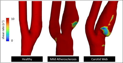 |
21 | Shear-rate values in the carotid bifurcation in subjects with carotid webs, subjects with atherosclerotic lesions, and healthy subjects.
Retta El Sayed1,2, Alireza Sharifi2, Charlie Park2, Diogo Haussen3, Jason Allen1,2,3, and John N Oshinski1,2
1Department of Biomedical Engineering, Georgia Institute of Technology & Emory University, Atlanta, GA, United States, 2Department of Radiology & Imaging Sciences, Emory University, Atlanta, GA, United States, 3Department of Neurology, Emory University, Atlanta, GA, United States
Carotid webs (CaWs) have been linked to cryptogenic strokes and therefore quantifying different hemodynamic parameters is essential to understand the mechanism of clot formation in patients. Computational fluid dynamics (CFD) based on patient-specific geometry and PCMR inlet flow conditions was conducted on five subjects with CaWs, five subjects with mild atherosclerosis (with a similar degree of luminal narrowing), and three healthy control subjects. The total area within the carotid bifurcation that contained low shear rate values associated with clot formation is significantly larger in CaW subjects compared to subjects with atherosclerotic lesions or healthy subjects.
|
||
1226 |
22 | Repeatability and reliability of 2D phase-contrast and 4D flow MRI when measuring cerebral arterial and venous pulsatility
Alasdair Graeme Morgan1, Michael Graeme Thrippleton1, Ning Jin2, Joanna Wardlaw1, and Ian Marshall1
1Centre for Clinical Brain Sciences, University of Edinburgh, Edinburgh, United Kingdom, 2Siemens Medical Solutions USA, Inc., Cleveland, OH, United States
We examined the test-retest repeatability and intraobserver reliability of 4D flow MRI while assessing the pulsatility and flow rates of a variety of cerebral arteries and veins in healthy volunteers. A subset of these vessels were also measured using 2D phase-contrast MRI, a more established method, to assess the level to which the lower-resolution (but higher-coverage) 4D method could compare to its 2D counterpart. Flow pulsatility appears to play a role in the development of cerebral small vessel disease and so testing the capabilities of 4D flow in this context is an important step before applying it to clinical studies.
|
||
1227 |
23 | 3D-Simulation of Flowing Spins using the Lattice Boltzmann Method with Maxwell Diffuse Reflection Boundaries
Ansgar Adler1, Philip Schaten2, Yong Wang3, and Martin Uecker4
1Insitut of Medical Engineering Graz, Graz, Austria, 2Institut Diagnostic and Interventional Radiologie, Göttingen, Germany, 3Max Planck Institute for Dynamics and Self-Organization, Göttingen, Germany, 4Institut of Medical Engineering, Graz, Austria
The lattice Boltzmann method (LBM) is a powerful technique to simulate complex fluid dynamical systems. The extension of the LBM to flow with a spin degree of freedom in external magnetic fields has shown promising results in 2D. Here, we describe first 3D numerical results utilizing extended kinetic-theory based boundary conditions. The simulation of a laminar pipe flow is verified on the Pouiseuille flow and shown to be able to predict signal changes caused by in-flow effects as expected from previous flow experiments.
|
||
1228 |
24 | Wall shear stress in abdominal aortic aneurysms: a 4D Flow MRI case-control study
Chiara Trenti1,2, Magnus Ziegler1,2, Niclas Bjarnegård1, Tino Ebbers1,2, Marcus Lindenberger1,3, and Petter Dyverfeldt1,2
1Department of Health, Medicine and Caring Sciences, Linköping University, Linköping, Sweden, 2Center for Medical Image Science and Visualization (CMIV), Linköping University, Linköping, Sweden, 3Department of Cardiology, Linköping University Hospital, Linköping, Sweden
Current guidelines for risk stratification of abdominal aortic aneurysm are based on vessel diameter and are not sufficient to prevent catastrophic events. Wall shear stress based parameters (WSS, OSI and RRT) are potential markers for AAA altered hemodynamics. Here, WSS vectors were computed from 4D flow MRI in the whole aorta of patients with AAA, age-matched elderly controls, and young normal controls. The aorta was divided in five segments and average values were computed in each segment. AAA had lower WSS and higher RRT in the IAA compared to proximal segments and to the age-matched controls, but not higher OSI.
|
||
1229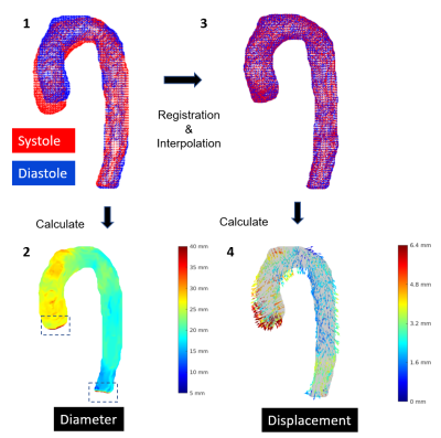 |
25 | Reproducibility of aortic diameter and displacement derived from free-breathing 3D balanced steady-state free precession CINE images at 3T
Renske Merton1, Eric M. Schrauben1, Gustav J. Strijkers2, Aart J. Nederveen1, and Pim van Ooij1,3
1Radiology and Nuclear Medicine, Amsterdam University Medical Centers, Amsterdam, Netherlands, 2Department of Medical Engineering & Physics, Amsterdam University Medical Centers, Amsterdam, Netherlands, 3University Medical Center Utrecht, Utrecht, Netherlands Capturing 3D aortic motion over the heart cycle may give insight into a new biomarker for aortic disease and potentially improve the measurement of aortic hemodynamic parameters. An isotropic, free-breathing, respiratory-corrected 3D CINE balanced steady-state free precession imaging technique of the thoracic aorta was developed to investigate scan-rescan reproducibility of aortic diameter and displacement measures in nine healthy volunteers. Scan-rescan diameter was highly reproducible (CV<10% , ICC=0.85-0.86 and Pearson’s ρ=0.87) while displacement was more variable (CV=34-42%, ICC=0.34-0.50, ρ=0.59-0.72). These results are encouraging for future studies investigating aortic motion in health and aortopathy. |
||
1230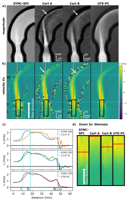 |
26 | Towards fast but accurate velocity quantification with 3D ultra-short TE phase contrast imaging
Katja Degenhardt1, Christoph Stefan Aigner1, Simon Schmidt2,3, Fabian J. Kratzer3, Max Müller4, Armin M. Nagel3,4, Jeanette Schulz-Menger5,6, and Sebastian Schmitter1,2,3
1Physikalisch-Technische Bundesanstalt (PTB), Braunschweig and Berlin, Germany, 2Center for Magnetic Resonance Research, University of Minnesota, Minneapolis, MN, United States, 3Division of Medical Physics in Radiology, German Cancer Research Center (DKFZ), Heidelberg, Germany, 4Institute of Radiology, University Hospital Erlangen, Friedrich-Alexander-Universität Erlangen-Nürnberg (FAU), Erlangen, Germany, 5Department of Cardiology and Nephrology, Experimental and Clinical Research Center, a joint cooperation between the Charité Medical Faculty and the Max-Delbrueck Center for Molecular Medicine and HELIOS Hospital Berlin Buch, Berlin, Germany, 6DZHK (German Center for Cardiovascular Research), partner site Berlin, Berlin, Germany
The aim of this work is to develop and investigate an accurate sequence for MR flow quantification. We utilized a 3D ultra-short echo time (UTE) flow sequence to minimize the displacement artifact that frequently occurs in MR flow imaging. In UTE acquisitions, the position is encoded at the beginning of the readout. Thus, the time difference between velocity encoding and spatial position encoding is minimized. This leads to an improved accuracy of the velocity quantification. The sequence was tested and validated against a reference sequence and a conventional 4D flow sequence in a flow experiment in vitro at 3T.
|
||
1231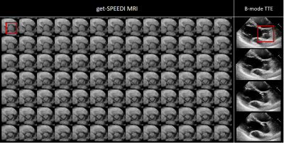 |
27 | Imaging of aortic valve dynamics: comparison between sub-millisecond MRI and echocardiography Video Permission Withheld
Alessandro M Scotti1, Qingfei Luo1, Zheng Zhong2, Noreen T Nazir3, Karen L Xie4, and Xiaohong Joe Zhou1,4,5,6
1Center for MR Research, University of Illinois at Chicago, Chicago, IL, United States, 2Department of Radiology, Stanford University, Stanford, CA, United States, 3Division of Cardiology, Department of Medicine, University of Illinois at Chicago, Chicago, IL, United States, 4Department of Radiology, University of Illinois at Chicago, Chicago, IL, United States, 5Department of Neurosurgery, University of Illinois at Chicago, Chicago, IL, United States, 6Department of Biomedical Engineering, University of Illinois at Chicago, Chicago, IL, United States
The aortic valve dynamics is traditionally examined using ultrasound echocardiography in clinical practice. Recently, a novel MRI technique capable of sub-millisecond temporal resolution, coined get-SPEEDI MRI, was successfully applied to visualize the aortic valve dynamics. We have evaluated both get-SPEEDI and echocardiography on healthy human subjects and found a substantial agreement between both techniques in the characterization of the aortic valve opening and closing phases. The sub-millisecond temporal resolution of get-SPEEDI allows for the measurement of steep AVA variations and the visualization of detailed features that may reflect the cardiac function and physiology.
|
||
 Back to Meeting Home
 Back to the Program-at-a-Glance
The International Society for Magnetic Resonance in Medicine is accredited by the Accreditation Council for Continuing Medical Education to provide continuing medical education for physicians.
 Back to Meeting Home
 Back to the Program-at-a-Glance
 View the Presentation