Digital Poster
Cardiac Anatomy & Tissue Characterization III
Joint Annual Meeting ISMRM-ESMRMB & ISMRT 31st Annual Meeting • 07-12 May 2022 • London, UK

Digital Poster
Cardiac Anatomy & Tissue Characterization III
| Computer # | ||||
|---|---|---|---|---|
1564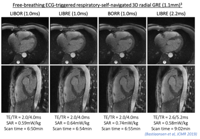 |
19 | Fat-free MRI with fast and power-optimized off-resonant LIBOR pulses
Jessica AM Bastiaansen1,2, Davide Piccini3, and Adele Mackowiak4
1Department of Diagnostic, Interventional and Pediatric Radiology (DIPR), Inselspital, Bern University Hospital, University of Bern, Bern, Switzerland, 2Translational Imaging Center, sitem-insel, Bern, Switzerland, 3Advanced Clinical Imaging Technology, Siemens Healthcare, Lausanne, Switzerland, 4Department of Radiology, Lausanne University Hospital (CHUV) and University of Lausanne (UNIL), Lausanne, Switzerland
Flexible off-resonant RF pulse designs such as LIBRE and BORR are inspired by conventional binomial 1-(180)-1 water excitation pulses and perform better in terms of fat suppression. To abbreviate scanning times, BORR and LIBRE pulse durations can be shortened by increasing the RF excitation frequency at the expense of increased RF power. To reduce RF power, we developed a novel off-resonant RF pulse design based on the conventional 1-(90)-1 water excitation pulse. LIBOR, compared with LIBRE and BORR, reduced the required RF power while maintaining short pulse durations and effective fat suppression in simulations, knee, and cardiac MRI at 3T.
|
||
1565 |
20 | Balanced-force shim system for correcting field inhomogeneities due to implanted cardioverter-defibrillators (ICDs): validation in phantoms
Ehud Jeruham Schmidt1, Mirko Hrovat2, Henry R Halperin1, and Aravindan J Kolandaivelu1
1Medicine (Cardiology), Johns Hopkins University, School of Medicine, Baltimore, MD, United States, 2Mirtech Inc., Boston, MA, United States
A resistive-shim system was constructed to correct the >100ppm magnetic-field inhomogeneity caused by implanted ICDs, which leads to signal loss and blurring in cardiac MRI. A plastic platform, held to the MRI stretcher, allows human-subject insertion, and holds a sensitive 720 Ampere*turns dipole-approximating shim-coil on a movable-fixture above the chest. Shim-coil is electronically decoupled from gradients and body-coil-RF. ICD field-inhomogeneity is measured with B0 mapping, and a correction field computed (shim-coil-center-location, DC-current-intensity). Shim-coil is moved to shim-coil-center-location, and a power-supply sends DC-current to the shim-coil. System was tested on phantoms overlaid with ICDs and corrected >75ppm inhomogeneity over 50x50x50mm3 FOV.
|
||
1566 |
21 | In-vivo comparison of Active Tracking based cardiac triggering and ECG-triggering for cardiac MR-thermometry
Ronald Mooiweer1,2, Rainer Schneider3, Axel Joachim Krafft3, Katy Empanger4, Jason Stroup4, Alexander Paul Neofytou1, Rahul K Mukherjee1, Steven Williams1,5, Tom Lloyd4, Mark O'Neill1, Reza Razavi1, Tobias Schaeffter6, Radhouene Neji1,2, and Sébastien Roujol1
1School of Biomedical Engineering and Imaging Sciences, Faculty of Life Sciences and Medicine, King's College London, London, United Kingdom, 2MR Research Collaborations, Siemens Healthcare Limited, Frimley, United Kingdom, 3Siemens Healthcare GmbH, Erlangen, Germany, 4Imricor Medical Systems, Burnsville, MN, United States, 5University of Edinburgh, Edinburgh, United Kingdom, 6Physikalisch-Technische Bundesanstalt (PTB), Braunschweig and Berlin, Germany
Continuous AT was used for cardiac synchronization of MR-thermometry with on-line signal filtering, calibration, and triggering. Experiments in a porcine animal model, where AT-trig was compared to ECG triggering, showed that AT-trig could potentially serve as alternative cardiac triggering strategy in situations where ECG triggering is not effective.
|
||
1567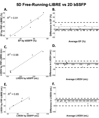 |
22 | 5D Free-Running-LIBRE MRI for 3D whole-heart Cine: A pilot study in patients at 3T
Katerina Eyre1, Michael Chetrit1, Lisa Leroi2, Jérôme Yerly3,4, Mitchel Benovoy2, Davide Piccini3,5, Matthias Stuber3,4, Matthias Friedrich1,2,6,7,8, and Jessica AM Bastiaansen3,9,10
1Radiology, McGill University Health Centre (MUHC), Montreal, QC, Canada, 2Circle Cardiovascular Imaging Inc., Calgary, AB, Canada, 3Diagnostic and Interventional Radiology, Le Centre Hospitalier Universitaire Vaudois (CHUV), Lausanne, Switzerland, 4Center for BioMedical Imaging (CIBM), Lausanne, Switzerland, 5Advanced Clinical Imaging Technology, Siemens Healthcare AG, Lausanne, Switzerland, 6Radiology, Université de Montréal, Montreal, QC, Canada, 7Cardiac Sciences, University of Calgary, Calgary, AB, Canada, 8Heidelberg University, Heidelberg, Germany, 9Diagnostic, Interventional and Pediatric Radiology (DIPR), Inselspital, Bern University Hospital, Bern, Switzerland, 10Translational Imaging Cente, Bern, Switzerland
Cardiac magnetic resonance imaging is often limited by long scan times and the need for multiple breath holds. A Free-Running sequence may overcome this limitation by providing 3-D cine data in a free-breathing non-ECG-triggered acquisition of 7 minutes. The clinical applicability of this method was tested in a cohort of 9 patients with varying pathologies. Myocardial blood-pool volumes and ejection fractions were compared between standard 2-D protocols and 5-D Free-Running-LIBRE. Free-Running-LIBRE successfully quantified volumes and ejection fractions with results correlating strongly to reference measurements. The method demonstrated additional potential to visualize irreversible myocardial injury in these patients.
|
||
1568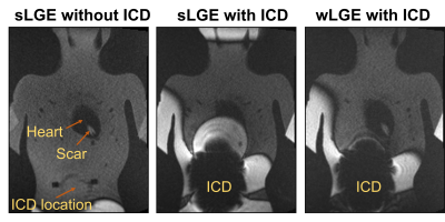 |
23 | Wideband Pulse Sequences for Suppressing Image Artifacts Induced by an ICD: A study in a Pediatric Anthropomorphic Phantom
Oluyemi Aboyewa1,2, KyungPyo Hong2, Fuchang Jiang1, Bhumi Bhusal2, Giorgio Bonmassar3, Andrada Popescu4, Gregory Webster5, Laleh Golestanirad1,2, and Daniel Kim1,2
1Department of Biomedical Engineering, McCormick School of Engineering, Northwestern University, Evanston, IL, United States, 2Department of Radiology, Feinberg School of Medicine, Northwestern University, Chicago, IL, United States, 3AA. Martinos Center Massachusetts General Hospital Harvard Medical School, Charlestown, MA, United States, 4Department of Medical Imaging, Ann & Robert H. Lurie Children's Hospital, Chicago, IL, United States, 5Division of Cardiology, Department of Pediatrics, Ann & Robert H. Lurie Children's Hospital, Chicago, IL, United States
This study investigated whether wideband late-gadolinium enhancement (LGE) and T1 mapping pulse sequences can suppress image artifacts induced by an ICD positioned 12cm inferior to the heart in a pediatric anthropomorphic phantom. The ICD induced a peak-to-peak center frequency shift of 970Hz across the heart. The normalized mean signal intensity within the heart was 1.91±1.20 and 0.98±0.39, for standard and wideband LGE with ICD, respectively. The mean myocardial T1 was 1486.1±607.7 and 1728.3±332.3msec for standard and wideband T1 mapping, respectively. Our results demonstrate that wideband pulse sequences can suppress image artifacts induced by an ICD in a pediatric anthropomorphic phantom.
|
||
1569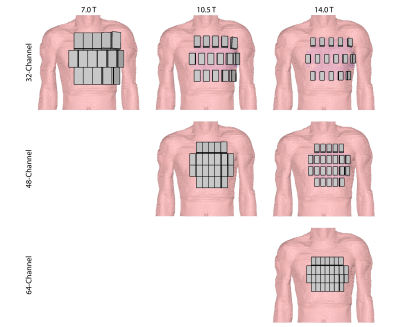 |
24 | Radiofrequency Array Concepts for Cardiac MRI at 10.5 T and 14.0 T: Does an Increased Channel Count Really Help?
Thomas Wilhelm Eigentler1,2, Bilguuun Nurzed1, Andre Kuehne3, and Thoralf Niendorf1,3,4
1Berlin Ultrahigh Field Facility (B.U.F.F.), Max Delbrück Center for Molecular Medicine in the Helmholtz Association, Berlin, Germany, 2Technische Universität Berlin, Chair of Medical Engineering, Berlin, Germany, 3MRI.TOOLS GmbH, Berlin, Germany, 4Experimental and Clinical Research Center (ECRC), a joint cooperation between the Charité Medical Faculty and the Max Delbrück Center for Molecular Medicine, Berlin, Germany
The gain in signal-to-noise ratio is driving the ultrahigh field and extreme field MRI. This work highlights the opportunities and electromagnetic constraints of realistic radiofrequency array concepts tailored for cardiovascular MRI at 7.0T, 10.5T, and 14.0T. For this purpose electromagnetic field simulations were performed using 32, 48 and 64-channel RF transceiver array configurations at 7.0T, 10.5T, and 14.0T. Our findings demonstrate, that a higher channel count increases the degrees of freedom of B1+ shaping. Furthermore, the preliminary results provide a strong mandate for static PTX or even dynamic PTX tailored at the heart at 10.5T and 14.0T.
|
||
1570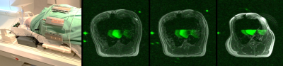 |
25 | Scalable and Modular 8-Channel Transmit and 8-Channel Flexible Receive Coil Array for 19F-MRI of Myocardial Infarction Studies in Large Animals
Ali Caglar Özen1, Johannes Fischer1, Simon Reiss1, Thomas Lottner1, Alexander Maier2, Waldemar Schimpf3, Timo Heidt2, Constantin von zur Mühlen2, and Michael Bock1
1Dept. Radiology, Medical Physics, Medical Center - University of Freiburg, Freiburg, Germany, 2Cardiology and Angiology I, Heart Center Freiburg University and Faculty of Medicine, Freiburg, Germany, 3Neurozentrum, Wissenschaftliche Werkstätten, Medical Center - University of Freiburg, Freiburg, Germany
In this study, we have developed an 8-channel transmit/ 8-channel receive coil array system for cardiac and thoracic MRI in pigs. Transmit array is scalable in size to increase transmit efficiency in pigs with smaller abdomen size. Receive array is semi-flexible to fit tightly on the dome-shaped thorax of pigs. We present a detailed transmit efficiency analysis of the Tx array including tolerance to mismatches at the input ports. We also present 19F MRI results from a cadaver measurement.
|
||
1571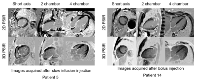 |
26 | 3D Whole Heart Grey-blood PSIR Imaging After Slow Infusion and Bolus Injection for High-resolution Isotropic LGE Imaging
Alina Schneider1, Reza Hajhosseiny1, Camila Munoz1, Giorgia Milotta2, Karl P Kunze1,3, Radhouene Neji1,3, Amedeo Chiribiri1, Pier Giorgio Masci1, René M Botnar1, and Claudia Prieto1
1School of Biomedical Engineering and Imaging Sciences, King's College London, London, United Kingdom, 2Wellcome Centre for Human Neuroimaging, UCL Queen Square Institute of Neurology, University College London, London, United Kingdom, 3MR Research Collaborations, Siemens Healthcare Limited, Frimley, United Kingdom 3D whole-heart free breathing water/fat non-rigid motion corrected grey-blood PSIR LGE technique has been recently proposed showing good agreement with conventional 2D grey-blood LGE MRI. Due to the relatively long scan time of ~10 minutes, contrast washout may influence scar detection and limit spatial resolution. Here we compare the proposed 3D grey-blood PSIR LGE after slow infusion and bolus injection at high isotropic spatial resolution to evaluate the benefit of the slow infusion injection. Both pipelines were compared to the conventional breath-held 2D grey blood PSIR-LGE imaging technique. Overall higher quality scores were observed in images acquired after slow infusion. |
||
1572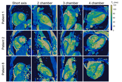 |
27 | Preliminary Clinical Validation of Non-rigid Motion-compensated 3D Whole-heart T2 mapping in a hybrid PET-MR system
Alina Schneider1, Camila Munoz1, Alina Hua1, Karl P Kunze1,2, Radhouene Neji1,2, Tevfik F Ismail1, René M Botnar1, and Claudia Prieto1
1School of Biomedical Engineering and Imaging Sciences, King's College London, London, United Kingdom, 2MR Research Collaborations, Siemens Healthcare Limited, Frimley, United Kingdom
Simultaneous 18F-FDG PET-MRI has shown promise for improved diagnostic accuracy of inflammatory cardiac diseases. However, respiratory motion and mis-registration between free-breathing 3D PET and 2D breath-held MR images remain challenges that have hindered its clinical adoption. We have recently proposed a free-breathing non-rigid motion-compensated 3D whole-heart T2-mapping that enables detection of myocardial inflammation and provides non-rigid respiratory motion fields to correct simultaneously acquired PET data in a 3T PET-MR system. Here, we perform a preliminary clinical validation of this approach in 8 patients with suspected cardiovascular disease. Good image quality with T2 values comparable to clinical 2D T2-mapping was achieved.
|
||
1573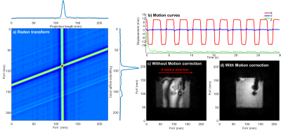 |
28 | Interventional continuous cardiac thermometry via simultaneous catheter tracking and undersampled radial golden angle acquisition
Maxime Yon1,2, Marylène Delcey1,2, Pierre Bour1,2, William Grissom3, Bruno Quesson1,2, and Valéry Ozenne1,2
1Electrophysiology and Heart Modeling Institute, Pessac, France, 2U1045, Centre de Recherche Cardio-Thoracique de Bordeaux Inserm, Bordeaux, France, 3Department of Biomedical Engineering, Vanderbilt University, Nashville, TN, United States Interventional MR thermometry has the potential to increase the success rate of the radio-frequency cardiac ablative procedure by allowing real-time monitoring of the lesion growth. Available methods are dependent on ECG-gating, which is poorly reliable in arrhythmic conditions. We propose a new approach based on radial continuous gradient echo acquisition combined with concomitant 2D intra-scan motion correction and direct estimation of the temperature from k-space data which could be a robust alternative to the more classical but ECG-triggered and artifact-prone EPI methods. |
||
1574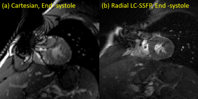 |
29 | Interleaved radial linear combination SSFP for improved cine imaging with implanted cardiac devices
Jie Xiang1, Jerome Lamy2, Rachel Lampert3, and Dana C. Peters1,2
1Department of Biomedical Engineering, Yale University, New Haven, CT, United States, 2Department of Radiology and Biomedical Imaging, Yale University, New Haven, CT, United States, 3Department of Medicine, Cardiovascular Division, Yale University, New Haven, CT, United States
Interleaved undersampled radial linear-combination balanced SSFP (LC-SSFP) was investigated as a method for improving cardiac cine images, acquired in subjects with metallic implants. Phantoms and healthy volunteer studies were performed, in the presence of metallic artifacts (generated by a stainless steel bolt). We found the proposed method eliminated some artifacts seen using standard Cartesian bSSFP. These findings may lead to improved cardiac cine imaging for patients with a pacemaker or implantable cardioverter defibrillator (ICD).
|
||
1575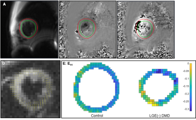 |
30 | Reproducibility of Left Ventricular CINE DENSE Strain in Pediatric Subjects with Duchenne Muscular Dystrophy
Zhan-Qiu Liu1,2, Nyasha Maforo3, Pierangelo Renella3, Nancy Halnon3, Holden Wu3, and Daniel Ennis4
1Cardiovascular Institute, Stanford University, Mountain View, CA, United States, 2Department of Radiology, Stanford University, Stanford, CA, United States, 3University of California, Los Angeles, Los Angeles, CA, United States, 4Stanford University, Mountain View, CA, United States
Left ventricular (LV) peak mid-wall circumferential strain (Ecc) and twist are early biomarkers for evaluating the subtle and highly variable onset and progression of cardiomyopathy in pediatric subjects with Duchenne muscular dystrophy (DMD). Cine Displacement Encoding with Stimulated Echoes (DENSE) has proven sensitive to changes in Ecc and twist, but has not been reported in a DMD cohort. We show that free-breathing DENSE CMR at 3T provides highly reproducible middle-ventricular Ecc, radial strain (Err), and twist measurements in healthy boys and boys with DMD without evidence of late gadolinium enhancement.
|
||
1576 |
31 | Biophysical-Model Based Synthesis of Cine CMR of Healthy and Pathological Left-Ventricular Function Video Permission Withheld
Stefano Buoso1, Thomas Joyce1, and Sebastian Kozerke1
1Institute of Biomedical Engineering, ETH Zurich and University of Zurich, Zurich, Switzerland
We propose to augment MRXCAT with left-ventricular anatomies and function, representing realistic population statistics, to enable design and validation of CMR imaging and inference approaches. A variational autoencoder is deployed to identify parametric anatomical representations from a cohort of healthy and diseased anatomies to allow for the generation of new synthetic anatomies. Cardiac function is simulated using a biophysical model also incorporating local tissue defects. The resulting ventricular tissue masks are enriched with background information from XCAT. Synthetic MR images are generated using MRXCAT, making available paired data of CMR images and (patho)physiological parameters including ground-truth ventricular strains and displacements.
|
||
1577 |
32 | The left atrial function in hypertension patients with left ventricular diastolic dysfunction:A myocardial MR strain study Video Not Available
Jinyang Wen1, Xin Zhang1, Lianggeng Gong1, Jiankun Dai2, and Long Qian2
1Radiology, The Second Affiliated Hospital of Nanchang University, Nanchang, China, 2MR Research, GE Healthcare, Beijing, China Heart failure with preserved ejection fraction(HFpEF)was evaluated by echocardiography commonly. In this study, we try to use Cardiovascular Magnetic Resonance Tissue-Tracking (CMR-TT) to assess left atrial (LA) function in hypertension patients (HP) with left ventricular diastolic dysfunction (LVDD). The results demonstrated that LA myocardial function decreased in HP with LVDD. LA myocardial strain parameters could detect sensitively the myocardial deformation of LVDD in HP. Total strain(εs) parameter was an excellent parameter for diagnose HFpEF and evaluating the degree of LVDD. |
||
1578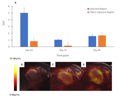 |
33 | Hybrid PET/MRI Dual-Condition Framework for Response Monitoring in Myocardial Infarction
Fayez Habach1,2, Jennifer Barry3, Melissa Larsen3, Marne Jamieson4, Amit Singnurkar5, Michael Laflamme6, and Nilesh Ghugre1,2
1Medical Biophysics, University of Toronto, Toronto, ON, Canada, 2Physical Sciences, Sunnybrook Research Institute, Toronto, ON, Canada, 3Schulich Heart Research Program, Sunnybrook Research Institute, Toronto, ON, Canada, 4Nuclear Medicine, Sunnybrook Health Sciences Centre, Toronto, ON, Canada, 5Medical Imaging, Sunnybrook Health Sciences Centre, Toronto, ON, Canada, 6Mcewen Stem Cell Institute, University Health Network, Toronto, ON, Canada
Hybrid PET/MRI is a promising tool that can characterize both metabolic and structural information and has been used extensively in the context of MI-related heart failure. We propose a novel dual-condition protocol that can be used to characterize metabolic state of cardiac tissue in vivo following MI, under different substrate conditions. Our results show significant metabolic changes under fasting and glucose loading conditions, and further longitudinal differences in MI-related metabolic remodeling within both the infarct zone and surrounding myocardium. This framework has potential to be used in testing therapeutic efficacy following MI in both preclinical and clinical contexts.
|
||
 Back to Meeting Home
 Back to the Program-at-a-Glance
The International Society for Magnetic Resonance in Medicine is accredited by the Accreditation Council for Continuing Medical Education to provide continuing medical education for physicians.
 Back to Meeting Home
 Back to the Program-at-a-Glance
 View the Presentation