Digital Poster
Vascular Imaging: Vessel Wall
Joint Annual Meeting ISMRM-ESMRMB & ISMRT 31st Annual Meeting • 07-12 May 2022 • London, UK

| Computer # | ||||
|---|---|---|---|---|
1744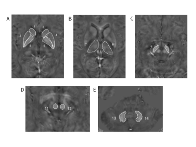 |
14 | Increased Iron Deposition in Gray Matter Nuclei in Patients with Intracranial Artery Stenosis Evaluated by Quantitative Susceptibility Mapping Video Not Available
Huimin Mao1, Weiqiang Dou2, Xinyi Wang1, Kunjian Chen1, and Yu Guo1
1Radiology, The First Affiliated Hospital of Shandong First Medical University, Jinan, China, 2MR Research China, GE Healthcare, Beijing, China
This study aimed to use quantitative susceptibility mapping (QSM) to systematically investigate iron content changes of gray matter (GM) nuclei in patients with long-term anterior circulation artery stenosis (ACAS) and posterior circulation artery stenosis (PCAS). The differences of iron-related susceptibility in GM nuclei, including bilateral caudate nucleus, putamen (PU), globus pallidus (GP), thalamus, substantia nigra (SN), red nucleus and dentate nucleus (DN) were explored in 25 ACAS and 25 PCAS patients. Compared with healthy controls, mean susceptibility of bilateral PU, GP, and SN in ACAS and PCAS patients, and of extra bilateral DN in PCAS patients were significantly increased.
|
||
1745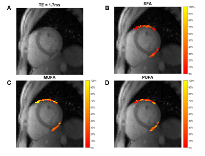 |
15 | Fatty Acid Composition MRI of Human Epicardial Adipose Tissue
Jack Echols1, Soham Shah1, and Frederick Epstein1
1Biomedical Engineering, University of Virginia, Charlottesville, VA, United States Epicardial adipose tissue (EAT) fatty acid composition (FAC) plays an important role in coronary vascular inflammation due to obesity and diabetes. We show for the first time the feasibility of quantifying human EAT FAC using radial multi-echo gradient-echo MRI with IDEAL mapping. This method may enable further investigations into the relationship between EAT fatty acid composition and cardiovascular disease. |
||
1746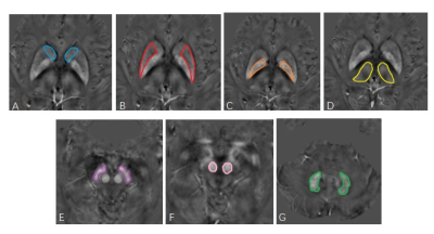 |
16 | Brain iron deposition in patients with unilateral middle cerebral artery stenosis: an in vivo quantitative susceptibility mapping study Video Not Available
Huimin Mao1, Weiqiang Dou2, Xinyi Wang1, Kunjian Chen1, and Yu Guo1
1Radiology, The First Affiliated Hospital of Shandong First Medical University, Jinan, China, 2MR Research China, GE Healthcare, Beijing, China
The main purpose was to quantitatively evaluate iron alterations in gray matter (GM) nucleus of patients with unilateral middle cerebral artery stenosis (MCAS)-related ischemic stroke using quantitative susceptibility mapping (QSM). Forty-six unilateral MCAS patients and thirty-eight healthy controls underwent QSM examination. Iron-related susceptibility of GM nucleus, including bilateral caudate nucleus, putamen (PU), globus pallidus (GP), thalamus, substantia nigra (SN), red nucleus, and dentate nucleus were assessed. Compared with healthy controls, PU, GP, and SN regions at lesion side presented significantly increased susceptibility in patients, indicating that abnormal iron metabolism may present in the brain after ischemic stroke.
|
||
1747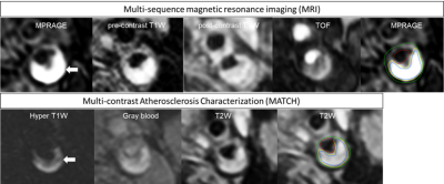 |
17 | MULTI-CONTRAST ATHEROSCLEROSIS CHARACTERIZATION (MATCH) AND MULTI-SEQUENCE MRI IN QUANTIFYING CAROTID PLAQUE COMPOSITION
Mohamed Kassem1,2, Ellen Eline Boswijk1,2, Jochem van der Pol1,2, Rik PM Moonen2, Jan Bucerius3, Werner H Mess4, Robert Jan van Oostenbrugge1,5, Zhaoyang Fan6, and M Eline Kooi1,2
1CARIM School for Cardiovascular Diseases, Maastricht University, Maastricht, Netherlands, Maastricht, Netherlands, 2Department of Radiology and Nuclear Medicine, Maastricht University medical center (MUMC+), Maastricht, Netherlands, 3Department of Nuclear Medicine, Georg-August University Göttingen, Gottingen, Germany, 4Department of Clinical Neurophysiology, Maastricht University medical center (MUMC+), maastricht, Netherlands, 5Department of Neurology, Maastricht University Medical Center+ (MUMC+), maastricht, Netherlands, 6Department of Radiology, University of Southern California, Los Angeles, CA, United States
Multi-contrast Atherosclerosis Characterization (MATCH) was developed to quantify of carotid atherosclerotic plaque composition within 5 minutes’ scan time. Twenty symptomatic patients with ≥2 mm carotid plaque underwent 3.0 Tesla conventional multi-sequence and MATCH MRI. Excellent agreement was obtained for scoring a Lipid-rich necrotic core (LRNC) intraplaque hemorrhage (IPH) on the MATCH images while fair for calcifications. No significant differences between MATCH and multi-sequence MRI were found in volume of LRNC, IPH and calcifications. We demonstrated excellent agreement between MATCH and multi-sequence MRI for the identification and quantification of LRNC and IPH within significant shorter time.
|
||
1748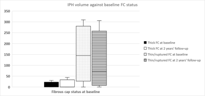 |
18 | THE ASSOCIATION BETWEEN PLAQUE SURFACE DISRUPTION AND FIBROUS CAP STATUS AT BASELINE AND VOLUME CHANGE OF INTRAPLAQUE HEMORRHAGE OVER TWO YEARS
Mohamed Kassem1,2, Tahnee Gorissen1, Mohamed AlBenwan1, Dianne van Dam-Nolen3, Madieke I Liem4, Rob J van der Geest5, Jeroen Hendrikse6, Werner H Werner Mess1,7, Paul J Nederkoorn4, Daniel Bos3,8, Patty Nelemans9, Robert Jan van Oostenbrugge1,10, and M Eline Kooi1,2
1CARIM School for Cardiovascular Diseases, Maastricht University, Maastricht, Netherlands, Maastricht, Netherlands, 2Department of Radiology and Nuclear Medicine, Maastricht University medical center (MUMC+), maastricht, Netherlands, 3Department of Radiology and Nuclear Medicine, Erasmus MC, University Medical Center Rotterdam, Rotterdam, Netherlands, 4Department of Neurology, Amsterdam UMC, location AMC, Amsterdam, Amsterdam, Netherlands, 5Division of Image Processing, Department of Radiology, Leiden University Medical Center, Leiden, Netherlands, 6Radiology, University Medical Center Utrecht, Utrecht, Utrecht, Netherlands, 7Department of Clinical Neurophysiology, Maastricht University Medical Center+ (MUMC+), maastricht, Netherlands, 8Epidemiology, Erasmus MC, Erasmus MC, University Medical Center Rotterdam, Rotterdam, Netherlands, 9Department of Epidemiology, Maastricht University, Maastricht, The Netherlands, maastricht, Netherlands, 10Department of Neurology, Maastricht University Medical Centre, maastricht, Netherlands
The factors that contribute to intraplaque hemorrhage (IPH) development within the carotid atherosclerotic plaque are incompletely understood. Previously, we demonstrated that IPH is associated with a thin/ruptured fibrous cap (TRFC) and disruption of the plaque surface on in a cross-sectional study. Baseline and 2 year’s follow-up carotid MR images of 110 patients from the Plaque at Risk (PARISK) study were analyzed. IPH volume change in TRFC at baseline was significant different (p=0.04) than thick fibrous cap group. Baseline IPH volumes are larger in patients with TRFC and disrupted plaque surface, but didn’t increase during the follow-up.
|
||
1749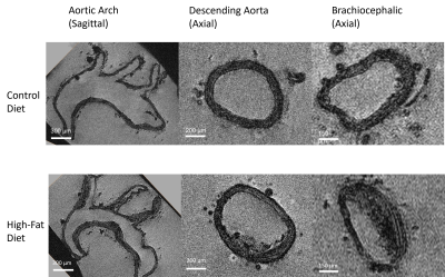 |
19 | High-Field Magnetic Resonance Microscopy of Aortic Plaques in a Mouse Model
Sean Gullette1, Stephen Andrews2, Courtney Whalen2, Floyd J. Mattie2, Ximing Ge2, Catharine Ross2, Rita Castro2,3,4, and Thomas Neuberger1,5
1Huck Institutes of The Life Sciences, The Pennsylvania State University, State College, PA, United States, 2Department of Nutritional Sciences, The Pennsylvania State University, State College, PA, United States, 3Research Institute for Medicines (iMed.ULisboa), Universidade de Lisboa, Portugal, Lisboa, Portugal, 4Department of Pharmaceutical Sciences and Medicines, Universidade de Lisboa, Portugal, Lisboa, Portugal, 5Department of Biomedical Engineering, The Pennsylvania State University, State College, PA, United States Currently, histological methods of plaque analysis in aortas from murine models of atherosclerosis do not allow for a holistic view of the vascular lesions. Using high-field MRI, plaque volume in Apoe-/- mice can be analyzed, and when compared to traditional histology, has a higher throughput and is non-destructive. Using a 3D gradient echo sequence at 14 tesla, high-resolution images of the aortas were achieved. The images were segmented and quantified providing a 3-dimensional view of plaque distribution and volume. Overall, using a high-field MRI system allowed for a detailed and complete view of vascular components. |
||
1750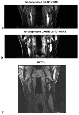 |
20 | Evaluation of flow suppression methods for multi-echo gradient echo carotid wall imaging
Junmin Liu1, Omer Oran2, and Maria Drangova1,3
1Robarts Research Institute, Schulich School of Medicine & Dentistry, Western University, London, ON, Canada, 2Siemens Healthcare Canada, Oakville, ON, Canada, 3Medical Biophysics, Schulich School of Medicine & Dentistry, Western University, London, ON, Canada
By adding long TEs to the Dixon acquisition, the chemical-shift-based T2*-weighted multi-echo GRE (CS-T2*-mGRE) techniques have gained attention, because they are able to generate co-registered multi-contrast images and quantitative maps. However, flow-suppressed CS-T2*-mGRE sequences have not been evaluated for carotid wall imaging. We compared the performance of simultaneous quantification of fat fraction (FF) and R2* from CS-T2*-mGRE sequences with and without saturation slabs, and with the incorporation of DANTE pulses for black blood imaging (DANTE-CS-T2*-mGRE); we used the FF for fat-suppression to better visualize the vessel wall from DANTE-CS-T2*-mGRE as well as the carotid artery lumen from CS-T2*-mGRE.
|
||
1751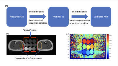 |
21 | Plaque to Myocardium Ratio in T1-weighted Magnetic Resonance Imaging: Analysis of Variabilities and Method for Standardization
Meng Lu1, Hui Han1, Debiao Li2,3, and Yibin Xie1
1Biomedical Imaging Research Institute, Cedars-Sinai Medical Center, Los Angeles, CA, United States, 2Cedars-Sinai Medical Center, Los Angeles, CA, United States, 3Department of Bioengineering, University of California, Los Angeles, Los Angeles, CA, United States
Plaque to Myocardium Ratio (PMR) based on T1-weighted MRI is an important quantitative biomarker to classify high-risk coronary plaque. However, it is calculated based on relative signal intensities, therefore, sensitive to variations in physiological and acquisition conditions. In this work, Bloch simulations were performed to explore the impact of heart rate, echo spacing, flip angle, and the number of readout segments on PMR. A computational model was further proposed to effectively reduce the acquisition-related PMR variations demonstrated in simulations and in vivo studies. The proposed method may potentially improve the precision of PMR and facilitate its comparison in longitudinal studies.
|
||
1752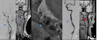 |
22 | Diagnosis and Follow-up of cervical vascular dissection using 3D MR vessel wall imaging: A Single Centre Experience of 65 Cases Video Not Available
Jin Zhang1, Beibei Sun2, Shenghao Ding2, Jiaqi Tian2, Jieqing Wan2, Jilei Zhang3, Weibo Chen3, Xihai Zhao4, Yuan Chun5, Jianrong Xu2, and Huilin Zhao2
1Radiology, Renji Hospital, School of Medicine, Shanghai Jiaotong University, Shanghai, China, 2Renji Hospital, School of Medicine, Shanghai Jiaotong University, Shanghai, China, 3Philips Healthcare, Shanghai, China, 4Biomedical Engineering & Center for Biomedical Imaging Research, Tsinghua University, Beijing, China, 5University of Washington, Seattle, WA, United States
Cervical artery dissection (CAD) is one of the important causes of ischemic stroke in young and middle-aged people[1; 2]. DSA remains the gold standard for identifying and characterizing carotid vascular dissections[3]. However, this imaging approach is known to have certain limitations, including invasiveness, ionizing radiation exposure, uneconomic, and inconvenience in follow-up. Recently, rapid three-dimensional MR vessel wall imaging (3D MR-VWI) techniques have been developed to visualize arterial lumen and outer wall boundaries non-invasively and without contrast administration. Non-invasive 3D MR-VWI has the potential to be a powerful tool for follow-up of cervical artery dissection.
|
||
1753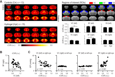 |
23 | Intraocular pressure elevation induces vascular and functional brain changes: A relative cerebrovascular reactivity resting-state fMRI Study
Russell W. Chan1,2, Yixi Xue1, Ji Won Bang1, Muneeb A. Faiq1, Thajunnisa A. Sajitha1, Royce P. Lee1, Peiying Liu3, Christopher K. Leung4, Gadi Wollstein1, Joel S. Schuman1, and Kevin C. Chan1,5
1Department of Ophthalmology, New York University Grossman School of Medicine, New York, NY, United States, 2Neuroscience Institute, New York University Grossman School of Medicine, New York, NY, United States, 3Department of Diagnostic Radiology and Nuclear Medicine, University of Maryland School of Medicine, Baltimore, MD, United States, 4Department of Ophthalmology, The University of Hong Kong, Hong Kong, Hong Kong, 5Department of Radiology, New York University Grossman School of Medicine, New York, NY, United States
Recently, we used a novel resting-state fMRI method to map relative cerebrovascular reactivity (rCVR) without gas challenge, and demonstrated decreased rCVR in the visual cortex and increased rCVR in the basal forebrain in glaucoma patients relative to healthy subjects. However, the underlying mechanisms remain unclear. Here, we applied a hydrogel-induced glaucoma mouse model to chronically elevate intraocular pressure, mapped rCVR using resting-state fMRI, and measured optomotor responses. Our results showed similar patterns of rCVR changes along with visual impairments, indicating a role of chronic intraocular pressure elevation on the widespread vascular and functional brain changes in experimental glaucoma.
|
||
 Back to Meeting Home
 Back to the Program-at-a-Glance
The International Society for Magnetic Resonance in Medicine is accredited by the Accreditation Council for Continuing Medical Education to provide continuing medical education for physicians.
 Back to Meeting Home
 Back to the Program-at-a-Glance
 View the Presentation