Digital Poster
Cardiac Anatomy & Tissue Characterization IV
Joint Annual Meeting ISMRM-ESMRMB & ISMRT 31st Annual Meeting • 07-12 May 2022 • London, UK

Digital Poster
Cardiac Anatomy & Tissue Characterization IV
| Computer # | ||||
|---|---|---|---|---|
1646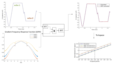 |
15 | Trajectory correction enables Free running cardiac DIXON at 3T
Pierre Daudé1,2, Thomas Troalen3, Adèle L C Mackowiak4,5, Davide Piccini4,6, Jerôme Yerly4,5, Josef Pfeuffer7, Frank Kober1,2, Sylviane Confort Gouny1,2, Monique Bernard1,2, Matthias Stuber4,5, Jessica A M Bastiaansen4,5,8,9, and Stanislas Rapacchi1,2
1Aix-Marseille Univ, CNRS, CRMBM, Marseille, France, 2APHM, Hôpital Universitaire Timone, CEMEREM, Marseille, France, 3Siemens Healthcare SAS, Saint-Denis, France, 4Department of Diagnostic and Interventional Radiology, Lausanne University Hospital, Lausanne, Switzerland, 5Center for Biomedical Imaging, Lausanne, Switzerland, 6Advanced Clinical Imaging Technology, Siemens Healthcare, Lausanne, Switzerland, 7Siemens Healthcare, MR Application Development, Erlangen, Germany, 8Department of Diagnostic, Interventional and Pediatric Radiology, Bern University Hospital (Inselspital), Bern, Switzerland, 9Translational Imaging Center, sitem-insel, Bern, Switzerland
Free-running cardiac DIXON-MRI has potential for T2*/PDFF quantification to explore cardiac fat accumulation and alteration in metabolic diseases. DIXON at 3T suffers from rapid fat-water phase accrual and inhomogeneous B0. Consequently, bipolar echoes, as opposed to monopolar, are required to achieve equal/shorter than in-phase/out-of-phase echo spacing. However, distortions occur between even and odd echoes due to gradients imperfections. Thus, the existing framework was extended with a k-space trajectory correction based on gradient impulse response function(GIRF). Both monopolar or bipolar echoes without GIRF correction resulted in fat-water swaps and unreliable quantitative maps, that were resolved using GIRF-corrected bipolar free-running DIXON.
|
||
1647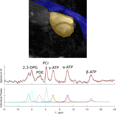 |
16 | SLAM reconstruction of 31P Cardiac MRS Improves PCr/ATP Repeatability Without Requiring Cardiac Gating
Andrew Tyler1,2, Moritz Hundertmark1, Jack J. Miller1,2,3,4, Oliver J. Rider1, Damian J. Tyler1,2, and Ladislav Valkovic1
1OCMR, University of Oxford, Oxford, United Kingdom, 2Department of Physiology, Anatomy & Genetics, University of Oxford, Oxford, United Kingdom, 3Department of Physics, University of Oxford, Oxford, United Kingdom, 4The MR Research Centre, Aarhus University, Aarhus, Denmark
Using the SLAM algorithm to reconstruct non-gated acquisition weighted 31P cardiac CSI data-sets improved repeatability by 35% and reduced variation by 23%, compared to a Fourier based reconstruction of the same data-set, without requiring a change to acquisition protocol.
|
||
1648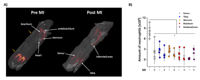 |
17 | A Cross-Species Approach for Noninvasive in Vivo Tracking of Neutrophil Dynamics upon Cardiovascular Injury by 1H/19F MRI
Pascal Bouvain1, Sebastian Temme1, Maria Grandoch1, and Ulrich Flögel1
1Heinrich Heine University, Düsseldorf, Germany
Intravenous 19F tracer application for in vivo labelling of neutrophils prior to injury allowed the non-invasive 3D visualization of neutrophils within their different hematopoietic niches over the entire body and the subsequent monitoring of their egress into affected tissues. Stimulated murine/human neutrophils exhibited enhanced labelling which could be exploited as an in vivo readout for their activation state in both sterile and nonsterile cardiovascular inflammation. In summary, the present study demonstrates that both human and murine neutrophils can be specifically targeted to track their dynamic trafficking by non-invasive 1H/19F MRI in vivo.
|
||
1649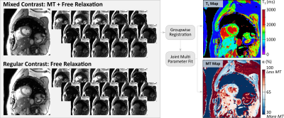 |
18 | Inversion Recovery Based Magnetization Transfer Mapping in the Human Myocardium In the presence of Incomplete Bound Pool Saturation at 3T
Sebastian Weingärtner1, Ömer Burak Demirel2, Qian Tao1, Joao Tourais1, and Mehmet Akcakaya2
1Department of Imaging Physics, Delft University of Technology, Delft, Netherlands, 2Department of Electrical and Computer Engineering and Center for Magnetic Resonance Research, University of Minnesota, Minneapolis, MN, United States
Quantitative imaging of magnetization transfer (MT) may enable non-contrast assessment of myocardial scar and fibrosis. However, its application at 3T has so far been limited. In this work we investigate an inversion recovery based method for joint quantification of T1 and MT maps. A numerical binary spin-bath model is used to account for incomplete bound pool saturation in the presence of limited preparation durations. In vivo images were obtained with high visual map quality for both T1 and MT quantification, and comparable, low CoV across the two parameters (T1: 5.5+-0.6% and MT: 5.8+-0.9%).
|
||
1650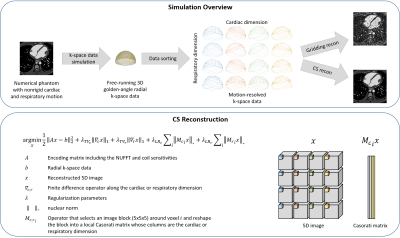 |
19 | Numerical optimization of 5D cardiac and respiratory motion-resolved CMR imaging for the assessment of left ventricular function
Jérôme Yerly1,2, Christopher W Roy1, Bastien Milani1, Davide Piccini1,3, Aurélien Bustin1,4,5, Mariana B.L. Falcão1, Ruud B. van Heeswijk1, and Matthias Stuber1,2,4
1Radiology, Lausanne University Hospital (CHUV) and University of Lausanne (UNIL), Lausanne, Switzerland, 2CIBM Center for Biomedical Imaging, Lausanne, Switzerland, 3Advanced Clinical Imaging Technology, Siemens Healthcare, Lausanne, Switzerland, 4Electrophysiology and Heart Modeling Institute, IHU LIRYC, Bordeaux, France, 5Cardiovascular Imaging, Hôpital Cardiologique du Haut-Lévêque, CHU de Bordeaux, Bordeaux, France
The free-running framework (FRF) was recently proposed to address the limitations of current techniques to assess left ventricular (LV) ejection fraction (LVEF). However, the accuracy of FRF to assess LVEF has yet to be quantitatively examined. This work rigorously quantifies and optimizes the effect of the regularization weights on LVEF and several image quality metrics using a numerical phantom with well-controlled boundary conditions, and validates the results in in-vivo 5D FRF data. The results demonstrated that the combination of regularization weights that are optimal in terms of image quality do not correspond to the optimal weights for LVEF assessment.
|
||
1651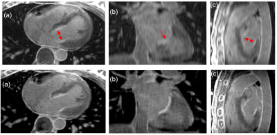 |
20 | Isotropic fixed time 3D late gadolinium enhancement MRI for left atrial fibrosis imaging
Ganesh Adluru1,2, Jason Mendes1, Ravi Ranjan3, Eugene Kholmovski1,4, and Edward V.R. DiBella1,2
1Radiology & Imaging Sciences, University of Utah, Salt Lake City, UT, United States, 2Biomedical Engineering, University of Utah, Salt Lake City, UT, United States, 3Internal Medicine, University of Utah, Salt Lake City, UT, United States, 4Biomedical Engineering, Johns Hopkins University, Baltimore, MD, United States
Left atrial late gadolinium enhancement imaging is a promising tool to identify scar and fibrosis. Existing acquisition and reconstruction methods can suffer from long scan time, poor image quality, and inability to accurately quantify the fibrosis. Here we propose a fixed time isotropic imaging of the left atrium with a resolution of 1.25mm3. We use retrospective respiratory navigation to remove inconsistent data due to motion and use a constrained reconstruction framework with total variation and 3D block-matching regularizers to remove the data undersampling artifacts. Promising results showing gains from the isotropic acquisitions are presented in canine and human studies.
|
||
1652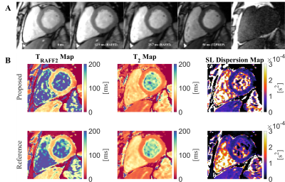 |
21 | Single Breath-hold Simultaneous T2 and TRAFF2 mapping for approximate Spin-lock Dispersion Mapping in the Myocardium at 3T
Joao Tourais1, Omer Burak Demirel2, Qian Tao3, Iain Pierce4, George Thornton4, Thomas Treibel4, Sebastian Weingärtner1, and Mehmet Akcakaya2
1Imaging Physics, Delft University of Technology (TU Delft), Delft, Netherlands, 2Department for Electrical and Computer Engineering, and Center for Magnetic Resonance Research, University of Minnesota, Minnesota, MN, United States, 3Delft University of Technology (TU Delft), Delft, Netherlands, 4Barts heart centre, Barts Health NHS Trust, London, United Kingdom
Spin-lock dispersion is a promising biomarker for the assessment of myocardial infarction. However, at 3T, the required range of T1⍴ maps acquired at different amplitudes suffers from specific absorption rate (SAR) limitations and off-resonance artifacts. Relaxation Along a Fictitious Field (RAFF) is an alternative to SL preparations with lower SAR requirements while still sampling relaxation in the rotating frame. Thus, a single breath-hold simultaneous TRAFF2 and T2 mapping sequence is proposed for sl dispersion mapping at 3T. High visual quality maps and accurate T2, TRAFF2, and SL dispersion values are achieved with the proposed sequence in phantoms and in vivo.
|
||
1653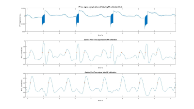 |
22 | Enabling Pilot Tone cardiac triggering for complete cardiac examinations using an RF calibration procedure
Peter Speier1, Yan Tu Huang2, Carmel Hayes1, Randall Kroeker1, Manuela Rick1, Michael Schwertfeger3, and Mario Bacher1
1Siemens Healthcare, Erlangen, Germany, 2Siemens Healthcare, Shenzen, China, 3ASTRUM IT GmbH, Erlangen, Germany
Contactless cardiac triggering using Pilot Tone was initially demonstrated for steady-state triggered cine-type sequences that, per definition, are performed with continuous, uniform RF pulses. Here we describe a method that stabilizes Pilot Tone cardiac triggering in the presence of RF artefacts, thereby allowing for pilot tone triggering of complete cardiac MR examinations with a range of sequence flavors including those applying RF pulses intermittently. The method uses an additional RF calibration measurement and avoids the RF artefact subspace in a PCA-based multi-channel coil combination calculation. Signal examples at 1.5T demonstrate effective RF suppression.
|
||
1654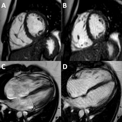 |
23 | Free-breathing pseudo-golden-angle bSSFP cine cardiac MRI for evaluation of biventricular function in patients with congenital heart disease
Dmitrij Kravchenko1, Alexander Isaak1, Shuo Zhang2, Narine Mesropyan1, Leon M. Bischoff1, Thomas Vollbrecht1, Oliver Weber2, Claus Christian Pieper1, Daniel Kuetting1, Christopher Hart3, Ulrike Attenberger1, and Julian Luetkens1
1Diagnostic and interventional radiology, University Hospital Bonn, Bonn, Germany, 2Philips GmbH DACH, Hamburg, Germany, 3Pediatric Cardiology, University Hospital Bonn, Bonn, Germany Cardiac MRI is currently the standard imaging modality for patients with congenital heart disease (CHD). However, the requirement for breath holds remains a big challenge in clinical practice and image quality is often significantly degraded by respiratory motion artifacts. We employed respiratory-triggered pseudo-golden-angle bSSFP imaging for free-breathing cine MRI and compared cardiac volumetry, function, and image quality with the standard breath hold technique. The proposed method showed good agreement with comparable diagnostic image quality and advantage in patients with limited breath hold capability. These demonstrated promise for a wider clinical routine application in patients with CHD. |
||
1655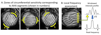 |
24 | Rapid and Robust Assessment of Circumferential Strain Recovery via Tagged MRI of Infarcted Pig Hearts After Human Cardiomyocyte Transplantation Video Not Available
Anna V Naumova1 and William S Kerwin1
1Radiology, University of Washington, Seattle, WA, United States
A new straight forward approach for rapid and robust quantification of myocardial circumferential strain has been developed. The key to this method is placement of the linear tags in 60-degree pattern offsets and alignment of the tags with the AHA segments for optimized segmental analysis. The approach has been implemented for the first time for evaluation of the human cardiomyocyte transplantation benefits in the infarcted pig heart. We have shown the temporal changes in circumferential strain and strain rate of the infarcted segments of myocardium and recovery of myocardial strain followed human cardiomyocyte transplantation in comparison with untreated control group.
|
||
1656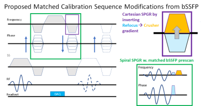 |
25 | An SSFP-SPGR Multi-sequence Cardiac Cine-Tag Acquisition Approach in One Breath-hold
Jacob P. Goes1, Vivian S. Nguyen1, Donovan Gorre2, Narutoshi Hibino3, Hui Wang4,5, Marcella K. Vaicik1, Amit R. Patel6, and Keigo Kawaji1,6
1Biomedical Engineering, Illinois Institute of Technology, Chicago, IL, United States, 2Radiology, University of Chicago Medical Center, Chicago, IL, United States, 3Surgery, University of Chicago Medical Center, Chicago, IL, United States, 4Philips, Gainesville, FL, United States, 5Department of Radiology, Cincinnati Children's Hospital Medical Center, Cincinnati, OH, United States, 6Medicine - Cardiology, University of Chicago Medical Center, Chicago, IL, United States
Multi-Contrast single breath-hold acquisition for cardiac MRI is highly desirable but is rate-limited by the need for a dedicated pre-scan per each acquisition. We revisit the notion of the necessary conditions for repeated preparation calibration steps, with the intent of loosening or selectively bypassing them in consecutive scan acquisitions. Under matched geometric conditions that account for sensitivities especially to off-resonance, we propose a simple bSSFP-SPGR based dual-acquisition method with a single intensive Cine calibration followed by a truncated SPGR calibration. This approach is demonstrated as Cine-Tag SSFP-SPGR CMR method that allows for one breath-hold functional acquisition of both scan sequences.
|
||
 Back to Meeting Home
 Back to the Program-at-a-Glance
The International Society for Magnetic Resonance in Medicine is accredited by the Accreditation Council for Continuing Medical Education to provide continuing medical education for physicians.
 Back to Meeting Home
 Back to the Program-at-a-Glance
 View the Presentation