Digital Poster
Flow Head to Toe II
Joint Annual Meeting ISMRM-ESMRMB & ISMRT 31st Annual Meeting • 07-12 May 2022 • London, UK

| Computer # | ||||
|---|---|---|---|---|
0895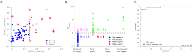 |
15 | Analysis of Fluid-Structure Stability from 4D Flow MRI to Predict Aortic Dilation
Tom Y Zhao1, Guy Elisha1, Ethan M I Johnson2, Sourav Halder1, Ben C Smith2, Bradley D Allen2, Michael Markl2, and Neelesh A Patankar1
1Northwestern University, Evanston, IL, United States, 2Northwestern University, Chicago, IL, United States
We present a novel analysis of 4D flow MRI in the thoracic aorta that identifies fluid structure instabilities, which are elevated in a cohort of 117 suspected-aortopathy patients as compared to 100 healthy subjects, and which are predictive of later complications shown in a follow-up analysis for 72 patients in the cohort.
|
||
0896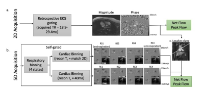 |
16 | Free-Running 5D Flow MRI: Impact of Cardiac Temporal Resolution on Flow Quantification
Elizabeth K. Weiss1,2, Cynthia K. Rigsby3, Joshua D. Robinson3, Justin Baraboo1,2, Liliana Ma1,2, Mariana B. L. Falcão4, Christopher W. Roy4, Matthias Stuber4, and Michael Markl1,2
1Biomedical Engineering, Northwestern University, Evanston, IL, United States, 2Radiology, Feinberg School of Medicine, Chicago, IL, United States, 3Medical Imaging, Lurie Children's Hospital, Chicago, IL, United States, 4Radiology, Lausanne University Hospital (CHUV) and University of Lausanne (UNIL), Lausanne, Switzerland
The flexible reconstruction of 5D flow MRI data may be particularly beneficial in pediatric cases where cardiac cycle length varies greatly. We compared flow measurements from 5D flow and 2D phase contrast. 5D flow data was reconstructed using either a cardiac bin width of 40ms or that matching the 2D phase contrast reconstruction. We find excellent correlation of both 5D flow reconstructions with 2D phase contrast. There is overestimation of arterial flow measures by 5D flow, with most overestimation by the short temporal resolution reconstruction. Further study should additionally compare with 4D flow MRI.
|
||
0897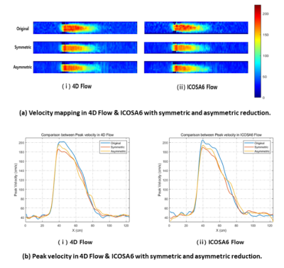 |
17 | Investigation of asymmetric undersampling scheme in accelerated flow imaging for improving velocity and turbulence kinetic energy estimation
Kyoung-Jin Park1,2, Ho-Jin Ha3, Dong-Hyun Yang2, Kang-Hyun Ryu4, Jae-Hun Lee1, and Dong-Hyun Kim1
1Electrical & Electronic Engineering, Yonsei university, Seoul, Korea, Republic of, 2Radiology (Cardiovascular Imaging), Asan Medical Center, University of Ulsan College of Medicine, Seoul, Korea, Republic of, 3Department of Mechanical and Biomedical Engineering, Kangwon National University, GANGWON-DO, Korea, Republic of, 4Radiology, Stanford University, Stanford, CA, United States
Compressed sensing (CS) technique has recently been used to accelerate long acquisition time in 4D Flow. However, it aggravates underestimation in velocity calculation and overestimation in turbulent kinetic energy (TKE) estimation, when under-sampling is applied. An asymmetric undersampling scheme method of the reference image and velocity encoding images could alleviate problems. The purpose of this paper is investigation of improving velocity and turbulence kinetic energy estimation with an asymmetric reduction for two motion encoding schemes (i.e., conventional 4D Flow vs ICOSA6 Flow)
|
||
0898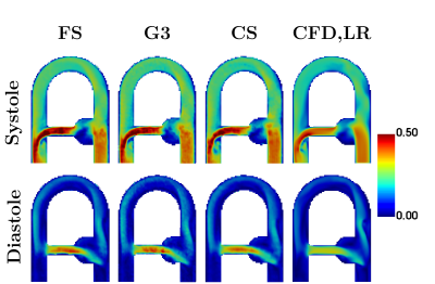 |
18 | COMPARISON OF CONVENTIONAL AND ACCELERATED 4D FLOW MRI ON A FLOW PHANTOM
Morgane Garreau1,2, Thomas Puiseux2,3, Ramiro Moreno3,4, Solenn Toupin5, Daniel Giese6, Simon Mendez1, and Franck Nicoud1
1IMAG, Univ. Montpellier, CNRS UMR 5149, Montpellier, France, 2Spin Up, Toulouse, France, 3I2MC, INSERM/UPS UMR 1297, Toulouse, France, 4ALARA Expertise, Strasbourg, France, 5Siemens Healthcare France, Saint-Denis, France, 6Siemens Healthcare, Erlangen, Germany
Fully-sampled and accelerated (GRAPPA R=3 and compressed sensing R=7.6) 4D Flow MRI acquisitions were compared for velocity profiles, flow rates and peak velocities in a rigid cardiovascular phantom under complex pulsatile flow conditions. Compatible CFD simulations were performed under the same flow conditions as a complementary modality. Good agreement was found between the three MR techniques, but discrepancies relative to the principle of mass conservation were noticed. Even though CFD presents some limitations as well, it appears to be a useful tool to investigate these differences.
|
||
0899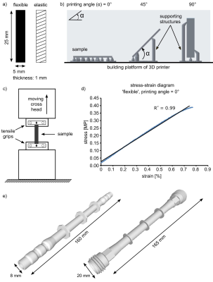 |
19 | Effect of wall elasticity on flow MRI in 3D printed models
Isil Unal1, Duygu Dengiz2, Eva Peschke1, Echard Quandt2, Mona Salehi Ravesh1, Mariya Pravdivtseva1, Jan-Bernd Hövener1, and Olav Jansen1
1Section Biomedical Imaging, Molecular Imaging North Competence Center (MOIN CC), Department for Radiology and Neuroradiology, University Medical Center Schleswig-Holstein, Kiel, Germany, 2Institute for Materials Science, Faculty of Engineering, University of Kiel, Kiel, Germany
2D phase-contrast (PC) MRI or 4D flow MRI allows to quantify the blood flow in vivo. However, the accuracy of these methods is influenced by many parameters. To optimize and validate these methods, patient-derived vascular models are key. Here, 3D printing is revolutionizing the manufacturing process, but realistic wall properties are essential for meaningful results. In this study, commercial elastic printing materials were used to fabricate vascular models. A tensile test was applied to all samples. Then the effect of wall elasticity on flow was investigated with 2D PC and 4D flow MRI. Wall motion was quantified using CINE MRI.
|
||
0900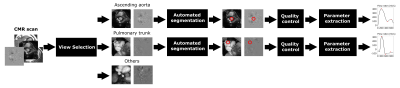 |
20 | Automatic analysis of cardiovascular magnetic resonance 2D phase contrast CMR imaging
Jorge Mariscal-Harana1, Ciaran O'Hanlon1, Carlota Asegurado Marquez1, Marwenie Petalcorin1, Reza Razavi1,2, Andrew P King1, Bram Ruijsink1,2,3, and Esther Puyol-Antón1
1School of Biomedical Engineering and Imaging Science, King's College London, London, United Kingdom, 2Department of Adult and Paediatric Cardiology, Guy’s and St Thomas’ NHS Foundation Trust, London, United Kingdom, 3Department of Cardiology, Heart and Lung Division, University Medical Center Utrecht, Utrecht, Netherlands
Cardiovascular magnetic resonance (CMR) based flow volume quantification in the great thoracic vessels is used in the assessment of several cardiovascular diseases such as valvular regurgitation, cardiac shunts and vascular health. Clinically, flow volume quantification is often performed based on semi-automatic segmentation of a vessel throughout the cardiac cycle in a manually positioned 2D phase-contrast (PC) CMR plane. In this work, we proposed a quality-controlled AI-based framework for automatic flow quantification from a full CMR scan that includes automated view selection. Results show high accuracy in view selection and excellent agreement between manual and automated flow quantification analysis.
|
||
0901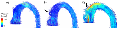 |
21 | 4D flow MRI: Ex-vivo swine model allows distinction between flow patterns induced by aortic valve replacement and surgical pathway alone.
Maren Friederike Balks1, Hiroyuki Saisho2, Buntaro Fujita2, Tim Schaller2, Najla Sadat2, Stephan Ensminger2, Jörg Barkhausen1, Alex Frydrychowicz1, and Thekla Helene Oechtering3
1Department of Radiology and Nuclear Medicine, Universität zu Lübeck, Lübeck, Germany, 2Department of Cardiac and Thoracic Vascular Surgery, Universität zu Lübeck, Lübeck, Germany, 3Department of Radiology, University of Wisconsin, Madison, WI, United States
Hemodynamic outcome after aortic valve replacement (AVR) seems dependent on the type of replacement. The impact of the surgical pathway (aortotomy) itself on hemodynamics is still unclear. We proposed an ex-vivo swine model for comprehensive evaluation of aortic hemodynamics after different types of AVR with 4D flow MRI. Postoperative flow changes could be attributed not only to the implanted valve but also to the aortotomy. The new neocuspidization Ozaki procedure compared favorably to a biological valve as it induced fewer secondary flow patterns. Among the three different AVR methods, the mechanical valve allowed for hemodynamics that most closely approached physiology.
|
||
0902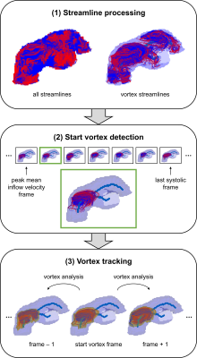 |
22 | Automated 4D flow-based blood vortex detection for estimation of mean pulmonary arterial pressure
Corina Kräuter1,2, Ursula Reiter1, Gabor Kovacs3,4, Clemens Reiter1, Marc Masana5, Horst Olschewski3,4, Michael Fuchsjäger1, Rudolf Stollberger2, and Gert Reiter1,6
1Department of Radiology, Medical University of Graz, Graz, Austria, 2Institute of Medical Engineering, Graz University of Technology, Graz, Austria, 3Department of Internal Medicine, Medical University of Graz, Graz, Austria, 4Ludwig Boltzmann Institute for Lung Vascular Research Graz, Graz, Austria, 5Institute of Computer Graphics and Vision, Graz University of Technology, Graz, Austria, 6Research and Development, Siemens Healthcare Diagnostics GmbH, Graz, Austria
Pulmonary hypertension is characterized by elevated mean pulmonary arterial pressure. Visual vortex analysis revealed a strong relation between elevated mean pulmonary arterial pressure and the duration of vortical blood flow along the pulmonary artery, however, automated pulmonary hypertension-related vortex detection methods are lacking. We propose a method for automated detection and tracking of the pulmonary hypertension-related vortex from 4D flow data and aim to compare it with visual analysis and to validate automatically estimated mean pulmonary arterial pressure against invasively measured results. The automated method agreed very well with visual vortex detection and accurately estimated elevated mean pulmonary arterial pressure.
|
||
 Back to Meeting Home
 Back to the Program-at-a-Glance
The International Society for Magnetic Resonance in Medicine is accredited by the Accreditation Council for Continuing Medical Education to provide continuing medical education for physicians.
 Back to Meeting Home
 Back to the Program-at-a-Glance
 View the Presentation