Digital Poster
Perfusion MRI
Joint Annual Meeting ISMRM-ESMRMB & ISMRT 31st Annual Meeting • 07-12 May 2022 • London, UK

| Computer # | ||||
|---|---|---|---|---|
0801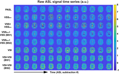 |
26 | Dual-module velocity-selective arterial spin labeling (dm-VSASL) dramatically improves the temporal signal-to-noise ratio of VSASL
Jia Guo1,2
1Bioengineering, University of California Riverside, Riverside, CA, United States, 2Center for Advanced Neuroimaging, UCR, Riverside, CA, United States
High temporal signal-to-noise ratio (tSNR) is desired for arterial spin labeling (ASL) methods. The tSNR of velocity-selective arterial spin labeling (VSASL) was limited in practice due to its sensitivity to error sources such as motion, eddy currents and diffusion effects. A novel VSASL strategy was invented to enable dual-module (dm-) VS saturation (VSS) and VS inversion (VSI) labeling for further improved SNR efficiency with VSASL. This study focused on the tSNR performance analysis and found the tSNR was doubled in gray matter using the new dm-VSASL. The explanation of such significant improvement was explored and validated by in vivo experiments.
|
||
0802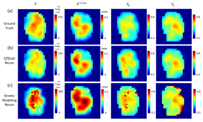 |
27 | Estimating perfusion and permeability using neural network with training data generated from vessel construction and transport simulation
Qihao Zhang1, Dominick Romano2, Thanh Nguyen2, Pascal Spincemaille3, and Yi Wang3
1Cornell University, Ithaca, NY, NY, United States, 2Cornell University, New York, NY, United States, 3Weill Cornell Medical College, New York, NY, United States
We propose to estimate perfusion parameters (perfusion $$$F$$$, permeability $$$K^{trans}$$$, vascular space volume $$$V_p$$$ and extravascular extracellular volume $$$V_e$$$) from contrast enhanced MRI using Quantitative Transport and Exchange network (QTEnet), a deep learning method that does not require an arterial input function. Training data were generated by solving the transport equation in simulated high-resolution vasculature and computing the corresponding 4D tracer propagation. A 3D U-net was trained to reconstruct perfusion parameters from the tracer propagation images. Tracer propagation simulated in experimentally obtained tumor vasculature was used for valiation, and the method was then applied to glioma DCE MRI data.
|
||
0803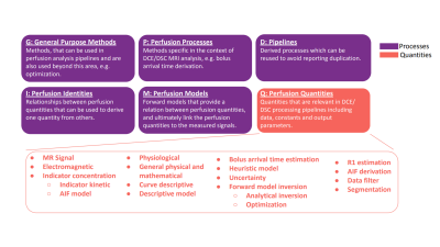 |
28 | Open Science Initiative for Perfusion Imaging (OSIPI): DCE/DSC lexicon for reporting perfusion analysis pipelines
Ben R Dickie1, Steven Sourbron2, Petra J van Houdt3, Laura Bell4, Rianne A van der Heijden5, Andrey Fedorov6, Jonathan Arvidsson7, Charlotte Debus8, Ingomar Gutmann9, Chad Quarles10, Ralf Floca11, Zaki ahmed12, David Buckley13, and Ina Kompan11
1Division of Informatics, Imaging and Data Sciences, The University of Manchester, Manchester, United Kingdom, 2University of Sheffield, Sheffield, United Kingdom, 3the Netherlands Cancer Institute, Amsterdam, Netherlands, 4Genentech, Inc, San Francisco, CA, United States, 5Erasmus MC University Medical Center, Rotterdam, Netherlands, 6Harvard Medical School, Boston, MA, United States, 7University of Gothenburg, Gothenburg, Sweden, 8Karlsruhe Institute for Technology, Karlsruhe, Germany, 9University of Vienna, Vienna, Austria, 10Barrow Neurological Institute, Phoenix, AZ, United States, 11German Cancer Research Center DKFZ, Heidelberg, Germany, 12Mayo Clinic, Rochester, MN, United States, 13The University of Leeds, Leeds, United Kingdom
The Open Science Initiative for Perfusion Imaging (OSIPI) aims to improve the reproducibility of perfusion MRI research through creation of acquisition and analysis standards. Specifically, Task Force 4.2 was established to develop a lexicon of variables and processes to facilitate standardised reporting of dynamic contrast-enhanced (DCE-) and dynamic susceptibility contrast (DSC-) MRI analysis pipelines. Here we report progress towards these objectives by presenting the current DCE/DSC lexicon structure, and provide a use-case to encode a simple analysis pipeline.
|
||
0804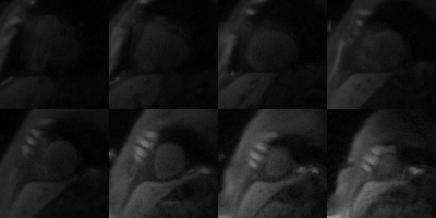 |
29 | High-resolution Spiral First-pass Myocardial Perfusion Imaging at 1.5 T with DEep learning-based rapid Spiral Image REconstruction (DESIRE)
Junyu Wang1, Patricia Rodriguez Lozano2, and Michael Salerno1,2,3,4
1Biomedical Engineering, University of Virginia, Charlottesville, VA, United States, 2Medicine, University of Virginia, Charlottesville, VA, United States, 3Radiology, University of Virginia, Charlottesville, VA, United States, 4Medicine, Stanford University, Stanford, CA, United States
First-pass contrast-enhanced myocardial perfusion imaging is useful for evaluating coronary artery disease (CAD). Spiral perfusion imaging techniques, using a motion-compensated (SMS-Slice-) L1-SPIRiT reconstruction, are capable of whole-heart high-resolution perfusion imaging. However, this reconstruction is performed off-line and takes ~30 minutes per slice. To address this, we developed a DEep learning-based rapid Spiral Image REconstruction technique (DESIRE) for spiral perfusion imaging at 1.5 T, for both single-slice (SS) and simultaneous multi-slice (SMS) MB=3 and MB=4 acquisitions, to provide rapid and high-quality reconstruction and make rapid online reconstruction feasible. High image quality was demonstrated using the proposed technique.
|
||
0805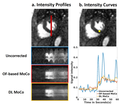 |
30 | Rapid Motion Correction with Deep Learning for First-Pass Cardiac Perfusion MRI
Lexiaozi Fan1,2, Huili Yang1,2, Li-Yueh Hsu3, Aggelos K Katsaggelos1,4,5, Bradley D Allen1, Daniel C Lee6, and Daniel Kim1,2
1Department of Radiology, Northwestern University Feinberg School of Medicine, Chicago, IL, United States, 2Department of Biomedical Engineering, Northwestern University, Evanston, IL, United States, 3Department of Radiology and Imaging Sciences, National Institutes of Health, Bethesda, MD, United States, 4Department of Electrical Engineering, Northwestern University, Evanston, IL, United States, 5Department of Computer Science, Northwestern University, Evanston, IL, United States, 6Division of Cardiology, Internal Medicine, Northwestern University Feinberg School of Medicine, Chicago, IL, United States
Motion correction (MoCo) is an important pre-processing step for pixel-by-pixel myocardial blood flow (MBF) quantification from cardiac perfusion MRI. It may also improve throughput of visual evaluation of perfusion images. One commonly used method for MoCo is optical flow (OF), which requires a moderate level of computational demand. In this study, we sought to perform rapid MoCo of respiratory motion on cardiac perfusion images using deep learning (DL). Our results show that the proposed DL MoCo performs 418-times faster than the reference OF approach without loss in accuracy.
|
||
0806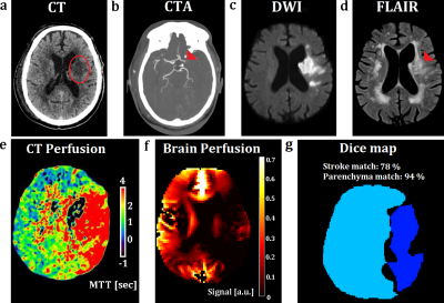 |
31 | Feasibility of Flow-related Enhancement Brain Perfusion MRI
Julian Glandorf1, Filip Klimes1, Andreas Voskrebenzev1, Marcel Gutberlet1, Agilo Luitger Kern1, Norman Kornemann1, Nima Mahmoudi2, Mike Peter Wattjes2, Frank Wacker1, and Jens Vogel-Claussen1
1Institut für Diagnostische und Interventionelle Radiologie, Hannover Medical School, Hannover, Germany, 2Institut für Diagnostische und Interventionelle Neuroradiologie, Hannover Medical School, Hannover, Germany
To test the feasibility of brain perfusion imaging exploiting flow-related enhancement during fast gradient echo sequences. Imaging was performed using Fast Low-Angle Shot (FLASH) and balanced steady-state free precession (bSSFP) sequences of a single axial slice with various imaging parameters at 1.5T and 3T. Perfusion-weighted maps were generated based on the Fourier decomposition method “Phase-REsolved FUnctional Lung” (PREFUL) MRI. The derived imaging protocol was evaluated in patients with metastases and a stroke showing high spatial overlap to post-contrast MRI and CT-perfusion. A cohort of 19 healthy individuals presented moderate, but significant regional correlation and high spatial Dice overlap to pCASL-MRI.
|
||
0807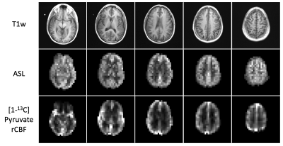 |
32 | Investigating Cerebral Perfusion with High Resolution Hyperpolarized [1-13C]Pyruvate MRI
Jasmine Y Hu1,2, Sana Vaziri1, Nikolaj Bogh3, Yaewon Kim1, Adam W Autry1, Robert A Bok1, Yan Li1,2, Christoffer Laustsen3, Peder EZ Larson1,2, Duan Xu1,2, Daniel B Vigneron1,2, and Jeremy W Gordon1
1Radiology and Biomedical Imaging, University of California San Francisco, San Francisco, CA, United States, 2Bioengineering, University of California Berkeley, Berkeley, CA, United States, 3MR Research Center, Clinical Medicine, Aarhus University, Aarhus, Denmark Acquiring high resolution 7.5 mm2 hyperpolarized [1-13C]pyruvate brain MRI allows for finer spatial delineation of brain structures and can be used to obtain cerebral perfusion parameters. In the healthy volunteers studied, pyruvate rCBV and rCBF were positively correlated to ASL perfusion values. Hyperpolarized pyruvate MRI can be used to assess cerebral metabolism and perfusion within the same study. |
||
0808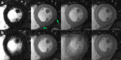 |
33 | Inline Implementation of Motion-Compensated, High-Resolution Myocardial Perfusion Imaging: Initial Experience
Karl Philipp Kunze1, Nina Mellor2, Tracy Moon2, Kuberan Pushparajah3, Radhouene Neji1, and Amedeo Chiribiri3
1MR Research Collaborations, Siemens Healthcare Limited, Frimley, United Kingdom, 2Evelina London Children’s Hospital, Guy’s and St Thomas’ NHS Foundation Trust, London, United Kingdom, 3Biomedical Engineering and Imaging Science, King's College London, London, United Kingdom
This abstract describes a feasibility study for a high-resolution perfusion imaging framework, including a dedicated sampling pattern and a motion compensation framework integrated into temporal regularization and implemented in-line on the scanner with GPU support. The approach was employed successfully in patients with suspected myocarditis at rest, showing superior ability to handle both respiratory and cardiac motion at an image resolution of 1.3 mm2, as compared to a clinical reference perfusion sequence and supported by Late Gadolinium Enhancement imaging findings.
|
||
0809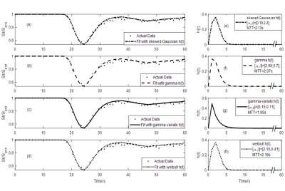 |
34 | Model-based deconvolution for DSC-MRI: A comparison of accuracy, precision, and computational complexity of transit time distributions
Rashed Sobhan1, Glyn Johnson1, and Donnie Cameron1,2
1Norwich Medical School, University of East Anglia, Norwich, United Kingdom, 2C.J. Gorter Centre for High Field MRI, Department of Radiology, Leiden University Medical Centre, Leiden, Netherlands To identify the optimal model-based deconvolution process for DSC-MRI, four models of transit time distribution (TTD) were compared in terms of goodness and stability of fit, consistency of perfusion estimates, computational complexity, and robustness against noise. Although all models gave similar fits, the gamma function converged faster and more consistently to the global minimum, regardless of the initial guess. Moreover, it gave more accurate and precise perfusion estimates in the presence of noise. We conclude that the gamma function is the most suitable TTD model for perfusion analysis, and may prove useful in urgent clinical situations and multi-centre studies. |
||
0810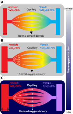 |
35 | Arterio-venous transit and oxygen extraction fraction before and after blood transfusion in sickle cell disease
Tonner DeBeer1, Lori C Jordan2, Spencer Waddle1, Chelsea A Lee2, Niral J Patel2, Maria Garza3, Larry Taylor Davis4, Sumit Pruthi4, Randall Sky Jones2, and Manus Joseph Donahue3
1Vanderbilt University, Nashville, TN, United States, 2Pediatrics, Vanderbilt University Medical Center, Nashville, TN, United States, 3Neurology, Vanderbilt University Medical Center, Nashville, TN, United States, 4Radiology and Radiological Sciences, Vanderbilt University Medical Center, Nashville, TN, United States
Most persons with sickle cell disease (SCD) lack conventional stroke risk factors, yet nearly 50% have evidence of brain infarcts by age 30 years, indicating alternative etiologies for ischemia. We investigated whether accelerated red cell transit affects oxygen extraction and improves following transfusion-induced increases in hemoglobin. Findings suggest that evidence of accelerated capillary transit is present on arterial spin labeling (ASL) MRI, and reduces following transfusion-induced increases in hemoglobin. Furthermore, the relationship between dural ASL signal and brain oxygen extraction evolves following transfusion, suggesting that oxygen delivery is complexly dependent on blood oxygen content and capillary dynamics.
|
||
 Back to Meeting Home
 Back to the Program-at-a-Glance
The International Society for Magnetic Resonance in Medicine is accredited by the Accreditation Council for Continuing Medical Education to provide continuing medical education for physicians.
 Back to Meeting Home
 Back to the Program-at-a-Glance
 View the Presentation