Digital Poster
New CMR Methods for Anatomy & Function (Micro- to Macro-) II
Joint Annual Meeting ISMRM-ESMRMB & ISMRT 31st Annual Meeting • 07-12 May 2022 • London, UK

Digital Poster
New CMR Methods for Anatomy & Function (Micro- to Macro-) II
| Computer # | ||||
|---|---|---|---|---|
1657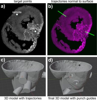 |
26 | Sample Extraction of ex vivo Myocardial Tissue: Combination of High Resolution MRI with 3D-printed Guides
Simon Reiss1, Johannes Fischer1, Julien Thielmann2, Thomas Lottner1, Timo Heidt2, Constantin von zur Mühlen2, and Michael Bock1
1Dept. of Radiology, Medical Physics, Medical Center University of Freiburg, Faculty of Medicine, University of Freiburg, Freiburg, Germany, 2Dept. of Cardiology and Angiology I, University Hospital Freiburg and Faculty of Medicine, University of Freiburg, Freiburg, Germany
Magnetic resonance provides a multitude of imaging techniques to detect and characterize myocardial infarction (MI). Terminal animal studies are often performed in which CMR findings after MI are correlated to histology. However, precise manual extraction of tissue samples from a specific myocardial region defined by CMR is difficult. Here, we propose the use of 3D-printed guides for extraction of tissue samples from myocardial regions pre-defined on high resolution ex vivo CMR. Using the presented technique, histology findings can be correlated to the exact position within the MRI data set with a precision of < 1 mm.
|
||
1658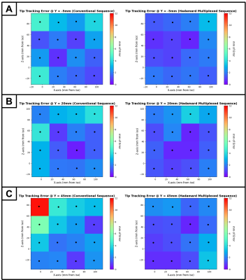 |
27 | Active Catheter Tracking Error Characterization for Minimally Invasive MR-Guided Cardiac Interventions
Arjun Gupta1, Labonny Biswas2, Jay Soni2, Brandon Coles1, Sebastian Ferguson2, Graham A. Wright3, and Nilesh R. Ghugre3
1Department of Medical Biophysics, University of Toronto, Toronto, ON, Canada, 2Physical Science Platform, Sunnybrook Research Institute, Toronto, ON, Canada, 3Schulich Heart Program, Sunnybrook Research Institute, Toronto, ON, Canada
Minimally invasive cardiac interventions, such as ablation treatment for ventricular tachycardia and therapy delivery involving biologics (cells, genes, biomaterials) for the treatment of myocardial infarction, require a tracking accuracy of <5mm. Our study aimed to characterize the spatial error associated with MR-guided active catheter tracking under static conditions. In a 1.5T MR scanner, we tracked an ablation catheter at 48 different positions, all confined within the boundaries of where a typical human heart would be located. We found that the error was both minimized and maintained below our 5mm target when using a Hadamard multiplexed active tracking sequence.
|
||
1659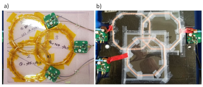 |
28 | Optimization of an RF array for cardiac magnetic resonance at 8.5 MHz
Gareth R Davies1, Robert S Stormont2, Lionel M Broche1, Dana K Dawson1, David J Lurie1, and P James Ross1
1Aberdeen Biomedical Imaging Centre, University of Aberdeen, Aberdeen, United Kingdom, 2GE Healthcare, Waukesha, WI, United States
The SNR and inter coil coupling properties of a range of litz wire coils, in conjunction with the circuitry to match the coils to a low impedance pre-amplifier, were examined. The results of these studies were combined with observations of SNR and coil coupling found in the images of phantoms, obtained with various array designs, to construct an RF array optimized for 0.2 T (8.5 MHz). The array has demonstrated its capability in acquiring cardiac images from human volunteers in our field cycling imager.
|
||
1660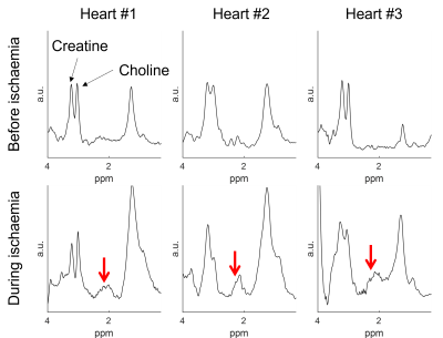 |
29 | Effects of warm ischemia on myocardial metabolism: a normothermic perfused heart MRS study
Belinda Ding1, Liam A.J. Young1,2, Margaret Huang3,4, Aravinda Page5, Simon Messer5, Mike Murphy3, Justin Perkins6, Elizabeth Tunnicliffe2, Stephen Large5, Christopher T Rodgers1, and Jonathan R Weir-McCall5,7
1Wolfson Brain Imaging Centre, University of Cambridge, Cambridge, United Kingdom, 2Oxford Centre for Magnetic Resonance, University of Oxford, Oxford, United Kingdom, 3MRC Mitochondrial Unit, University of Cambridge, Cambridge, United Kingdom, 4Department of Surgery, University of Cambridge, Cambridge, United Kingdom, 5Royal Papworth Hospital, Cambridge, United Kingdom, 6Royal Veterinary College, London, United Kingdom, 7Department of Radiology, University of Cambridge, Cambridge, United Kingdom
Ischaemia-reperfusion injury is a key pathophysiological process driving cardiac injury in myocardial infarction, heart surgery and transplants. Succinate accumulation is a specific marker of ischaemic injury, and a potential therapeutic target to reduce reperfusion injury.Single-voxel proton spectra were acquired during normal perfusion and during ischaemia in three machine-perfused porcine hearts. During ischaemia, a peak was evident in the expected region of succinate, which was absent during normal perfusion. This was further examined in a phantom model which confirmed the peak location with concentration-signal correlation of R>0.999. MRS detection of succinate holds potential for the detection of ischaemia.
|
||
1661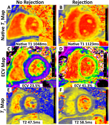 |
30 | Texture Analysis Identifies Significant Pattern Differences in Pediatric Heart Transplant Recipients with and without Acute Rejection
Margaret Samyn1, Ke Yan2, Kristen George-Durrett3, Justin Godown3, Kimberly Crum3, Maryanne Christ4, Kak-Chen Chan4, Kak-Chen Chan4, David Bearl3, Debra Dodd3, Lazaro Hernandez4, Jonathan Soslow3, and Bruce Damon5
1Pediatrics, Medical College of Wisconsin, Milwaukee, WI, United States, 2Biostatistics, Medical College of Wisconsin, Milwaukee, WI, United States, 3Pediatrics, Division of Pediatric Cardiology, Vanderbilt University Medical Center, Nashville, TN, United States, 4Pediatric Cardiology, Joe DiMaggio Children’s Hospital at Memorial Healthcare System, Hollywood, FL, United States, 5Stephens Family Clinical Research Institute Carle Foundation Hospital, Urbana, IL, United States
Acute rejection (AR) continues to cause significant morbidity and mortality in pediatric heart transplant recipients (PHTx). Endomyocardial biopsy is the standard-of-care for diagnosis of AR, but it is invasive and associated with morbidity and mortality and can miss patchy AR. Cardiac magnetic resonance (CMR) was performed using parametric mapping and novel texture analysis to detect patterns of myocardial edema and fibrosis. Patients with AR had significant differences in texture analysis compared with patients without AR. CMR with texture analysis has potential as a non-invasive method for detection of AR in PHTx.
|
||
1662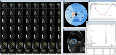 |
31 | Cine Image Based Cardiac Disease Classification Using Random Forest Classifier and Graph Based Deep Learning Approach
Akos Varga-Szemes1, Teodora Chitiboi2, U. Joseph Schoepf1, Athira J Jacob2, Sayan Ghosal3, Fei Xiong4, Puneet Sharma2, Jonathan Aldinger1, and Tilman Emrich1
1Medical University of South Carolina, Charleston, SC, United States, 2Siemens Healthcare, Princeton, NJ, United States, 3Johns Hopkins University, Baltimore, MD, United States, 4Siemens Healthcare, Charleston, SC, United States
Existing work demonstrates the value of image-based cardiovascular diagnosis with AI. We aimed to develop and test a machine learning algorithm for cardiovascular disease classification based on cine image datasets of 570 consecutive patients. Disease classification was performed using a random forest (RF) classifier and a disease classification network based on graph attention networks. The fully automated deep learning algorithm showed high accuracy for cardiac disease classification based on cine images only. Such algorithm has the potential to improve the efficiency of the reading process, especially by identifying and filtering out patients with normal cardiac anatomy and function.
|
||
1663 |
32 | Evaluation of diastolic (dys-)function using CMR feature tracking in all four cardiac chambers
Ana Fehrmann1, David Maintz1, and Bettina Baeßler2
1Department of Radiology, University Hospital of Cologne, Cologne, Germany, 2Department of Radiology, University Hospital of Würzburg, Würzburg, Germany Due to demographic changes, the prevalence of cardiac conditions predisposing to diastolic dysfunction (DD) is rising steadily. The main focus of non-invasive assessment of parameters for DD has been on left ventricular strain alterations. In this prospective study, we performed strain analysis of all 4 cardiac chambers using cardiac magnetic resonance feature tracking in patients with echocardiographically diagnosed DD and compared the results to a healthy control group. Left atrial strain parameters proved to be the most potent predictors of DD and allowed for a clear separation between patients and age-matched controls. |
||
1664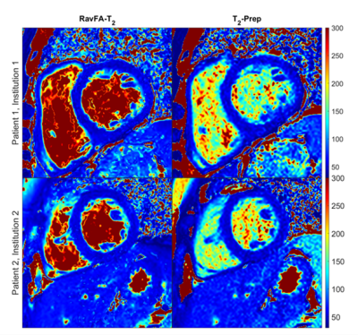 |
33 | Rapid Variable Flip-angle T2 Quantification (RavFa-T2) of the Myocardium using Steady-state Free Precession: A Two-Center Study Video Permission Withheld
Céline Marquet1,2,3, Jihye Jang1,2,4, Andrew J. Powell1,2, Brian Fonseca5,6, Lorna P. Browne5,6, and Mehdi H. Moghari5,6
1Harvard Medical School, Boston, MA, United States, 2Boston Children’s Hospital, Boston, MA, United States, 3Technical University of Munich, Munich, Germany, 4Philips Healthcare, Boston, MA, United States, 5University of Colorado, Aurora, CO, United States, 6Children’s Hospital Colorado, Aurora, CO, United States We developed a novel method (RavFa-T2) for T2 quantification of myocardium using transient steady-state free precession (SSFP) with a variable flip angle scheme. RavFa-T2 was systematically analyzed based on a phantom and a two-center patient study. We show that RavFa-T2 yields accurate T2 estimates for the myocardium compared to the state-of-the-art method T2-Prep and reduces scan time to ≤4 seconds. |
||
1665 |
34 | Strain-free in-vivo time dependent diffusion measures in the myocardium: A potential new biomarker ?
Ignasi Alemany1,2, Jan N Rose1, Pedro F. Ferreira2,3, Sonia Nielles-Vallespin2,3, Denis J. Doorly1, and Andrew D. Scott2,3
1Department of Aeronautics, Imperial College of London, London, United Kingdom, 2Cardiovascular Magnetic Resonance Unit, Royal Brompton Hospital, London, United Kingdom, 3National Heart and Lung Institute, Imperial College London, London, United Kingdom
Diffusion tensor cardiovascular magnetic resonance (DT-CMR) has proved to be a unique non-invasive method to investigate the myocardial microstructure. The time dependence of the diffusion tensor provides insights into the barriers that restrict free diffusion. We present a method to assess the time dependence of diffusion in the beating heart along with numerical simulations that demonstrate the sensitivity of this potential new biomarker to changes in myocardial microstructure including membrane permeability, extracellular volume fraction and cell hypertrophy.
|
||
1666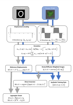 |
35 | Theoretical Considerations on Joint Inference of Cardiac Diffusion and Strain Tensors from second-order motion compensated cDTI Data
Jonathan Weine1, Stefano Buoso1, and Sebastian Kozerke1
1Institute for Biomedical Engineering, University and ETH Zurich, Zürich, Switzerland
Cardiac diffusion tensor imaging (cDTI) as well as cardiac strain imaging both offer invaluable information on the functional state of the heart. Acquiring both, takes too long to be clinically feasible. Free breathing, cDTI acquisition with second-order motion compensated diffusion encoding waveforms display residual phase variations, that encode higher orders of contractile motion during the diffusion encoding process. This work presents a theoretical framework on how to jointly infer strain and diffusion tensors from complex-valued cDTI data with varying trigger delays.
|
||
1667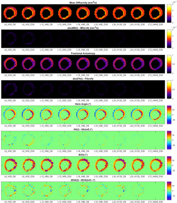 |
36 | In Vivo Clinical Cardiac DTI in 5 minutes Video Permission Withheld
Irvin Teh1, Christopher Kelly1, David Shelley2, Ana-Maria Poenar1, Sven Plein1, Erica Dall'Armellina1, Christopher Nguyen3,4, and Jürgen E. Schneider1
1Leeds Institute of Cardiovascular and Metabolic Medicine, University of Leeds, Leeds, United Kingdom, 2Leeds Teaching Hospitals Trust, Leeds, United Kingdom, 3Massachusetts General Hospital, Harvard Medical School, Cardiovascular Research Center, Martinos Center for Biomedical Imaging, Charlestown, MA, United States, 4Health Science Technology, Harvard-MIT, Cambridge, MA, United States
There is strong interest in characterising the cardiac microstructure using in vivo cardiac diffusion tensor imaging (CDTI). However, its use in larger clinical studies is often hampered by long scan times. We sought to rationalise the scan parameters needed for a clinically feasible CDTI protocol, by comparing carefully subsampled data against a 24-minute reference dataset. A design strategy was identified based on maximising the number of diffusion-weighting (DW) directions, subject to minimum SNR requirements. Feasibility of a 5-minute protocol was demonstrated where NRMSE(MD) = 5.2±0.2%, NRMSE(FA) = 12.8±0.2%, RMSE(HA) = 5.5±0.4°, RMSE(absE2A) = 15.7±1.9°.
|
||
1668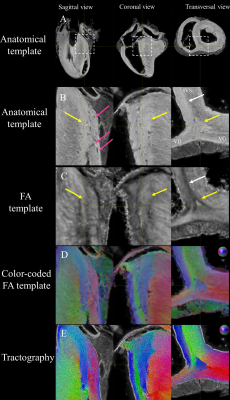 |
37 | A groupwise registration & tractography framework for cardiac architecture description by diffusion MRI: application to ventricular junctions
Julie Magat1,2,3, Maxime Yon1,2,3, Yann Bihan-Poudec4, and Valéry Ozenne1,2,3,5
1IHU Liryc, Electrophysiology and Heart Modeling Institute, Foundation Bordeaux Université, Bordeaux, France, 2Univ. Bordeaux, Centre de recherche Cardio-Thoracique de Bordeaux, U1045, Bordeaux, France, 3INSERM, Centre de recherche Cardio-Thoracique de Bordeaux, U1045, Bordeaux, France, 4Institut des Sciences Cognitives Marc Jeannerod, CNRS UMR 5229, Université Claude Bernard Lyon I, Bron, France, 5Centre de Résonance Magnétique des Systèmes Biologiques, UMR 5536, CNRS/Université de Bordeaux, Bordeaux, France
In this study, we developed a groupwise registration and tractography framework to investigate the global myofiber arrangement of large mammalian sheep hearts. To demonstrate the potential application of the proposed method, a novel description of sub-regions in the intraventricular septum (IVS) is presented. The study focuses on one fiber-bundle in the posterior junction and three fiber-bundles in the anterior junction.
|
||
1669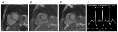 |
38 | Accuracy of left ventricular function measurements using retro-gated real-time cine bSSFP cardiovascular magnetic resonance
Ben K Statton1, Alaine Berry1, Peter Kellman2, Hui Xue2, Stuart Cook3, and Declan O'Regan3
1MRC, London Institute of Medical Sciences, Imperial College London, London, United Kingdom, 2National Heart, Lung, and Blood Institute, National Institute of Health, Bethesda, MD, United States, 3MRC, London Institute of Medical Sciences, UKRI, London, United Kingdom
Cardiac magnetic resonance with a retrospective ECG-gated breath-hold cine stack is the reference standard for assessing LV volumes, however this technique is unsuitable for patients who cannot hold their breath or suffer arrhythmia. A potential solution is a “retro-gated” real-time sequence which is prospectively triggered to acquire 120 frames over multiple heartbeats in each slice. Images from a beat most closely matching the median beat length for the entire stack are then temporally interpolated to 30 output phases for each slice. This retro-gated method showed good agreement with the reference standard sequence in a cohort of healthy volunteers.
|
||
1670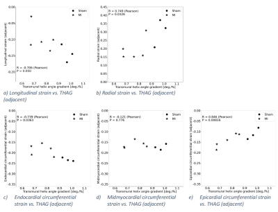 |
39 | The relationship between cardiac fiber structure and regional function after myocardial infarction in rats
Bård Andre Bendiksen1,2,3, Einar Sjaastad Nordén1,2,3, Ivar Sjaastad1,2, Lili Zhang1,2, and Emil Knut Stenersen Espe1,2
1Institute for Experimental Medical Research, University of Oslo and Oslo University Hospital, Oslo, Norway, 2KG Jebsen Center for Cardiac Research, University of Oslo, Oslo, Norway, 3Oslo New University College, Oslo, Norway
Little is known about the relationship between cardiac function and myocardial fiber structure after myocardial infarction (MI). We hypothesized that post-MI remodeling of left ventricular fiber structure would be reflected in changes in the regional strains. Strains and cardiac fiber structure were measured using MRI in five rats with MI, and three sham-operated rats. Transmural variation in fiber helix angles was reduced adjacent to the infarct. This reduction was associated with reduced radial, longitudinal, and endocardial circumferential strain, and improved epicardial circumferential strain in the adjacent zone. Our findings emphasize the relevance of cardiac fiber structure remodeling after myocardial infarction.
|
||
 Back to Meeting Home
 Back to the Program-at-a-Glance
The International Society for Magnetic Resonance in Medicine is accredited by the Accreditation Council for Continuing Medical Education to provide continuing medical education for physicians.
 Back to Meeting Home
 Back to the Program-at-a-Glance
 View the Presentation