Digital Poster
Flow Head to Toe IV
Joint Annual Meeting ISMRM-ESMRMB & ISMRT 31st Annual Meeting • 07-12 May 2022 • London, UK

| Computer # | ||||
|---|---|---|---|---|
1312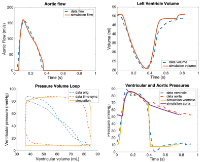 |
18 | Towards non-invasive assessment of cardiovascular physiology by combining cardiac MRI with predictive biomechanical modeling
Rebecca Waugh1,2, Maria Gusseva3,4, Gerald Greil2, Dominique Chapelle3,4, Tarique Hussain2, and Radomir Chabiniok2
1UT Dallas, Dallas, TX, United States, 2Pediatrics, UT Southwestern Medical Center, Dallas, TX, United States, 3Inria, Palaiseau, France, 4LMS, Ecole Polytechnique, Palaiseau, France
Biomechanical models coupled with cardiovascular MRI (CMR) have the potential to provide non-invasively physiological parameters of clinical interest. The approach is presented and validated on a group of 10 patients who underwent CMR and invasive catheterization. Patient-specific models are built based on CMR data and maximum/minimum arterial pressure. The stroke work, derived from ventricular pressure-volume (P-V) loops, was comparable between the measured and simulated P-V loops, suggesting the high quality of the in silico loops. An excellent correlation of the model-derived myocardial contractility with maximum time-derivative of ventricular pressure gives an opportunity for the non-invasive characterization of patients’ inotropic state.
|
||
1313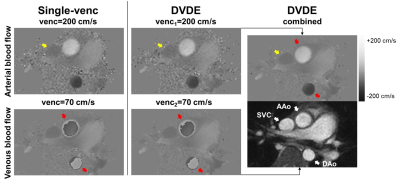 |
19 | Dual-venc Dual-echo (DVDE) Phase-contrast MRI for Simultaneous Measurement of Arterial and Venous Blood Flow
Jihye Jang1, Yansong Zhao1, Jouke Smink2, Andrew J Powell3, Lorna Browne4, and Mehdi H Moghari4
1Philips Healthcare, Gainsville, FL, United States, 2Philips Healthcare, Best, Netherlands, 3Boston Children’s Hospital and Harvard Medical School, Boston, MA, United States, 4Children's Hospital Colorado and University of Colorado, Aurora, CO, United States
Phase-contrast (PC) MRI enables non-invasive quantification of blood velocity and flow. The velocity encoding factor (venc) selected by the operator is chosen to maximize the velocity-to-noise ratio (VNR) while preventing aliasing artifacts. To minimize the acquisition time increase associated with an additional venc, a dual-venc dual-echo (DVDE) technique has been proposed where both venc images are acquired within a single TR as dual-echo images. Compared to the single-venc PC-MRI, DVDE allows simultaneous imaging of arterial and venous blood flow with an enhanced VNR and similar blood flow and velocity.
|
||
1314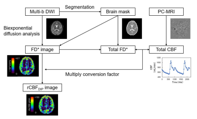 |
20 | Quantification of regional cerebral blood flow using diffusion imaging with phase-contrast (DIP)
Genki Nambu1, Naoki Ohno1, Tosiaki Miyati1, Fumiki Sugita1, Yuki Makino2, Momoka Ikeda1, Noam Alperin3, Toshifumi Gabata1, and Satoshi Kobayashi1
1Division of Health Sciences, Graduate School of Medical Sciences, Kanazawa University, Kanazawa, Ishikawa, Japan, 2Department of Radiology, Kanazawa University Hospital, Kanazawa, Ishikawa, Japan, 3Department of Radiology, University of Miami, Miami, FL, United States
Perfusion-related diffusion coefficient (D*) in intravoxel incoherent motion analysis is closely correlated with the regional cerebral blood flow (rCBF). However, the D* is only a semiquantitative relative value of rCBF, which makes absolute rCBF quantification challenging. To solve this problem, we developed a novel method of diffusion imaging with phase contrast (DIP), in which the total CBF (tCBF) from phase-contrast (PC)-MRI was used to convert the perfusion-related parameters in the brain to absolute rCBF. Using this method, we measured rCBF in gray matter and white matter. Each value was consistent with literature values assessed using [15O]-water positron emission tomography.
|
||
1315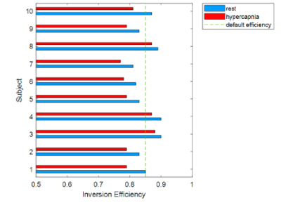 |
21 | A simplified method to estimate blood flow velocity and inversion efficiency in the major cerebral arteries using multi-PLD pCASL
Freya Allery1,2, Daniel P. Bulte1, Embla Hocking1, Nicholas P. Blockley3,4, James W. Harkin5, and Joana Pinto1
1Institute of Biomedical Engineering, Department of Engineering Science, University of Oxford, Oxford, United Kingdom, 2Institute of Health Informatics, University College London, London, United Kingdom, 3Sir Peter Mansfield Centre, School of Medicine, University of Nottingham, Nottingham, United Kingdom, 4School of Life Sciences, University of Nottingham, Nottingham, United Kingdom, 5Poole Hospital NHS Foundation Trust, Poole, United Kingdom
ASL inversion efficiency is often assumed as a pre-defined value or estimated by acquiring an additional phase-contrast MR image. While the former approach might be inaccurate in certain conditions, the latter can be time-consuming. Here, we propose a simplified method that estimates blood flow velocity and inversion efficiency solely using the ASL data. This novel strategy was tested using multi-PLD pCASL datasets with two different blood flow dynamics (normo- and hypercapnia). Our results show the feasibility of this approach and highlight the need for subject/condition-specific inversion efficiency estimation for a more accurate quantification of CBF.
|
||
1316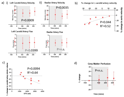 |
22 | A multi-organ assessment of the acute effects of haemodialysis using intradialytic multiparametric magnetic resonance imaging
Eleanor F Cox1, Venkata Rukmini Latha Gullapudi2,3, Charlotte E Buchanan1, Kelly White3, Sebastian Coleman1, Daniel Cocking1, Isma Kazmi2,3, Katrin Köhler4, Bernard Canaud4, Maarten W Taal2,3, Nicholas M Selby2,3, and Susan T Francis1
1Sir Peter Mansfield Imaging Centre, University of Nottingham, Nottingham, United Kingdom, 2Centre for Kidney Research and Innovation, University of Nottingham, Derby, United Kingdom, 3Renal Unit, University Hospitals of Derby and Burton NHS Foundation Trust, Derby, United Kingdom, 4Center of Excellence Medical Europe, Middle East and Africa, Fresenius Medical Care, Bad Homburg, Germany Twelve patients underwent serial cardiac, cerebral and renal multiparametric 3T MRI scans before, during and after haemodialysis. During dialysis, there was a significant decline in cardiac and stroke index and myocardial strain, with increased diastolic dysfunction. This was accompanied by a reduction in blood flow to both the heart and brain. White matter T1 increased during dialysis suggesting fluid shifts increasing water content resulting in local oedema. Total kidney volume, renal cortex T1 and T2* all reduced during dialysis likely reflecting reduced renal blood volume and change in renal tissue water. These results highlight the acute multi-organ effects of dialysis. |
||
1317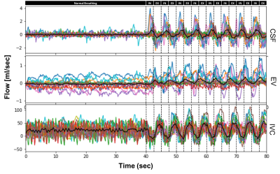 |
23 | Exploring coupling of venous blood and CSF fluid flow using flow sensitive Real-time phase-contrast MRI (PC-MRI) during forced breathing Video Permission Withheld
Prativa Sahoo1, Jost M Kollmeier2, and Steffi Dreha Kulaczewski1
1University Medicine Göttingen, Göttingen, Germany, 2Max Planck Institute for Biophysical Chemistry, Göttingen, Germany
Venous system pathologies have increasingly been linked to disorders of CSF circulation whereas the exact coupling mechanisms still remain unknown. Using real-time phase-contrast MRI fluid dynamics of both systems were studied during normal and forced breathing in healthy subjects at spinal lumbar level L3. The aim was to provide experimental evidence that respiration couples CSF and venous flow. A correlation between CSF and venous flow could be observed during forced breathing. The findings point to a dynamic interplay between the fluid systems to regulate intracranial volume. Further insights will expand our understanding of the pathophysiology of different forms hydrocephalus.
|
||
1318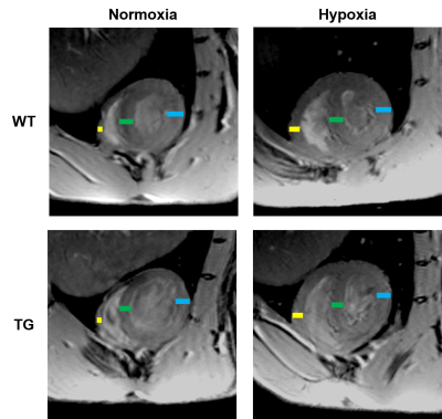 |
24 | Characterization of cardiac alterations in transgenic rat model of pulmonary arterial hypertension exposed to chronic hypoxia
Dounia El Hamrani1, Julie Magat1, Jérôme Naulin1, Céline Ayez1, Marilyne Campagnac2, Frédéric Perros3, Christelle Guibert2, David Benoist1, and Bruno Quesson1
1IHU Liryc, Bordeaux, France, 2CRCTB-INSERM U1045, Bordeaux, France, 3Université Paris–Saclay, AP-HP, INSERM UMR_S 999, Le Kremlin Bicêtre, France
Transgenic rats with Bmpr2 mutation is a model of heritable pulmonary arterial hypertension (PAH). This study aims to evaluate cardiac alterations of Bmpr2-mutants associated with a risk factor (hypoxia) by MRI and 31P-MRS. Transgenic rats exposed to chronic hypoxia showed a drastic dilatation of the RV along with an increased wall thickness and no variation of ejection fraction, thus resembling right heart failure with preserved ejection fraction. The cardiac energy balance is disturbed in Bmpr2 rats under hypoxia indicating a metabolic insult. Bmpr2 mutation associated with chronic hypoxia can aggravate right heart failure and thereby accentuated the onsets of PAH.
|
||
1319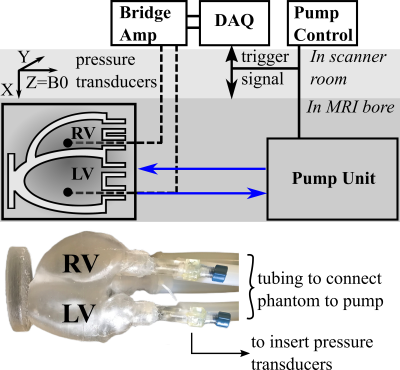 |
25 | Impact of cardiac geometry segmentation on MRI-driven estimates of myocardial stiffness in an in vitro synthetic heart model
Fikunwa Kolawole1,2,3, Mathias Peirlinck4, Tyler E Cork1,2,5, Marc E Levenston1,3, Ellen Kuhl3, and Daniel Ennis1,2
1Radiology, Stanford University, Stanford, CA, United States, 2Radiology, Veterans Administration Health Care System, Palo Alto, CA, United States, 3Mechanical Engineering, Stanford University, Stanford, CA, United States, 4Biomechanical Engineering, Technische Universiteit Delft, Delft, Netherlands, 5Bioengineering, Stanford University, Stanford, CA, United States MRI-driven computational constitutive modeling can be used to obtain subject-specific myocardial passive stiffness. Verifying the accuracy and precision of this technique requires overcoming the challenge of obtaining ground-truth in vivo myocardial stiffness. We developed a controllable in vitro diastolic filling setup which incorporates a soft heart phantom of known myocardium-mimicking mechanical stiffness and MRI properties. Using the setup we demonstrate that uncertainties in quantifying cardiac reference geometry can lead to errors in estimating myocardial passive stiffness. The in vitro setup is designed to enable us to achieve our overarching goal: to extensively validate in vivo MRI-based myocardial passive stiffness estimation. |
||
 Back to Meeting Home
 Back to the Program-at-a-Glance
The International Society for Magnetic Resonance in Medicine is accredited by the Accreditation Council for Continuing Medical Education to provide continuing medical education for physicians.
 Back to Meeting Home
 Back to the Program-at-a-Glance
 View the Presentation