Digital Poster
Arterial Spin Labelling
Joint Annual Meeting ISMRM-ESMRMB & ISMRT 31st Annual Meeting • 07-12 May 2022 • London, UK

| Computer # | ||||
|---|---|---|---|---|
0903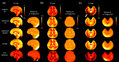 |
23 | Optimization and evaluation of inversion pulses for background suppressed pseudo-Continuous Arterial Spin Labeling at 7T
Chenyang Zhao1, Kai Wang1, Christina Graf2, Rudolf Stollberger2, and Danny JJ Wang1,3
1Laboratory of FMRI Technology (LOFT), Mark & Mary Stevens Neuroimaging and Informatics Institute, Keck School of Medicine, University of Southern California, Los Angeles, CA, United States, 2Institute of Medical Engineering, Graz University of Technology, Graz, Austria, 3Department of Neurology, Keck School of Medicine, University of Southern California, Los Angeles, CA, United States
Background suppression (BS) of pseudo-Continuous Arterial Spin Labeling (pCASL) needs to be optimized at 7T to ameliorate the ASL signal loss caused by the low inversion efficiency (IE) of BS due to B1 inhomogeneity. In this study, the original HS pulse used as BS at 3T was assessed at 7T, and four non-selective inversion pulses, including HS, WURST, OPTIM, and pTx adiabatic, were optimized and evaluated in phantom and in-vivo studies. Less than 80% IE achieved by the original HS pulse was improved to 85% by optimized pulses. Markedly higher tSNR was reported in pCASL with the optimized pulses.
|
||
0904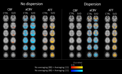 |
24 | Multi-delay ASL perfusion imaging: impact of modeling dispersion and interaction with denoising strategies
Sara Pires Monteiro1, Joana Pinto2, Michael Chappell3, Ana Fouto1, Miguel Viana-Baptista4, Pedro Vilela5, and Patrícia Figueiredo1
1Department of Bioengineering, ISR-Lisboa/LARSyS and Department of Bioengineering, Instituto Superior Técnico – Universidade de Lisboa, Lisbon, Portugal, 2Institute of Biomedical Engineering, Department of Engineering Science, University of Oxford, Oxford, United Kingdom, 3Sir Peter Mansfield Imaging Centre, School of Medicine, University of Nottingham, Nothingam, United Kingdom, 4Neurology Department, Hospital Egas Moniz, Centro Hospitalar de Lisboa Ocidental; CEDOC - Nova Medical School, New University of Lisbon, Lisbon, Portugal, 5Imaging Department, Hospital da Luz, Lisbon, Portugal
Arterial spin labeling (ASL) acquisitions at multiple post-labeling delays allow for appropriate kinetic models to be fitted to the data, potentially providing more accurate perfusion quantification. We investigated the influence of denoising strategies based on repetition averaging and ICA, together with the choice of an extended kinetic model with or without dispersion, on multi-delay ASL measurements from a group of small vessel disease patients and their age-matched controls. While ICA denoising generally improved model fitting, repetition averaging interacted with modeling dispersion and subject group, significantly impacting the estimation of perfusion and macrovascular contributions, mostly in arterial locations and with pathology.
|
||
0905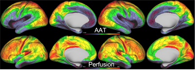 |
25 | The minimal processing pipeline for arterial spin labeling data from the Human Connectome Project Lifespan studies of Aging and Development
Flora Kennedy McConnell1,2,3, Jack Toner1,2, Thomas Kirk4,5, Yuriko Suzuki5, Martin Craig1,2, Timothy S. Coalson6, Michael P. Harms7, Matthew F. Glasser6,8, and Michael A. Chappell1,2,3,5
1Radiological Sciences, Mental Health and Clinical Neurosciences, School of Medicine, University of Nottingham, Nottingham, United Kingdom, 2Sir Peter Mansfield Imaging Centre, School of Medicine, University of Nottingham, Nottingham, United Kingdom, 3Nottingham Biomedical Research Centre, Queen's Medical Centre, University of Nottingham, Nottingham, United Kingdom, 4Institute of Biomedical Engineering, University of Oxford, Oxford, United Kingdom, 5Wellcome Centre for Integrative Neuroimaging, FMRIB, Nuffield Department of Clinical Neurosciences, University of Oxford, Oxford, United Kingdom, 6Department of Neuroscience, Washington University School of Medicine, Washington University in St Louis, St Louis, MO, United States, 7Department of Psychiatry, Washington University School of Medicine, Washington University in St Louis, St Louis, MO, United States, 8Department of Radiology, Washington University School of Medicine, Washington University in St Louis, St Louis, MO, United States
The arterial spin labelling (ASL) data from the Human Connectome Project (HCP) Lifespan studies can provide a source of unusually high-resolution hemodynamic measures from >2500 individuals with ages 5-21 and 37-100+. This work presents a summary of the minimal ASL processing pipeline used to provide pre-processed calibrated perfusion and arterial arrival time measurements for the cortical surface and subcortical volumes, from these individuals. The pipeline accounts for slice-wise image intensity variations resulting from the simultaneous multi-slice acquisition used to achieve high spatial resolution. These measures and this pipeline will be made available to the global neuroscience and neuroimaging communities.
|
||
0906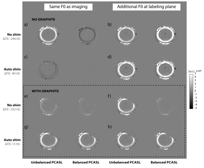 |
26 | F0 determination at the labeling plane: the neglected factor for successful pCASL perfusion MRI
Lena Vaclavu1, Kim van de Ven2, and Matthias van Osch1
1C.J. Gorter Center for High Field MRI, Department of Radiology, Leiden University Medical Center, Leiden, Netherlands, 2Philips Healthcare, Best, Netherlands
In perfusion imaging with arterial spin labeling (ASL), the labeling plane is located further away from the isocenter of the scanner, than the imaging volume. This can result in offsets in resonance frequencies which can lead to imperfect inversion and hence lower labeling efficiency. In this study, a perfusion phantom is used to study the influence of off-resonance on two arterial spin labeling schemes, in order to determine whether an additional F0 determination at the labelling plane is the redeeming factor for successful ASL MRI independent of the labeling scheme used.
|
||
0907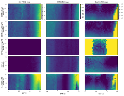 |
27 | Combined optimisation of delays and echo times for blood-brain barrier permeability measurements using multi-TE pCASL MRI
Logan X Zhang1 and Michael A Chappell2,3,4
1Department of Engineering Science, Institute of Biomedical Engineering, Nottingham, United Kingdom, 2Nuffield Department of Clinical Neurosciences, University of Oxford, Wellcome Centre for Integrative Neuroimaging, FMRIB, Oxford, United Kingdom, 3School of Medicine, University of Nottingham, Radiological Sciences, Division of Clinical Neurosciences, Nottingham, United Kingdom, 4School of Medicine, University of Nottingham, Sir Peter Mansfield Imaging Centre and Mental Health & Clinical Neurosciences, Nottingham, United Kingdom
Blood-brain barrier permeability to water has been measured as the exchange time (Texch) between intra- and extra-vascular spaces using multi-TE arterial spin labelling (ASL). This relies on small signal changes associated with labelled blood water delivery, making it challenging to achieve high accuracy in short yet clinically-desirable scan durations. In this study, we used an optimal sampling framework to generate pseudo-continuous ASL protocols that were jointly optimised for delays and echo times. Simulation showed that while optimised protocols had overall improvements in parameter estimation compared to conventionally even-sampled protocols, accurately estimating Texch could not be achieved without sacrificing CBF accuracy.
|
||
0908 |
28 | Open Science Initiative for Perfusion Imaging (OSIPI): Arterial Spin Labeling Imaging and Analysis Lexicon and Reporting Recommendations
Yuriko Suzuki1, Patricia Clement2, Weiying Dai3, Sudipto Dolui4, Maria Fernández-Seara5, Thomas Lindner6, Henk JMM Mutsaerts7, Jan Petr8, Xingfeng Shao9, Manuel Taso10, and David L Thomas11
1Wellcome Centre for Integrative Neuroimaging, FMRIB, Nuffield Department of Clinical Neurosciences, University of Oxford, Oxford, United Kingdom, 2Ghent Institute for Functional and Metabolic Imaging, Ghent University, Ghent, Belgium, 3State University of New York at Binghamton, Binghamton, NY, United States, 4University of Pennsylvania, Philadelphia, PA, United States, 5Department of Radiology, Clínica Universidad de Navarra, Pamplona, Spain, 6University Hospital Hamburg-Eppendorf, Hamburg, Germany, 7Department of Radiology and nuclear medicine, Amsterdam neuroscience, Amsterdam University Medical Center, Amsterdam, Netherlands, 8Helmholtz-Zentrum Dresden-Rossendorf, Dresden, Germany, 9University of Southern California, Los Angeles, CA, United States, 10Department of Radiology, Beth Israel Deaconess Medical Center and Harvard Medical School, Boston, MA, United States, 11Dept of Brain Repair and Rehabilitation, UCL Queen Square Institute of Neurology, University College London, London, United Kingdom
As part of the Open Science Initiative for Perfusion Imaging (OSIPI), the aim of this work is to develop a Lexicon and Reporting Recommendations for arterial spin labeling (ASL) perfusion MR imaging and analysis. The lexicon describes standardized nomenclature and terminology for ASL acquisition techniques, parameters and physiological constants that are required for quantitative analysis. Additionally, a community-endorsed recommendation for reporting acquisition parameters in publications is provided. Overall, this project aims to improve the clarity and consistency of ASL terminology, which in turn will facilitate comparisons between different studies.
|
||
0909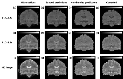 |
29 | Correction of artefacts in simultaneous multi-slice multi-PLD arterial spin labelling data using Gaussian Process regression
Jack Toner1,2, Flora Kennedy McConnell1,2,3, Yuriko Suzuki4, Timothy S. Coalson5, Michael P. Harms6, Matthew F. Glasser5,7, and Michael A. Chappell1,2,3,4
1Radiological Sciences, Mental Health & Clinical Neurosciences, School of Medicine, University of Nottingham, Nottingham, United Kingdom, 2Sir Peter Mansfield Imaging Centre, School of Medicine, University of Nottingham, Nottingham, United Kingdom, 3Nottingham Biomedical Research Centre, Queens Medical Centre, University of Nottingham, Nottingham, United Kingdom, 4Wellcome Centre for Integrative Neuroimaging, FMRIB, Nuffield Department of Clinical Neurosciences, University of Oxford, Oxford, United Kingdom, 5Department of Neuroscience, Washington University School of Medicine, St Louis, MO, United States, 6Department of Psychiatry, Washington University School of Medicine, St Louis, MO, United States, 7Department of Radiology, Washington University School of Medicine, St Louis, MO, United States
Simultaneous multi-slice (SMS) acquisitions enable higher resolutions to be achieved for arterial spin labelling (ASL) images. However, SMS acquisitions can introduce a banded pattern of intensity within the images. This reduces the quality of motion estimation as the algorithm aligns the bands in preference to the brain structures. We introduce a Gaussian Process model that can be used to correct the banding in SMS multiple post-labelling delay ASL data, which should improve motion correction. We demonstrate its effectiveness on 10 subjects from the Human Connectome Project Aging dataset. We anticipate that the model will generalise to other ASL datasets.
|
||
0910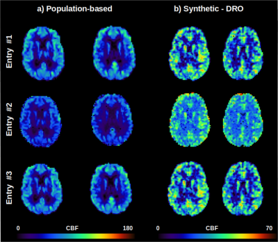 |
30 | The Open Science Initiative for Perfusion Imaging (OSIPI): Results from the ASL MRI Challenge Video Not Available
Udunna Anazodo1,2, Joana Pinto3, Flora Kennedy McConnell4,5,6, Cassandra Gould van Praag7,8, Henk Mutsaerts9, Aaron Oliver Taylor10, Jan Petr11, Diego Pineda-Ordóñez12, Maria-Eleni Dounavi13, Irène Brumer14, Wei Siang Marcus Chan14, Jack Toner15, Jian Hu15, Logan X. Zhang3, Laura Bell16, Joseph G. Woods17, Moss Y Zhao18, Paula Croal4,5, and Andre Monteiro Paschoal19
1Lawson Health Research Institute,, London, ON, Canada, 2Western University, London, ON, Canada, 3Institute of Biomedical Engineering, Department of Engineering Science, University of Oxford, Oxford, United Kingdom, 4Radiological Sciences, Division of Clinical Neuroscience, School of Medicine, University of Nottingham, Nottingham, United Kingdom, 5Sir Peter Mansfield Imaging Centre, School of Medicine, University of Nottingham, Nottingham, United Kingdom, 6Nottingham Biomedical Research Centre, Queens Medical Centre, Nottingham, United Kingdom, 7Wellcome Centre for Integrative Neuroimaging, University of Oxford, Oxford, United Kingdom, 8Department of Psychiatry, University of Oxford, Oxford, United Kingdom, 9Department of Radiology and Nuclear Medicine, Amsterdam Neuroscience, Amsterdam University Medical Center,, Amsterdam, Netherlands, 10Gold Standard Phantoms Limited, London, United Kingdom, 11Helmholtz-Zentrum Dresden-Rossendorf, Institute of Radiopharmaceutical cancer research, Dresden, Germany, 12Department of Radiology, Clinica Del Country, Bogotá, Colombia, 13Department of Psychiatry, University of Cambridge, Cambridge, United Kingdom, 14Computer Assisted Clinical Medicine, Mannheim Institute for Intelligent Systems in Medicine, Medical Faculty Mannheim, Heidelberg University, Heidelberg, Germany, 15Mental Health & Clinical Neurosciences, School of Medicine, University of Nottingham, Nottingham, United Kingdom, 16Genentech, Inc., South San Francisco, CA, United States, 17Center for Functional Magnetic Resonance Imaging, Department of Radiology, University of California San Diego, La Jolla, CA, United States, 18Department of Radiology, Stanford University, Stanford, CA, United States, 19Institute of Radiology and Department of Radiology and Oncology, University of Sao Paulo, Sao Paulo, Brazil
The OSIPI ASL MRI Challenge is an initiative of the ASL community aiming to characterize the variability of CBF quantification arising from different pipelines. The goal of this challenge is to establish best practice in ASL data processing, understand the sources of variability, make ASL analysis more reproducible, and enable fair comparison between studies. Here, we analyzed 3 submitted entries from 7 teams registered in the challenge. The preliminary results showed pipelines based in different programming languages and analysis tools, leading to important variability in the quantitative CBF maps compared to the ground-truth.
|
||
0911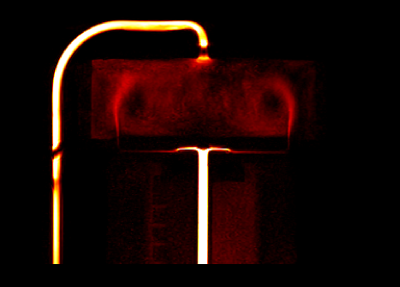 |
31 | A vortical phantom for ASL perfusion MRI
Aaron Oliver-Taylor1 and Xavier Golay1,2
1Gold Standard Phantoms, London, United Kingdom, 2Queen Square Institute of Neurology, University College London, London, United Kingdom
Most perfusion phantoms that have been created make use of porous media to simulate the microvasculature, however their characterisation introduces additional uncertainties into a perfusion measurement. We take inspiration from a recently published impinging jet perfusion phantom and presenting a phantom that makes use of vortices to mix and disperse the perfusion signal. Multi post labelling delay pCASL data was acquired, and fit to the general kinetic model. By assuming the measured perfusion equates to the equivalent perfusion from the systemic flow rate the labelling efficiency can be estimated, and the resultant measured and fitted signal curves closely match.
|
||
0912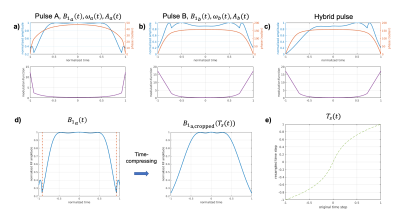 |
32 | SAR-reduced Asymmetric tr-FOCI for PICORE-ASL
Didi Chi1,2, Yasmin Blunck1,2, Rebecca Glarin2, Catherine E. Davey1,2, Josef Pfeuffer3, Daniel Staeb3, Jin Jin3, and Leigh A. Johnston1,2
1Department of Biomedical Engineering, University of Melbourne, Parkville, Australia, 2Melbourne Brain Centre Imaging Unit, University of Melbourne, Parkville, Australia, 3MR Research Collaborations, Siemens Healthcare Pty Ltd, Melbourne, Australia
PICORE-ASL using tr-FOCI pulses during labelling typically generates high power deposition at 7T. The current study introduces an asymmetric tr-FOCI pulse for PICORE-ASL to achieve SAR reduction. Results from simulations, phantom and in vivo experiments demonstrate the SAR reduction that can be achieved by using the asymmetric pulses without sacrificing inversion efficiency, slice profiles or CBF measurement.
|
||
0913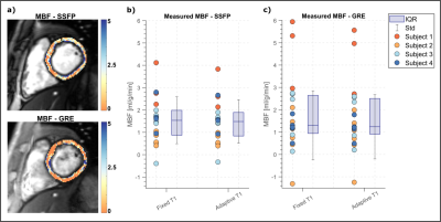 |
33 | Towards reproducible Arterial Spin Labelling in the myocardium: Impact of blood T1 time and imaging readout parameters
Masa Bozic-Iven1,2, Stanislas Rapacchi3, Iain Pierce4, George Thornton4, Qian Tao2, Lothar Schad1, Thomas Treibel4, and Sebastian Weingaertner2
1Computer Assisted Clinical Medicine, University Heidelberg, Mannheim, Germany, 2Delft University of Technology, Delft, Netherlands, 3University Aix-Marseille, Marseille, France, 4Barts Heart Centre, London, United Kingdom
Despite promising results, clinical translation of myocardial arterial spin labelling (myoASL) is hampered by insufficient reproducibility and robustness. We investigated the influence of physiological and sequence parameters on FAIR-myoASL in simulations as well as phantom experiments, and developed a correction method based on separately acquired T1 maps. Our simulation and phantom results show acquisition related MBF differences, potentially undermining the reproducibility of myoASL measurements. Inaccuracies between true and reconstruction blood T1, further render the sequence susceptible to heart rate variations, particularly for larger T1 mismatch. Using accurate blood T1 times in reconstruction may improve robustness and reproducibility.
|
||
0914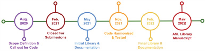 |
34 | The Open Science Initiative for Perfusion Imaging (OSIPI): ASL Code Library
María Guadalupe Mora Álvarez1, Li Zhao2, Sudipto Dolui3, Manuel Taso4, Yiming Wang5, Limin Zhou5, Ze Wang6, Azeez Adebimpe7, Henk Mutsaerts8, and Ananth J. Madhuranthakam5
1Department of Diagnostic and Interventional Neuroradiology, Technical University Munich (TUM), Munich, Germany, 2College of Biomedical Engineering & Instrument Science, Zhejiang University, Hangzhou, China, 3Department of Radiology, University of Pennsylvania, Philadelphia, PA, United States, 4Division of MRI Research, Department of Radiology, Beth Israel Deaconess Medical Center and Harvard Medical School, Boston, MA, United States, 5Department of Radiology, UT Southwestern Medical Center, Dallas, TX, United States, 6Department of Diagnostic Radiology and Nuclear Medicine, University of Maryland School of Medicine, Baltimore, MD, United States, 7Department of Psychiatry, University of Pennsylvania, Philadelphia, PA, United States, 8Amsterdam University Medical Center, Amsterdam, Netherlands
Task force 2.2 of the Open Science Initiative for Perfusion Imaging (OSIPI) is developing a library of open-source functions, and scripts for Arterial Spin Labeled (ASL) perfusion imaging preprocessing and analysis. This is aimed for developers of ASL perfusion methods looking for specific functionalities or development templates, or who want to share their own in-house code with others. The collected source code will be organized and documented so as to support the open library development.
|
||
 Back to Meeting Home
 Back to the Program-at-a-Glance
The International Society for Magnetic Resonance in Medicine is accredited by the Accreditation Council for Continuing Medical Education to provide continuing medical education for physicians.
 Back to Meeting Home
 Back to the Program-at-a-Glance
 View the Presentation