Digital Poster
Advanced Imaging of Stroke & Ischemia
Joint Annual Meeting ISMRM-ESMRMB & ISMRT 31st Annual Meeting • 07-12 May 2022 • London, UK

Digital Poster
Advanced Imaging of Stroke & Ischemia
| Computer # | ||||
|---|---|---|---|---|
1015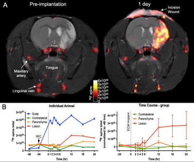 |
22 | Serial in vivo 19F MRI tracks the immune cell response to bioscaffolds implanted in a rat model of stroke
Mike Modo1, Harmanvir Ghuman2, Reem Azar2, Ryan Krafty3, Stephen Badylak4, and T. Kevin Hitchens5
1Radiology, University of Pittsburgh, Pittsburgh, PA, United States, 2Bioengineering, University of Pittsburgh, Pittsburgh, PA, United States, 3Biological Science, University of Pittsburgh, Pittsburgh, PA, United States, 4Surgery, University of Pittsburgh, Pittsburgh, PA, United States, 5Neurobiology, University of Pittsburgh, Pittsburgh, PA, United States
Regenerative medicine is increasingly investigating how an initial pro-inflammatory response convert to a pro-repair response. Implantation of bioscaffolds exploits this mechanism and allows tissue to regrow in large defects. Even the brain, which is known to not regrow tissue spontaneously, can regenerate lost tissue after implantation of a inductive bioscaffold. Little is known about how immune cells respond to the implantation of a bioscaffold. We here characterized the acute spatiotemporal dynamics of immune cell infiltration into the brain and a bioscaffold by tagging immune cells with perfluorocarbon nanoemulsions for detection by 19F MRI and anatomical context using 1H MRI.
|
||
1016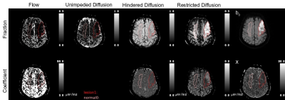 |
23 | Multimodal apparent diffusion model provides comprehensive analysis of clinical subacute stroke lesions
Frederick C. Damen1, Changliang Su2, Burce Ozgen Mocan1, Tibor Vályi-Nagy3, Rifeng Jiang4, and Kejia Cai1,5
1Radiology, University of Illinois at Chicago, Chicago, IL, United States, 2Medical Imaging, Sun Yat-sen University Cancer Center; State Key Laboratory of Oncology in South China; Collaborative Innovation Center for Cancer Medicine, Guangzhou, Guangdong, China, 3Pathology, University of Illinois at Chicago, Chicago, IL, United States, 4Radiology, Fujian Medical University Union Hospital, Fuzhou, Fujian, China, 5Biomedical Engineering, University of Illinois at Chicago, Chicago, IL, United States
We present our initial findings of applying Multimodal Apparent Diffusion (MAD) MRI, including b-values up to 10,000 s/mm2, to analyze subacute stroke lesions for the characterization of stroke lesions based on micro-environment biomarkers in order to guide treatment in clinics. MAD model is able to separate the acquired diffusion weighted signal into flow, and, unimpeded (fluid), hindered, and restricted apparent diffusion components. Significant differences were found in MAD parameters between lesions and normal appearing white matter. Fraction of unimpeded diffusion significantly increased. Hindered diffusion was significantly lower in lesions. On the other hand, restricted diffusion was significantly higher.
|
||
1017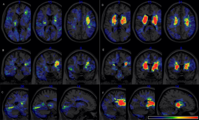 |
24 | Association between lesion locations and depressive symptoms in acute stroke patients using voxel-based lesion-symptom mapping Video Not Available
Peng Wang1, Jinjing Wang2, Yong Zhang3, Mengmeng Gu4, Shiyi Jiang1, Dawei Yin1, Wen Sun1, and Xinfeng Liu2
1The First Affiliated Hospital of University of Science and Technology of China, Hefei, China, 2Jinling Hospital, Nanjing, China, 3GE Healthcare, Shanghai, China, 4Nanjing First Hospital, Nanjing, China Post-stroke depression (PSD) is a common neuropsychiatric symptom after stroke. The purpose of this study was to investigate the association between lesion locations of brain stroke and PSD occurrence using Voxel-based Lesion-Symptom Mapping (VLSM). VLSM analysis identified clusters within anterior cingulate gyrus, left hippocampus, left lingual lobe where lesions were significantly associated with PSD. The lesion location showed the promise to be used as an independent risk factor of PSD in patients with acute ischemic stroke. |
||
1018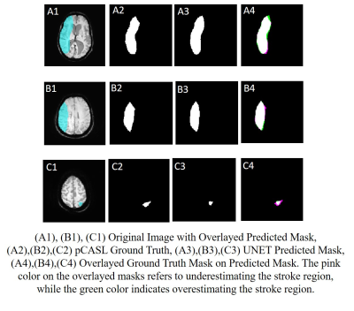 |
25 | Deep Learning-based Stroke Region Segmentation on Susceptibility Weighted Images in Acute Stroke
Ankit Kandpal1, Tanuja Jayas1, Rupsa Bhattacharjee1, Rakesh Kumar Gupta2, and Anup Singh1,3,4
1Centre for Biomedical Engineering, Indian Institute of Technology, Delhi, New Delhi, India, 2Department of Radiology, Fortis Memorial Research Institute, Gurugram, India, 3Department of Biomedical Engineering, All India Institute of Medical Sciences, New Delhi, India, 4School for Artificial Intelligence, Indian Institute of Technology, Delhi, New Delhi, India
SWI plays a critical role in stroke in demonstration of hemorrhagic transformation of stroke and demonstration of thrombus in the intracranial arteries. Recently it has been used to quantify the penumbra in acute stroke. It highlights venous vasculature in acute stroke due to hypoxia in the acute ischemic tissue without the need for any contrast injection and adding additional sequence that results in time penalty. The objective of this study was to develop an automatic framework for penumbra detection using only SWI images. Evaluation of segmentation results shows a dice similarity coefficient of 0.72 and a jaccard index of 0.60.
|
||
 Back to Meeting Home
 Back to the Program-at-a-Glance
The International Society for Magnetic Resonance in Medicine is accredited by the Accreditation Council for Continuing Medical Education to provide continuing medical education for physicians.
 Back to Meeting Home
 Back to the Program-at-a-Glance
 View the Presentation