Digital Poster
Cerebrovascular Anatomy
Joint Annual Meeting ISMRM-ESMRMB & ISMRT 31st Annual Meeting • 07-12 May 2022 • London, UK

| Computer # | ||||
|---|---|---|---|---|
1388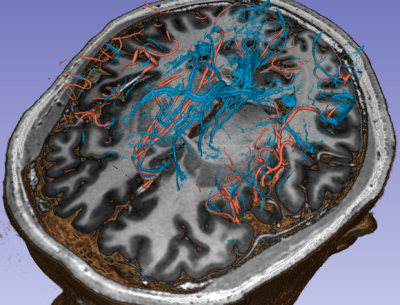 |
12 | High-resolution vascular imaging of the whole brain at 7T in 15 minutes
Arun Joseph1,2,3, Patrick Liebig4, Piotr Radojewski2,5, Roland Wiest2,5, Tobias Kober6,7,8, and Tom Hilbert6,7,8
1Advanced Clinical Imaging Technology, Siemens Healthcare AG, Bern, Switzerland, 2Translational Imaging Center, sitem-insel AG, Bern, Switzerland, 3Magnetic Resonance Methodology, Institute of Diagnostic and Interventional Neuroradiology, University of Bern, Bern, Switzerland, 4Siemens Healthcare GmbH, Erlangen, Germany, 5Support Center for Advanced Neuroimaging, Institute for Diagnostic and Interventional Neuroradiology, Inselspital, Bern University, Bern, Switzerland, 6Advanced Clinical Imaging Technology, Siemens Healthcare AG, Lausanne, Switzerland, 7Department of Radiology, Lausanne University Hospital and University of Lausanne, Lausanne, Switzerland, 8LTS5, École Polytechnique Fédérale de Lausanne (EPFL), Lausanne, Switzerland
Vascular disease such as arteriovenous malformations can be fatal and lead to brain damage if not detected early. To that end, time of flight angiography is an important tool to visualize vessels, especially the arteries with implications for treatment planning and follow-up. Similarly, susceptibility weighted imaging can be used to visualize veins due to its sensitivity to magnetic field inhomogeneities. However, high resolution TOF and SWI acquisitions can lead to clinically unfeasible scan times. Here, we propose to use compressed-sensing-accelerated high resolution TOF and SWI acquisitions at 7T in <15 minutes to visualize the vascular system of the whole brain.
|
||
1389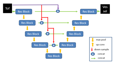 |
13 | Automatic vascular segmentation of neck TOF MRA images based on 3D CNN model Video Not Available
Wei Qiu1, Hanyu Wei1, Shuo Chen1, and Rui Li1
1Department of Medicine, CBIR, Tsinghua University, Beijing, China
Accurate and fast automatic Carotid artery segmentation of time of flight(TOF) MRA plays an important role in the auxiliary diagnosis of carotid artery disease. Considering the complexity and uncertainty of doctors’ manual segmentation of neck vessels, automatic segmentation algorithms are required in clinical practice. A segmentation model based on 3D Convolutional neural network (CNN) was proposed to segment carotid arteries from TOF MRA images. With innovative adjustment of the network architecture and parameters for carotid application, our model showed better performance than other baseline models on private dataset.
|
||
1390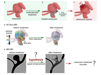 |
14 | Quantification and visualization of aneurysm treatment outcome with signal enhancement on Black Blood MRI.
Mariya S. Pravdivtseva1, Fritz Wodarg2, Jana Korte3, Philipp Berg3, Jan-Bernd Hövener1, Olav Jansen2, and Naomi Larsen2
1Department of Radiology and Neuroradiology, Section Biomedical Imaging, Molecular Imaging North Competence Center (MOIN CC), University Medical Center Schleswig-Holstein (UKSH), Kiel University, Kiel, Germany, 2Department of Radiology and Neuroradiology, University Medical Center Schleswig-Holstein (UKSH), Kiel, Germany, 3Research Campus STIMULATE, University of Magdeburg, Magderburg, Germany
Intracranial aneurysm is a life-threatening disease that can be treated with flow-modulating devices (FMDs) by reducing aneurysm blood flow. Aneurysms might rupture after treatment, thus biomarker of successful treatment is needed. Flow MRI can detect flow reduction, however, it is impaired by metal artifacts from FMDs. Black-blood (BB) MRI is less sensitive to metal artifacts and often results in poor slow-flow suppression. We hypothesize that BB contrast changes after aneurysm treatment. Here, after placing FMDs in 3D-printed aneurysm models, an enhanced signal was found on BB MRI, associated with reduced aneurysmal flow after flow modulation and thus possible treatment success.
|
||
1391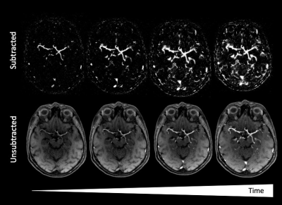 |
15 | Field Strength and Gadolinium Dose Dependency of Dynamic Time-Resolved 4D MRA of the Brain Using Golden-Angle Radial Sparse Parallel (GRASP) MRI
Adam Goldman-Yassen1,2, Vishal Patel3, Maria J. Borja3, Anna Derman4, Duan Chen5, Siddhant Dogra3, Roy Wiggins3, Kai Tobias Block3, and Seena Dehkharghani3,6
1Department of Radiology, Children's Healthcare of Atlanta, Atlata, GA, United States, 2Department of Radiology and Imaging Sciences, Emory University, Atlanta, GA, United States, 3Department of Radiology, New York University Langone Medical Center, New York, NY, United States, 4Department of Radiology, Maimonides Medical Center, Brooklyn, NY, United States, 5Department of Radiology, Weill Cornell Medical Center, New York, NY, United States, 6Department of Neurology, New York University Langone Medical Center, New York, NY, United States
GRASP MRI obtained as part of routine clinical care, without specific angiographic parameters, can be reconstructed post-hoc into dynamic 4D MR images and can be displayed with high spatial and temporal resolution. Additionally, contrast dose and magnet strength appear to have minimal effect on the diagnostic quality of angiographic images. Further exploration into diagnostic accuracy and the tolerance of varying field strength and dosing to further increasing degrees of acceleration is warranted.
|
||
1392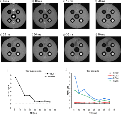 |
16 | In vivo, high-resolution Black Blood MRI at 7T Video Permission Withheld
Eva Peschke1, Mariya Pravdivtseva1, Olav Jansen2, Naomi Larsen2, and Jan-Bernd Hövener1
1Section Biomedical Imaging, Molecular Imaging North Competence Center (MOIN CC), Department of Radiology and Neuroradiology, University Medical Center Schleswig - Holstein, Kiel University, Kiel, Germany, Kiel, Germany, 2Department of Radiology and Neuroradiology, University Medical Center Schleswig-Holstein, Kiel University, Kiel, Germany, Kiel, Germany Vessel wall imaging (VWI) is a unique method to depict the vessel walls in vivo but was not applied in small animal models at 7T yet. Here, we developed a setup to emulate T1 contrast and flow to optimize Black Blood MRI in vitro. Using the results, we implemented a RARE sequence that allows in vivo Black Blood MRI with a high resolution of 0.156 x 0.156 mm² and successful flow suppression. Next, the model will be extended for VWI to measure the enhancement of Gadolinium contrast agent. |
||
| 1393 | 17 | White matter perivascular space enlargement in cerebral small vessel disease indicates cerebral amyloid angiopathy pattern changes Video Not Available
hui hong1 and minming zhang2
1the second affiliated hospital of zhejiang university, school of medicine, hangzhou, China, 2radiology, the second affiliated hospital of zhejiang university, school of medicine, hangzhou, China The enlargement of white matter perivascular space(PVS) is highly CAA related, however lack of studies investigated its role in stable pattern CSVD patients, we hypothesis that CSVD patients with white matter PVS enlargement indicates a CAA pattern changes. Here we compared patients with and without white matter PVS enlargement from imaging manifestation, imaging mechanism and clinical cognitive ability. We foud that patients with PVS enlargenmet showed a pattern similar to previous symptomatic CAA, that is more lobar microbleed, more interstitial fluid retention and significantly lower global cognitive ability. Extra dignosis and treament should be carried out to this pattern CSVD. |
||
1394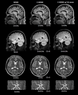 |
18 | Three-fold accelerated MRI and MR angiography on Moyamoya disease examination using deep learning constrained Compressed SENSE reconstruction
Yajing Zhang1, Jilei Zhang2, Maoxue Wang3, Kun Wang3, Xiance Zhao2, Peng Wu2, Weibo Chen2, Queenie Chan2, Johannes M. Peeters4, Marc Van Cauteren5, and Bing Zhang3
1BU-MR Clinical Science, Philips Healthcare, Suzhou, China, 2Philips Healthcare, Shanghai, China, 3Department of Radiology, Nanjing Drum Tower Hospital, The Affiliated Hospital of Nanjing University Medical School, Nanjing, China, 4BU-MR Clinical Science, Philips Healthcare, Best, Netherlands, 5BU-MR Clinical Science, Philips Healthcare, Tokyo, Japan
MR angiography (MRA) in moyamoya disease has been uniquely important in the evaluation of changes in vasculature and brain parenchyma, and clinical follow-up. In this work, we investigate the performance of accelerated MRA under a deep learning constrained Compressed SENSE reconstruction framework. Results on both volunteer and patients demonstrate a three-fold scan acceleration against the routine MRA with comparable image quality.
|
||
 Back to Meeting Home
 Back to the Program-at-a-Glance
The International Society for Magnetic Resonance in Medicine is accredited by the Accreditation Council for Continuing Medical Education to provide continuing medical education for physicians.
 Back to Meeting Home
 Back to the Program-at-a-Glance
 View the Presentation