Digital Poster
Cerebrovascular Function
Joint Annual Meeting ISMRM-ESMRMB & ISMRT 31st Annual Meeting • 07-12 May 2022 • London, UK

| Computer # | ||||
|---|---|---|---|---|
1466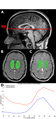 |
16 | Assessing Drug Effect on the Velocity Pulsatility in the Perforating Arteries of the Basal Ganglia at 3T-MRI in Pseudoxanthoma Elasticum
Stanley D.T. Pham1, Rick J. van Tuijl1, Jonas W. Bartstra1, Tim C. van den Beukel1, Geert Jan Biessels2, Pim A. de Jong1, Wilko Spiering3, Birgitta K. Velthuis1, and Jaco J.M. Zwanenburg1
1Radiology, UMC Utrecht, Utrecht, Netherlands, 2Neurology and Neurosurgery, UMC Utrecht, Utrecht, Netherlands, 3Vascular Medicine, UMC Utrecht, Utrecht, Netherlands
This feasibility study aimed to detect drug effects on perforating artery velocity as measured with two-dimensional phase-contrast (2D-PC) velocity measurements at 3T-MRI. Seventeen patients with pseudoxanthoma elasticum were included into a treatment group who received etidronate (n=9) and into a placebo group (n=8). No significant differences were found between both groups at baseline and one-year follow-up. In the etidronate group, mean velocity (Vmean) was significantly higher at follow-up (5.61 [4.77–6.45] cm/s) compared to baseline (4.80 [4.05–5.54] cm/s). In the placebo group, Vmean did not increase significantly. Measuring drug effects was feasible using 2D-PC measurements at 3T-MRI.
|
||
1467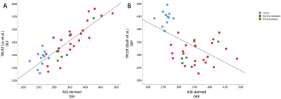 |
17 | Comparison of cerebral oxygen extraction fraction using ASE and TRUST methods in patients with sickle cell disease and healthy controls
Slim Fellah1, Chunwei Ying1, Kristin Guilliams1, Melanie Fields1, Yasheng Chen1, Joben Lewis1, Amy Mirro1, Rachel Cohen1, Nkemdilim Igwe1, Cihat Eldeniz1, Dengrong Jiang2, Hanzhang Lu2, Jin-Moo Lee1, Andria Ford1, and Hongyu An1
1Washington University School of Medicine, Saint Louis, MO, United States, 2Johns Hopkins University School of Medicine, Baltimore, MD, United States
We evaluated cerebral oxygen extraction fraction (OEF) in patients with sickle cell disease (SCD) and controls using two MR techniques: asymmetric spin echo (ASE) and T2-relaxation-under-spin-tagging (TRUST). We hypothesized that both methods would yield similar OEF and that TRUST-derived OEF would be elevated in patients with SCD vs. controls as previously found using ASE-derived OEF. ASE-derived and TRUST-derived OEF using the Lu 2012 calibration model showed a positive correlation (rho=0.823, p<0.001). Conversely, ASE-derived vs. TRUST-derived OEF using the Bush 2021 model showed a negative association (rho=-0.569, p<0.001). The directionality of these associations was heavily dependent on the calibration model used.
|
||
1468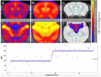 |
18 | MRI Estimates of Cerebral Blood Flow and Venous Oxygenation as Surrogate Markers in a Mouse Model for Sickle Cell Anemia
Yi-Fen Yen1, Joseph B Mandeville1, Yin-Ching Chan1, Suk-Tak Chan1, Erin E Hardy1, Jay Janz2, Steven Arkin2, Denis Rybin2, Kelly Knee2, Debra D Pittman2, and James A Goodman2
1Athinoula A. Martinos Center for Biomedical Imaging, Massachusetts General Hospital, Chalrestown, MA, United States, 2Pfizer, Inc, Cambridge, MA, United States
We have assessed cerebral blood flow (CBF), venous T2, and hematocrit (Hct) in mice to characterize the performance of MR markers of cerebral physiology in wildtype (WT) mice and the Townes transgenic mouse model of Sickle Cell Disease (SCD). SCD mice exhibited increased CBF, decreased venous T2, and decreased Hct compared to matched WT mice. Combining MR measures of CBF and venous T2 with Hct measurement improved the disease-specific differentiation. Test-retest variability was approximately 20% for CBF and venous T2 and 10% for Hct. Methods employed in this study are fully translational to the clinic.
|
||
1469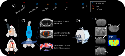 |
19 | Multi-modal Study Comparisons of Hemodynamics after a Traumatic Brain Injury (TBI) and Subarachnoid Hemorrhage (SAH)
Laurel Dieckhaus1, Ali Kamali1, Emily C Peters2, Collin A Preszler1, Christa M Sonderer1, Paulo Pires2, Kaveh Laksari1, and Elizabeth B Hutchinson1
1Biomedical Engineering, University of Arizona, Tucson, AZ, United States, 2Physiology, University of Arizona, Tucson, AZ, United States
To examine the first hours after brain injury, we utilized photoacoustic imaging (PAI), ultrasound (US), and color doppler (CD) alongside MRI metrics. We wanted to investigate the early pathomechanisms that involve hemodynamic response such as blood flow, oxygenation, and edema.
|
||
1470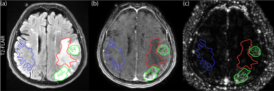 |
20 | QTM Quantifies Velocity of Tumor Vasculature in Gliomas
Dominick Jon Romano1, Qihao Zhang1, Ilhami Kovanlikaya2, Gloria Chia-Yi Chiang2, Pascal Spincemaille2, and Yi Wang3
1Biomedical Engineering, Cornell University, New York, NY, United States, 2Radiology, Weill Cornell Medical College, New York, NY, United States, 3Radiology, Cornell University, New York, NY, United States
A recently proposed perfusion analysis method, Quantitative Transport Mapping (QTM), was applied in malignant Glioma (grade III and IV) to obtain the flow speed map. Pathological lesions were separated into DCE-Enhancing, Darkening, and total affected ROIs. The average speed scalar for each ROI suggests that DCE-Enhancing tumor presents with significantly increased speed while non-enhancing tumor does not experience a speed change.
|
||
1471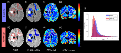 |
21 | Relative cerebral blood volume reduction in hyperintense brain regions of glioma patients treated with proton radio(chemo)therapy
Katharina Witzmann1,2, Felix Raschke1,2, Tim Wesemann3, Steffen Appold2,4, Mechthild Krause1,2,4,5,6, Jennifer Linn3, and Esther G.C. Troost1,2,4,5,6
1Helmholtz-Zentrum Dresden-Rossendorf, Institute of Radiooncology – OncoRay, Dresden, Germany, 2OncoRay - National Center for Radiation Research in Oncology, Faculty of Medicine and University Hospital Carl Gustav Carus, Technische Universität Dresden, Helmholtz-Zentrum Dresden-Rossendorf, Dresden, Germany, 3Institute of Neuroradiology, University Hospital Carl Gustav Carus and Medical Faculty of Technische Universität, Dresden, Germany, 4Department of Radiotherapy and Radiation Oncology, Faculty of Medicine and University Hospital Carl Gustav Carus, Technische Universität Dresden, Dresden, Germany, 5German Cancer Consortium (DKTK), Partner Site Dresden, and German Cancer Research Center (DKFZ), Heidelberg, Germany, 6National Center for Tumor Diseases (NCT), Partner Site Dresden, Germany: German Cancer Research Center (DKFZ), Heidelberg, Germany; Faculty of Medicine and University Hospital Carl Gustav Carus, Technische Universität Dresden, Dresden, Germany, and; Helmholtz Association / Helmholtz-Zentrum Dresden-Rossendorf (HZDR), Dresden, Germany
Imaging biomarkers capable of distinguishing between tumor recurrence and treatment effect are of high relevance for radiation therapy. In 14 glioma patients, we analyzed changes in relative cerebral blood volume (rCBV) in areas of hyperintensities on T2-weighted magnetic resonance imaging (MRI) appearing after irradiation with protons. rCBV values were evaluated comparing the baseline and the latest follow-up measurement both visually and based on histograms. A significant rCBV perfusion decrease was observed in those hyperintensities, which may be interpreted as treatment effect. Further work is needed correlating the rCBV changes with histology and patient outcome.
|
||
1472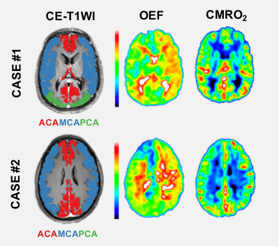 |
22 | Oxygen Extraction Fraction and Cerebral Metabolic Rate of Oxygen derived from DSC-MRI in Patients with Moyamoya Disease
Markus Fahlström1, Anders Lewén2, Per Enblad2, and Johan Wikström1
1Surgical Sciences, Uppsala University, Uppsala, Sweden, 2Neuroscience, Uppsala University, Uppsala, Sweden
Moyamoya disease is a cerebrovascular disease where changes to oxygen extraction fraction (OEF) and cerebral metabolic rate of oxygen (CMRO2) are important parameters to assess the risk of future ischemic stroke. By applying a microvasculature model to dynamic susceptibility contrast (DSC) perfusion data parametric maps of both OEF and CMRO2 can be generated. For both OEF and CMRO2 significant differences between affected and unaffected vascular territorial regions were found. OEF and CMRO2 derived from DSC perfusion shows promise in detecting cerebral hemodynamic impairment in patients with moyamoya disease. However, further investigations are needed to clarify the clinical potential.
|
||
1473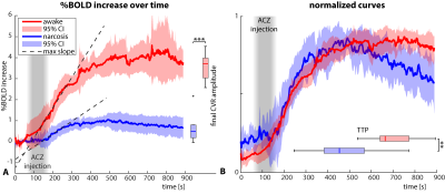 |
23 | Narcosis depresses the BOLD-CVR response to acetazolamide in pediatric moyamoya vasculopathy
Pieter T Deckers1, Jeroen CW Siero2,3, Annick Kronenburg1, Kees PJ Braun4, Bart van der Zwan1, and Alex A Bhogal2
1Neurosurgery, UMC Utrecht, Utrecht, Netherlands, 2Imaging, UMC Utrecht, Utrecht, Netherlands, 3Spinoza Centre for Neuroimaging, Amsterdam, Netherlands, 4Pediatric Neurology, UMC Utrecht, Utrecht, Netherlands
Measuring cerebrovascular reactivity (CVR) under narcosis is underreported, while narcosis is often necessary for pediatric or cognitively impaired patients. When acetazolamide is used in awake patients, maximum CBF increase reaches a plateau after ~12min. Using ASL- and BOLD-MRI with acetazolamide we showed that for pediatric moyamoya patients the response is different. Patients under narcosis show lower CVR and reach peak CBF earlier (after around six minutes), after which CBF decreases again without the plateau-phase. This shows the response to acetazolamide is distinctively different between awake and narcosis patients and caution is warranted during interpretation of narcosis CVR images.
|
||
 Back to Meeting Home
 Back to the Program-at-a-Glance
The International Society for Magnetic Resonance in Medicine is accredited by the Accreditation Council for Continuing Medical Education to provide continuing medical education for physicians.
 Back to Meeting Home
 Back to the Program-at-a-Glance
 View the Presentation