Digital Poster
Cerebrovascular Reactivity
Joint Annual Meeting ISMRM-ESMRMB & ISMRT 31st Annual Meeting • 07-12 May 2022 • London, UK

| Computer # | ||||
|---|---|---|---|---|
1671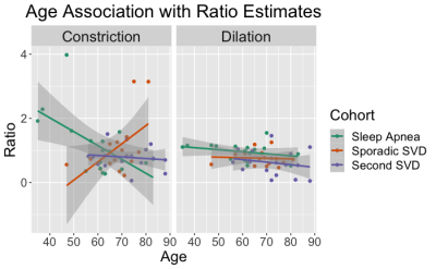 |
40 | Vascular supply affects hemodynamic responses to a hypercapnia cerebrovascular reactivity challenge assessed by BOLD-fMRI
Kayley Marchena-Romero1,2, Xiang Ji2, Andrew Centen2, Rosa Sommer2,3, Joel Ramirez2, Michael J Thrippleton4, Andrew Lim2,3, Joanna M Wardlaw4, Sandra E Black2, and Bradley J MacIntosh1,2
1Medical Biophysics, University of Toronto, Toronto, ON, Canada, 2Sunnybrook Research Institute, Toronto, ON, Canada, 3Institute of Medical Sciences, University of Toronto, Toronto, ON, Canada, 4Centre for Clinical Brain Sciences, University of Edinburgh, Edinburgh, United Kingdom
Cerebrovascular reactivity (CVR) is a dynamic assessment of hemodynamic response during hypercapnia. Conventional CVR metrics provide a single estimate to summarize the dilatory response to CO2. We propose to isolate dilation and constriction responses to a CVR challenge measured by BOLD-fMRI and assess the effect of vascular supply on vasoreactivity. We show that vascular supply affects the hemodynamic response to hypercapnia, and that this effect varies over time in different clinical cohorts. This work suggests that the extraction of individual features embedded in a CVR challenge can help characterize cerebrovascular physiology in unique clinical cohorts.
|
||
1672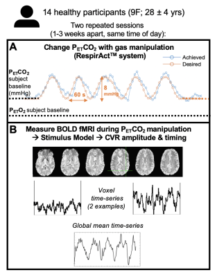 |
41 | Repeatability of cerebrovascular reactivity amplitude and timing estimates using a sinusoidal manipulation of end-tidal CO2
Rachael C Stickland1, Kristina M Zvolanek1,2, and Molly G Bright1,2
1Physical Therapy and Human Movement Sciences, Feinberg School of Medicine, Northwestern University, Chicago, IL, United States, 2Biomedical Engineering, McCormick School of Engineering, Northwestern University, Evanston, IL, United States
We assessed the repeatability of fMRI Cerebrovascular Reactivity (CVR) estimates using CO2 sinusoidal stimuli. We estimated BOLD-CVR amplitude and phase using a fast Fourier transform (FFT) analysis and a cross-correlation analysis (Rapidtide). Amplitude and delay maps showed sensible regional variations for both time-points, but participant variability was notable. CVR spatial correlations between time-points were higher after averaging within cortical parcels, compared to weak voxel-wise correlations. Qualitatively, Rapidtide achieved higher between-session correlations than FFT. GM average amplitudes agreed between time-points. At the correct spatial scale, with careful consideration of parameter scaling, this short stimulus is a viable CVR mapping option.
|
||
1673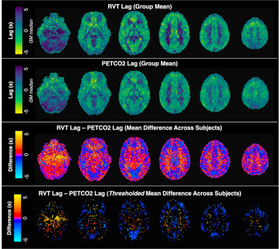 |
42 | Respiration volume per time (RVT) as a surrogate for end-tidal CO2 to map hemodynamic lag with BOLD fMRI
Kristina M. Zvolanek1,2, Stefano Moia3, Rachael C. Stickland1, César Caballero-Gaudes3, and Molly G. Bright1,2
1Physical Therapy and Human Movement Sciences, Northwestern University, Chicago, IL, United States, 2Biomedical Engineering, Northwestern University, Evanston, IL, United States, 3Basque Center on Cognition, Brain and Language, Donostia, [Gipuzkoa], Spain
Using a breath-hold task to modulate end-tidal CO2 during a BOLD fMRI scan is an established method to map hemodynamic lag. However, end-tidal CO2 measurements may not be reliable in less compliant subjects and require additional monitoring equipment. We investigated whether a related metric, respiration volume per time (RVT) can be used as a surrogate for end-tidal CO2 by directly comparing the lag values derived from each physiological measurement. Similar spatial variation was observed in healthy adult participants, albeit with some degree of proportional bias, indicating respiration belt data can be used as an alternative to map hemodynamic lag.
|
||
1674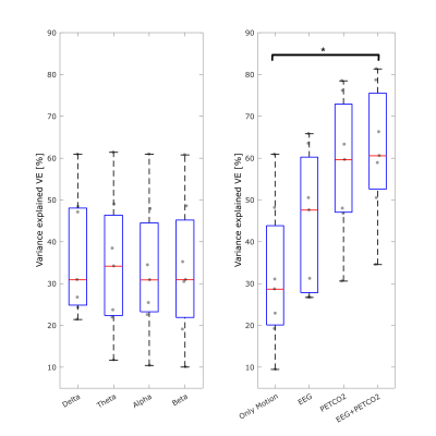 |
43 | Neuronal modulation in cerebrovascular reactivity measurements using a breath-hold task: a simultaneous EEG-fMRI study
Inês Esteves1, Beatriz Raposo1, Marta Xavier1, Ana R. Fouto1, Amparo Ruiz-Tagle1, Catarina Domingos1, Athanasios Vourvopoulos1, Nuno A. Silva2, Rita G. Nunes1, Raquel Gil-Gouveia3, Agostinho Rosa4, and Patrícia Figueiredo4
1ISR-Lisboa and Department of Bioengineering, Instituto Superior Técnico – Universidade de Lisboa, Lisboa, Portugal, 2Learning Health, Hospital da Luz, Lisboa, Portugal, 3Neurology Department, Hospital da Luz, Lisboa, Portugal, 4ISR-Lisboa and Department of Bioengineering, Instituto Superior Técnico – Universidade de Lisboa, Lisbon, Portugal
fMRI can be used to assess cerebrovascular reactivity by measuring BOLD changes during a breath hold task. This is assumed to be isometabolic so that BOLD changes cannot be attributed to neuronal activity. We performed simultaneous EEG-fMRI recordings during a breath-hold task to assess to what extent neuronal activity retrieved from the EEG may contribute to explain the BOLD signal variance that is usually interpreted as reactivity. We found that neuronal modulation measured by EEG band power did not explain significantly more BOLD variance across GM than PetCO2 changes, indicating that the associated fMRI measurements truly reflect cerebrovascular reactivity.
|
||
1675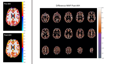 |
44 | Impact of Acute Intermittent Hypoxia on BOLD fMRI measures of cerebrovascular reactivity
Mark Andrew Hoggarth1, Rachael C Stickland1, Kimberly J Hemmerling2, Milap Sandhu3,4, and Molly G Bright1,2
1Physical Therapy and Human Movement Sciences, Northwestern University, Chicago, IL, United States, 2Biomedical Engineering, Northwestern University, Chicago, IL, United States, 3Physical Medicine and Rehabilitation, Northwestern University, Chicago, IL, United States, 4Shirley Ryan AbilityLab, Chicago, IL, United States
We assess the effects of an emerging intervention for rehabilitation, termed acute intermittent hypoxia (AIH), on cerebrovascular reactivity (CVR) as measured with BOLD fMRI. Lag-optimized CVR was measured pre- and post-AIH using a breath-hold task paradigm in 8 healthy participants. Five participants achieved the target drop in oxygen saturation (SpO2) to 85%; 3 did not. Overall changes in group mean CVR were varied following AIH, slightly increasing for those who achieved the targeted SpO2, and decreasing in those who did not. This work motivates continued study of the effects of AIH interventions on CVR.
|
||
1676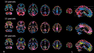 |
45 | Self organization of breath-hold induced cerebrovascular BOLD fMRI responses reveals physiologically-driven brain parcellation and networks
Stefano Moia1, Molly G Bright2,3, and César Caballero-Gaudes1
1Basque Center on Cognition, Brain and Language, Donostia, Spain, 2Physical Therapy and Human Movement Sciences, Feinberg School of Medicine, Northwestern University, Chicago, IL, United States, 3Biomedical Engineering, McCormick School of Engineering, Northwestern University, Evanston, IL, United States
Recent literature brought up the existence of physiological and vascular brain networks, i.e. areas showing synchronous responses to cardiac and respiratory fluctuations or hypercapnia. In this work, we show how breath-hold-induced BOLD fMRI time series from a dense mapping dataset can be parcellated into physiologically-driven networks, showing evidence of vascular contributions to the default mode network, in individuals and at the group level.
|
||
1677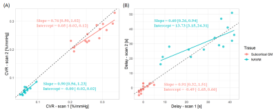 |
46 | Repeatability of the BOLD-CVR experiment at 3 T with fixed inspired CO2 concentration gas stimulus
Emilie Sleight1,2, Michael S Stringer1,2, Madeleine Murphy3, Isla Mitchell3, Ian Marshall1,2, Joanna M Wardlaw1,2, and Michael J Thrippleton1,2
1Centre for Clinical Brain Sciences, University of Edinburgh, Edinburgh, United Kingdom, 2UK Dementia Research Institute, University of Edinburgh, Edinburgh, United Kingdom, 3Edinburgh Imaging Facility Royal Infirmary of Edinburgh, University of Edinburgh, Edinburgh, United Kingdom The BOLD response to a gas with fixed inspired CO2 concentration is commonly used to assess the health of the cerebral vasculature. Cerebrovascular reactivity (CVR) magnitude repeatability has been assessed in previous studies, but without reporting potential physiological confounders such as heart and respiration rates. Furthermore, less is known about CVR delay repeatability. In this work, we assessed the within-day test-retest repeatability of both quantities in healthy volunteers at 3 T while measuring the heart and respiration rates. We found that both show good repeatability, though CVR delay estimates are unreliable in tissues with low contrast to noise ratio. |
||
1678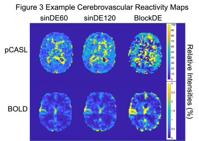 |
47 | Improving measures of cerebrovascular reactivity using a sinusoidal hypercapnic challenge and dual-echo pseudocontinuous ASL
Colette Clare Milbourn1 and Nicholas Paul Blockley2
1School of Life Sciences, University of Nottingham, Nottingham, United Kingdom, 2The School of Life Sciences, University of Nottingham, Nottingham, United Kingdom
New clinical tools are needed for the diagnosis and prognosis of cerebrovascular diseases. Cerebrovascular Reactivity (CVR) is a potential marker for brain health and can be induced using stressors to the brain like high carbon dioxide (hypercapnia) challenges. These challenges can be used to map CVR. Dual-echo pseudocontinuous Arterial Spin Labelling (pCASL) and Blood Oxygen Level Dependent (BOLD) weighted imaging was combined with sinusoidally modulated and conventional block hypercapnic challenges to map CVR. Preliminary results show qualtitative improvements to pCASL based CVR maps when using sinusoidal hypercapnic challenges compared with the conventional block.
|
||
 Back to Meeting Home
 Back to the Program-at-a-Glance
The International Society for Magnetic Resonance in Medicine is accredited by the Accreditation Council for Continuing Medical Education to provide continuing medical education for physicians.
 Back to Meeting Home
 Back to the Program-at-a-Glance
 View the Presentation