Digital Poster
Muscle I
Joint Annual Meeting ISMRM-ESMRMB & ISMRT 31st Annual Meeting • 07-12 May 2022 • London, UK

| Computer # | ||||
|---|---|---|---|---|
1019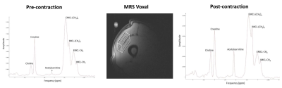 |
26 | Contraction-Induced Changes in Acetylcarnitine in the Human Vastus Lateralis Muscle Detected by 1H MRS at 3T
Rajakumar Nagarajan1, Christopher Hayden2, Samantha Gilmore2, and Jane A Kent2
1Human Magnetic Resonance Center, Institute for Applied Life Sciences, University of Massachusetts Amherst, Amherst, MA, United States, 2Kinesiology, University of Massachusetts Amherst, Amherst, MA, United States
Using 1H-MRS at 3T, vastus lateralis muscle acetylcarnitine was measured in 8 young male subjects at rest and in response to incremental, dynamic knee extension contractions. Intramyocellular [acetylcarnitine] increased >3-fold in response to the contraction protocol, thus buffering mitochondrial acetyl coenzyme A and thereby supporting oxidative metabolism of glucose and fat in the citric acid cycle. Measurement of acetylcarnitine has the potential to be a useful tool for the investigation of muscle substrate use and metabolic flexibility. This work is the first demonstration of dynamic changes in muscle [acetylcarnitine] at 3T.
|
||
1020 |
27 | Ultrashort Echo Time Magnetization Transfer Ratio (UTE-MTR) Measurements of Paraspinal Muscles Correlate Well with Lumbar Disc Herniation Video Not Available
Jia-Xin Feng1, Jin Liu1, Jian-Wei Liao1, Wei Li1, Xiao-Jun Chen1, Lin Yao1, Pan-Hui Huang1, Long Qian2, Ya-Jun Ma3, and Shao-Lin Li1
1Department of Radiology, The Fifth Affiliated Hospital of Sun Yat-Sen University, Zhuhai, China, 2MR Research, GE Healthcare, Beijing, China, 3Department of Radiology, University of California San Diego, San Diego, CA, United States
Ultrashort echo time magnetization transfer (UTE-MT) is suggested to a non-invasive technique to assess collagenous matrix indirectly. In this study, we utilized UTE-MT sequence to investigate the relationship between UTE-MT ratio (UTE-MTR) measurements of paraspinal muscles and disc herniation in patients with disc degeneration. We found that the UTE-MTR showed a good performance in differentiation of patients with and without disc herniation, thus potentially valuable in predicting disc termination.
|
||
1021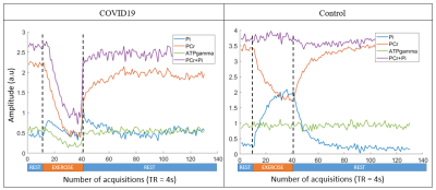 |
28 | Assessment of skeletal muscle energy metabolism by 31P MRS in COVID19 and Multiple Sclerosis patients: technical and clinical insights
Antoine Naëgel1,2, Jabrane Karkouri1,2,3, Hélène Ratiney1, Djahid Kennouche1,4, Nicolas Royer1,4, Guillaume Millet1,4, Jill Slide5, Jérôme Morel6, Pierre Croisille1, and Magalie Viallon1
1Université de Lyon, INSA-Lyon, Université Claude Bernard Lyon 1, UJM-Saint Etienne, CNRS, Inserm, CREATIS UMR 5220, U1206, Lyon, France, 2Siemens Healthineers, Saint-Denis, France, 3Wolfson Brain Imaging Center, University of Cambridge, Cambridge, United Kingdom, 4LIBM - Laboratoire Interuniversitaire de Biologie de la Motricité,Université Jean Monnet Saint Etienne, Université Claude Bernard Lyon 1, Université Savoie Mont Blanc, Saint-Etienne, France, 5Department of Radiology, Michigan State University, East Lansing, MI, United States, 6anaesthetics and intensive care department, UJM-Saint-Etienne, Centre Hospitalier Universitaire de Saint-Étienne, Saint-Etienne, France Dynamic 31P MRS was performed during a standardized exercise of the lower leg, in patients with chronic fatigue enrolled in 2 clinical studies: multiple sclerosis patients and COVID19 patients that were hospitalized in intensive care unit and requiring respiratory assistance. In this work, we also revisit certain assumptions on the metabolite T1 and question shortcuts often made to shorten 31P protocol for a better patient’s compliance. |
||
1022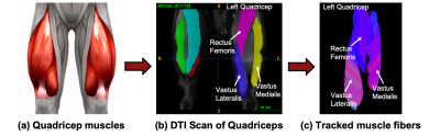 |
29 | A Diffusion Tensor Imaging approach to investigate the effects of exercise on quadricep muscle fiber lengths
Reese A. Dunne1,2, Garry E. Gold2, and Valentina Mazzoli2
1Department of Mechanical Engineering, Mississippi State University, Mississippi State, MS, United States, 2Department of Radiology, Stanford University, Stanford, CA, United States
There is currently no established method to monitor muscle architecture throughout exercise intervention. This work investigates the value of DTI and fiber tractography to detect changes in fiber length in the quadriceps caused by resistance training. Five human subjects participated in a three-month training regimen, and DTI scans of their quadriceps were acquired. Through DTI tractography, fiber lengths were measured and compared between quadricep muscle components. There were no statistically significant changes in fiber length after three months of exercise, showing further optimization is needed to utilize DTI tractography for monitoring fiber length changes due to exercise.
|
||
1023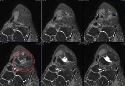 |
30 | Comparative Study on MR Imaging Quality of Anterior Talofibular Ligament based on Carotid Plaque Special Surface Coil
Yunjie Liao1 and Chen Thomas Zhao2
1Department of Radiology, the Third Xiangya Hospital, Central South University, Changsha, China, 2Philips Healthcare, Guangzhou, China
Ankle sprain is a common athletic injury. Most patients with acute ankle sprain could not be treated properly, without precise diagnosis. Conventional Magnetic Resonance Imaging (MRI) sequences, like T1WI and T2WI, could not provide diagnostic proof as accurately as possible. Proton density weighted imaging (PDWI) collaborating with fat saturation (FS) could be a better alternative to detect the lesions. A comparative study was established, to investigate the performance of 4 sequences, with 8-channel carotid special surface coil. The results showed that PDWI-FS may be the best choice on diagnosis of ankle injuries among 4 sequences
|
||
1024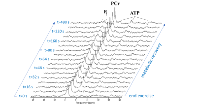 |
31 | 31P-MRS reveals oxidative capacities of red and white fibers in human muscle differ 7-fold in vivo
Jeroen A Jeneson1,2, Laura E Habets1, Melissa T Hooijmans2, Sandra M van den Berg2, Bart Bartels1, W Ludo van der Pol3, and Aart J Nederveen2
1Child Health, University Medical Center Utrecht, Utrecht, Netherlands, 2Radiology and Nuclear Medicine, Amsterdam University Medical Center|location AMC, Amsterdam, Netherlands, 3Neurology and Neurosurgery, University Medical Center Utrecht, Utrecht, Netherlands
31P MRS has been used to non-invasively diagnose muscular mitochondrial dysfunction in disease. However, shifts in muscle fiber type composition due to muscle disuse are potential confounders since mitochondrial and capillary densities differ between fiber types. We obtained new quantitative information on this subject matter on basis of dynamic in vivo 31P MRS recordings from upper arm muscle of fourteen healthy subjects performing a maximal arm-cycling test. Analysis of the post-exercise time course of Pi in red and white myofiber pools distinguished by myoplasmic pH revealed a sevenfold difference in oxidative capacity between these fiber types in vivo.
|
||
1025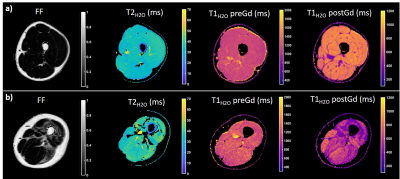 |
32 | Assessment of skeletal muscle extracellular volume fraction in Becker Muscular Dystrophy using MR fingerprinting with water and fat separation
Benjamin Marty1,2, Pierre-Yves Baudin1,2, Yves Fromes1,2, Karim Wahbi3, and Harmen Reyngoudt1,2
1NMR Laboratory, Institute of Myology, Neuromuscular Investigation Center, Paris, France, 2NMR Laboratory, CEA, DRF, IBFJ, MIRCen, Paris, France, 3Reference Center for Muscle Diseases Paris-Est, Institute of Myology, Paris, France
Becker muscular dystrophy (BMD) is a genetic neuromuscular disorder leading to muscle weakness and degeneration. Quantitative MRI has been widely proposed to characterize skeletal muscle tissue structure of BMD and revealed increased intramuscular fat fraction associated with functional decline. In this study, we measured the extracellular volume fraction (ECV) of skeletal muscle tissues using MR fingerprinting with water and fat separation. We evaluated this variable as an early imaging biomarker of skeletal muscle tissue alterations. Compared to healthy controls, we showed that the muscles of BMD subjects present elevated ECV before fatty replacement occurs.
|
||
1026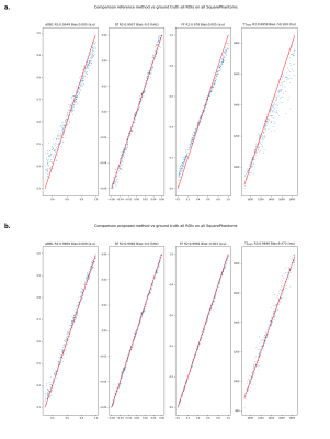 |
33 | Bi-component dictionary for efficient quantification of fat fraction and water T1 via MR Fingerprinting
Constantin Slioussarenko1,2, Pierre-Yves Baudin1,2, Harmen Reyngoudt1,2, and Benjamin Marty1,2
1NMR Laboratory, Institute of Myology, Neuromuscular Investigation Center, Paris, France, 2NMR Laboratory, CEA, DRF, IBFJ, MIRCen, Paris, France
In the field of neuromuscular disorders (NMD), fat fraction (FF) is an established biomarker of disease severity and water T1 (T1H2O) has been shown to be a potential biomarker of disease activity. Those can be efficiently estimated through Magnetic Resonance Fingerprinting. However for muscle studies water-fat separation and FF quantification increase drastically the size of the dictionary and pattern matching time. We introduce here a bicomponent dictionary where water and fat signals are stored separately. This approach allows improving MRF performance and increasing the accuracy of FF quantification.
|
||
1027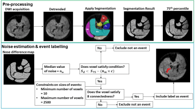 |
34 | Spontaneous motor unit activity in a healthy ageing population measured using motor unit MRI (MUMRI)
Matthew George Birkbeck1,2,3, Linda Heskamp1, Julie Hall1,4, Ian Schofield1, Avan Sayer1,3, Richard Dodds1,3, Roger Whittaker1,5, and Andrew Blamire1
1Translational and Clinical Research Institute, Newcastle University, Newcastle upon Tyne, United Kingdom, 2Northern Medical Physics and Clinical Engineering, Newcastle upon Tyne NHS FT, Newcastle upon Tyne, United Kingdom, 3NIHR Newcastle Biomedical Research Centre, Newcastle University, Newcastle upon Tyne, United Kingdom, 4Department of Neuroradiology, Newcastle Upon Tyne NHS FT, Newcastle upon Tyne, United Kingdom, 5Department of Neurophysiology, Newcastle upon Tyne NHS FT, Newcastle upon Tyne, United Kingdom
Sarcopenia is commonly associated with ageing, whereby individuals lose muscle mass and strength. One potential contributor to sarcopenia is the degeneration of motor units (MU), defined as a single motor neuron and the muscle fibres it innervates. We used motor unit MRI (MUMRI) to investigate changes to spontaneous MU activity and morphology in a cohort of healthy ageing adults. We found that MU activity and morphology did not appear to change with healthy ageing. We next aim to apply the MUMRI technique in patients with sarcopenia to look for evidence of accelerated MU loss compared to these healthy controls.
|
||
1028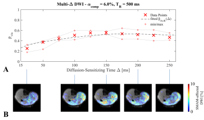 |
35 | Capability of Diffusion-Weighted Stimulated-Echo Imaging to Visualize Spontaneous Muscular Activities in Resting Musculature
Martin Schwartz1,2, Petros Martirosian1, Guenter Steidle1, Thorsten Feiweier3, Bin Yang2, and Fritz Schick1
1Section on Experimental Radiology, University Hospital of Tuebingen, Tuebingen, Germany, 2Institute of Signal Processing and System Theory, University of Stuttgart, Stuttgart, Germany, 3Siemens Healthcare GmbH, Erlangen, Germany
Visualization of spontaneous mechanical activities in resting musculature using diffusion-weighted stimulated-echo imaging is highly depending on the sequence parameter settings as well as on the physiological parameters of the muscular contraction. Therefore, a concept for estimation of the visualization probability by using two contraction models is given in this work. Furthermore, the proposed concept is validated using diffusion-weighted measurements with varying diffusion-sensitizing times. The determined visualization probability is in good accordance to the relative number of visible spontaneous mechanical activities in the resting lower leg musculature.
|
||
1029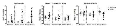 |
36 | Multi-modal MR in the upper arm muscles of patients with Spinal Muscular Atrophy
Melissa Tamara Hooijmans1, Laura E. Habets2, Sandra A.M. van den Berg-Faay1, Martijn Froeling3, Gustav J. Strijkers4, Fay-lynn Asselman5, Aart J. Nederveen1, Jeroen A.L Jeneson2, Bart Bartels2, and W. Ludo van der Pol5
1Department of Radiology and Nuclear Medicine, Amsterdam Movement Sciences, Amsterdam University Medical Center, Amsterdam, Netherlands, 2Center for Child Development, Exercise and Physical Literacy, Wilhelmina Children’s Hospital, University Medical Center Utrecht, Utrecht, Netherlands, 3Department of Radiology, University Medical Center Utrecht, Utrecht, Netherlands, 4Department of Biomedical Engineering & Physics, Amsterdam Movement Sciences, Amsterdam University Medical Center, Amsterdam, Netherlands, 5Department of Neurology and Neurosurgery, UMC Utrecht Brain Center, University Medical Center Utrecht, Utrecht, Netherlands
With upcoming therapeutic developments, quantitative MR is increasingly used to assess disease state and progression in neuromuscular disorders, including SMA. Yet, little is known about the disease state in the arm muscles in SMA. A multi-modal MR approach was used to examine fat fraction (FF), T2-relaxation time and diffusion properties, in the arm muscles of SMA patients and controls. Our results showed higher FF in the arm muscles of SMA patients which were negatively associated with the level of muscle strength. No clear changes were observed for qT2 while diffusion indices showed potential for mapping changes in muscle tissue itself.
|
||
1030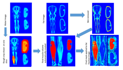 |
37 | Assessing Semi-Automatic processing of Muscle Mass and Fat Fractions from mDIXON whole body MRI
Rosemary Nicholas1, Paul Greenhaff1, and Susan Francis1
1UNIVERSITY OF NOTTINGHAM, Nottingham, United Kingdom
Muscle volume and fat fraction can be quantified from mDIXON scans either manually or using automated processes. Here we compare manual volumes of thigh and calf muscle ROIs with an automated pipeline created using FSL’s FAST segmentation, to compare muscle volume and fat fraction across subject groups and with their DXA values. Automatic volume segmentations correlated highly with manually drawn measures (r=.975) as well as DXA (r=0.840). Group comparisons show COPD and post-COVID patients had significantly lower muscle mass and higher fat fraction. Automatic segmentation performs well compared to manually derived volumes and is more time efficient.
|
||
1031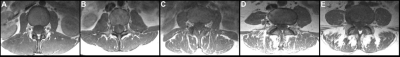 |
38 | Paraspinal muscles in low back pain: Comparison between standard parameters and chemical shift encoding-based water-fat MRI Video Permission Withheld
Nico Sollmann1,2,3, Noah B. Bonnheim4, Gabby B. Joseph1, Ravi Chachad1, Jiamin Zhou1, Jeannie F. Bailey4, Xiaojie Guo4, Ann A. Lazar5, Thomas M. Link1, Aaron J. Fields4, and Roland Krug1
1Department of Radiology and Biomedical Imaging, University of California San Francisco, San Francisco, CA, United States, 2Department of Diagnostic and Interventional Radiology, University Hospital Ulm, Ulm, Germany, 3Department of Diagnostic and Interventional Neuroradiology, Technical University of Munich, Munich, Germany, 4Department of Orthopaedic Surgery, University of California San Francisco, San Francisco, CA, United States, 5Department of Epidemiology and Biostatistics, University of California San Francisco, San Francisco, CA, United States
Low back pain (LBP) is a global health burden, and up to 90% of affected patients are diagnosed with non-specific LBP. A structure increasingly recognized as a potential contributor to LBP is paraspinal musculature (PSM). In this study we revealed that the fat fraction (FF) of PSM derived from chemical shift encoding-based water-fat MRI (CSE-MRI) is associated with the Goutallier classification (GC) at each lumbar level as well as with self-reported pain. In contrast, other parameters (muscle volume, lumbar indentation value [LIV], muscle-fat index [MFI]) do not significantly correlate to the FF, despite being frequently used to evaluate PSM quality.
|
||
 Back to Meeting Home
 Back to the Program-at-a-Glance
The International Society for Magnetic Resonance in Medicine is accredited by the Accreditation Council for Continuing Medical Education to provide continuing medical education for physicians.
 Back to Meeting Home
 Back to the Program-at-a-Glance
 View the Presentation