Digital Poster
Muscle II
Joint Annual Meeting ISMRM-ESMRMB & ISMRT 31st Annual Meeting • 07-12 May 2022 • London, UK

| Computer # | ||||
|---|---|---|---|---|
1130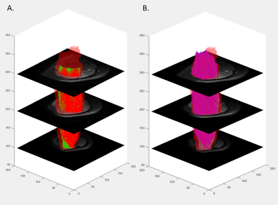 |
35 | Relationships Between Muscle Function, Muscle Fat, and Architecture Measured by MRI in Vivo
Joseph A. Gordon III1, Nicholas M. Remillard1, Luke R. Arieta1, Rajakumar M. Nagarajan1,2, Bruce M. Damon3,4, and Jane A. Kent1,2
1Kinesiology, University of Massachusetts Amherst, Amherst, MA, United States, 2Institute for Applied Life Sciences, University of Massachusetts Amherst, Amherst, MA, United States, 3Carle Clinical Imaging Research Program, Carle Health, Urbana, IL, United States, 4Stephens Family Clinical Research Institute, Carle Health, Urbana, IL, United States
The purpose of this work was to investigate the associations between skeletal muscle function and markers of fat content, diffusivity, and architecture. Combining MR techniques that measure muscle structure in vivo with muscle strength measures can elucidate mechanisms other than muscle size that may contribute to functional impairments. This study identified variables (e.g., fascicle length, muscle curvature, pennation angle, fat fraction) that are worthy of investigation in conjunction with functional parameters in future research, and highlighted assessment of muscle function across various contraction types and velocities.
|
||
1131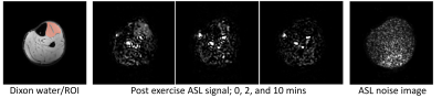 |
36 | Signal-to-noise requirements for accurate and precise perfusion parameter estimation with pulsed arterial spin labeling MRI in skeletal muscle
Donnie Cameron1,2, Thom T.J. Veeger1, Esther J. Schrama3, Celine Baligand4, Lydiane Hirschler1, Matthias J.P. van Osch1, and Hermien E. Kan1
1C.J. Gorter Center for High Field MRI, Department of Radiology, Leiden University Medical Center, Leiden, Netherlands, 2Norwich Medical School, University of East Anglia, Norwich, United Kingdom, 3Department of Neurology, Leiden University Medical Center, Leiden, Netherlands, 4Université Paris-Saclay, CEA, CNRS, MIRCen, Laboratoire des Maladies Neurodégénératives, Fontenay-aux-Roses, France
To develop perfusion applications in neuromuscular diseases, we examined the SNR and noise sensitivity of pulsed ASL for estimating microvascular reactivity and perfusion in skeletal muscle. We determined muscle blood flow and arterial transit time in volunteers after exercise and used these to inform simulations to establish the minimum ASL SNR required for accurate and precise perfusion parameter estimation. We observed a peak SNR of 10 in the tibialis anterior muscle. Simulations indicated that SNR>6 is sufficient for accurate parameter estimation, while SNR>12 is preferred for good precision. These findings will inform future exercise ASL protocol development in patient cohorts.
|
||
1132 |
37 | Evaluating Structural and Functional Lower-limb Asymmetries through MRI and Wearable Sensors
Andrew M Schmidt1, Elka Rubin1, Michael Ko1, Lauren E Watkins2, Marco Barbieri1, Laurel Hales3, Garry Gold1,2, Scott Delp2,4,5, Feliks Kogan1, Valentina Mazzoli1, and Akshay S Chaudhari1,6
1Radiology, Stanford University, Stanford, CA, United States, 2Bioengineering, Stanford University, Stanford, CA, United States, 3Electrical Engineering, Stanford University, Stanford, CA, United States, 4Mechanical Engineering, Stanford University, Stanford, CA, United States, 5Orthopaedic Surgery, Stanford University, Stanford, CA, United States, 6Biomedical Data Science, Stanford University, Stanford, CA, United States
Current evaluation methods of rehabilitation following acute musculoskeletal injuries are largely qualitative. MRI and biomechanics tools can provide sensitive, quantitative measures of knee joint and lower extremity muscle changes, but the relationship between MRI and gait markers is not well characterized. We combined an MRI protocol with wearable sensors in healthy participants to characterize the relationship between gait kinematic asymmetries and thigh muscle and cartilage morphology and composition. We show that vastus lateralis (VL) muscle microstructure assessed via Diffusion Tensor Imaging (DTI) may be sensitive to gait variations. Future work may further explore these correlations in patients with musculoskeletal injuries.
|
||
1133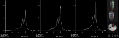 |
38 | Fast scan downfield MRS for NAD+ quantification in human skeletal muscle in vivo at 7 T
Ravi Prakash Reddy Nanga1, Mark Elliott2, Neil Wilson2, Sophia Swago3, Walter Witschey2, and Ravinder Reddy2
1Perelman School of Medicine at The University of Pennsylvania, Philadelphia, PA, United States, 2Radiology, Center for Advance Metabolic Imaging in Precision Medicine, Perelman School of Medicine at The University of Pennsylvania, Philadelphia, PA, United States, 3Bioengineering, University of Pennsylvania, Philadelphia, PA, United States
1H downfield MRS is an emerging technique for quantification of in vivo NAD+ concentrations in human tissue. Though NAD+ can be detected in the skeletal muscle downfield spectrum (>4.7 ppm) despite its low concentration (<1 mM), quantification of [NAD+] in muscle disorders is further challenged by depleted NAD+ and long scan times. To address this challenge, we developed and optimized a downfield MRS technique for rapid measurement of NAD+ targeting calf skeletal muscle in a large voxel approach. This optimized technique has allowed us rapid measurement of NAD+ and shortened scan time considerably compared to previously published MRS techniques.
|
||
1134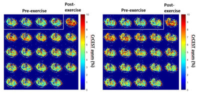 |
39 | Creatine chemical exchange saturation transfer (CrCEST) MRI reproducibility in healthy adults at 3T Video Not Available
Zane Coleman1, Ayaz Khan1, Shubo Wang1, Pat Hanby1, Donald Wallace1, Zoltan Patay1, and Puneet Bagga1
1Diagnostic Imaging, St. Jude Children's Research Hospital, Memphis, TN, United States
CrCEST MRI indirectly measures creatine as it exchanges amine (-NH2) protons with free water in the skeletal muscle. When performed on the calf after plantar flexion exercise, creatine recovery data can be obtained in all four major calf muscles. While 2D CrCEST has been performed in adults before, no studies showing its reproducibility have been published. Here, we show that the essential muscles in plantar flexion movement, gastrocnemius muscles, in the calf are candidates for reproducible CrCEST data.
|
||
1135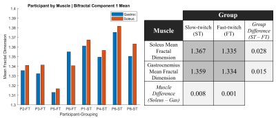 |
40 | Differentiating Skeletal Muscle Fiber Composition Using a Bi-Fractal Model of Resting-State Muscle BOLD
Joshua Ethan McGillivray1,2, Dinesh Kumbhare1,2,3, and Michael D Noseworthy1,2,4,5
1School of Biomedical Engineering, McMaster University, Hamilton, ON, Canada, 2Imaging Research Centre, St. Joseph's Healthcare, Hamilton, ON, Canada, 3Department of Medicine and Biomedical Engineering, University of Toronto, Toronto, ON, Canada, 4Electrical and Computer Engineering, McMaster University, Hamilton, ON, Canada, 5Department of Radiology, McMaster University, Hamilton, ON, Canada Lower leg resting-state BOLD images (n=8 males, 4 endurance, 4 power athletes), were acquired after 30min of rest, allowing for blood-flow normalization. BOLD images were motion corrected and the gastrocnemius and soleus manually segmented using an anatomical reference for their differing twitch fibre profiles. Voxel-wise BOLD mono- and bi-fractal dimension were computed using the scaled windowed variance approach, with linear detrending, removing scanner induced low-frequency variations. The bi-fractal dimension was significantly different between endurance and power groups in both muscles. Specific bi-fractal components more readily distinguished soleus and gastrocnemius for the endurance (I) and power (II) group, when at rest. |
||
1136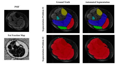 |
41 | Deep Learning and Fat Fraction Based Analysis of Calf MRI in Diabetic Patients Following Exercise Intervention
Jill T Shah1, Tanay T Shah2, Haresh R Rajamohan3,4, Katherine Medina3,5, Smita Rao6, Cem Deniz3,5, and Ryan Brown3,5
1NYU Grossman School of Medicine, New York University, New York, NY, United States, 2College of Arts and Sciences, New York University, New York, NY, United States, 3Center for Biomedical Imaging, Department of Radiology, New York University Grossman School of Medicine, New York, NY, United States, 4Center for Data Science, New York University, New York, NY, United States, 5Center for Advanced Imaging Innovation and Research (CAI2R), Department of Radiology, New York University Grossman School of Medicine, New York, NY, United States, 6Department of Physical Therapy, New York University, New York, NY, United States Diabetic peripheral neuropathy (DPN) is characterized by increased adiposity implicated in metabolic dysfunction. Proton-based Dixon MRI is an appropriate means to quantify adiposity, but analysis requires time-consuming manual image segmentation. To address this problem, we developed an automated segmentation algorithm based on convolutional neural networks that provided high dice similarity coefficient scores (>0.88) on multiple regions of interest (ROI) within the calf. We utilized the networks to analyze fat fraction trends in individuals with DPN following a 10-week supervised exercise program. We measured decreased adiposity in the combined calf interstitial and muscle space (P<0.1) but not in individual muscle ROIs. |
||
1137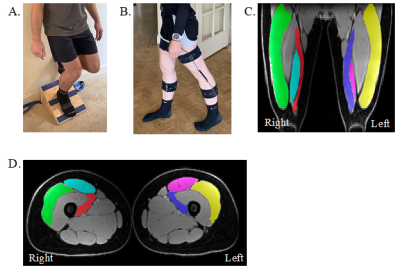 |
42 | Effect of Resistance Training on Lower-Extremity Gait Kinematics and Muscle Morphology
Michael Ko1, Andrew Schmidt1, Elka Rubin1, Lauren Watkins2, Marco Barbieri1, Laurel Hales3, Anthony Gatti1, Garry Gold1,2, Feliks Kogan1, Scott Delp2,4,5, Valentina Mazzoli1, and Akshay Chaudhari1,6
1Radiology, Stanford University, Stanford, CA, United States, 2Bioengineering, Stanford University, Stanford, CA, United States, 3Electrical Engineering, Stanford University, Stanford, CA, United States, 4Mechanical Engineering, Stanford University, Stanford, CA, United States, 5Orthopaedic Surgery, Stanford University, Stanford, CA, United States, 6Biomedical Data Science, Stanford University, Stanford, CA, United States
Gait retraining has been studied as an intervention to improve osteoarthritis symptoms but has a variety of limitations. An alternative approach may be muscle strengthening interventions that have known impacts on gait changes. However, limited studies have examined the quantitative relationships between muscle strengthening and gait. Here, we combine a rapid MRI protocol with wearable sensors to determine that a 12-week exercise intervention induced significant changes in both quadricep muscle morphology and gait kinematics. Morphological changes in different muscles were related to different kinematic changes, which may inform future strengthening interventions that aim to achieve specific changes in gait kinematics.
|
||
1138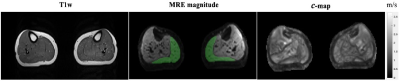 |
43 | Muscle stiffness quantified by multifrequency MR elastography in patients with Achilles tendinopathy
Mahsa Salimi Majd1, Markus Lerchbaumer1, Suchung Kim1, Tilman Hees1, Yang Yang1, Steffen Görner1, Thomas Fischer1, Ingolf Sack1, and Jing Guo1
1Charité - Universitätsmedizin Berlin, Berlin, Germany
Achilles tendinopathy is a common injury among athletes. It has been reported that the calf muscles are involved and affected by Achilles tendinopathy. In this study, tomoelastography, a multifrequency MR-elastography (MRE) technique, was applied to patients with Achilles tendinopathy for the characterization of the biomechanical properties of gastrocnemius muscle. Preliminary findings showed the stiffness of gastrocnemius muscle increased significantly as a result of tendinopathy.
|
||
1139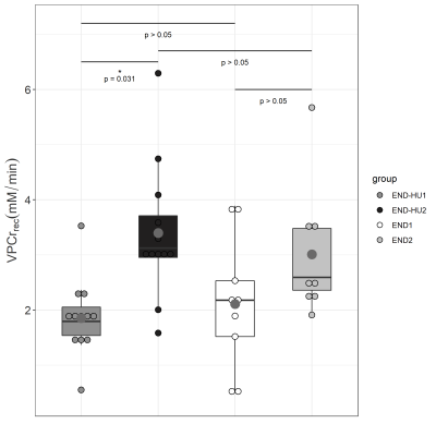 |
44 | Skeletal muscle energetics and function in sickle cell disease murine model: a synergetic effect of hydroxyurea and endurance training?
Constance P Michel1, Laurent A Messonnier2, Benoit Giannesini1, and David Bendahan1
1Aix-Marseille Univ, CNRS, CRMBM, Marseille, France, 2Laboratoire Interuniversitaire de Biologie de la Motricité, Chambéry, France
In sickle cell disease (SCD), hydroxyurea can increase foetal haemoglobin production and restore oxygen-carrying capacity of patients. As endurance training is also known to improve muscle aerobic capacity, one can wonder whether hydroxyurea and endurance training can have a synergetic effect. Townes mice (model of SCD) have been trained with and without hydroxyurea for 8 weeks. Muscle energetics and function were assessed in response to a rest-stimulation-recovery protocol before and after interventions. While training increased force production, mitochondrial function was improved when training was combined to hydroxyurea supplementation. In SCD, this combination would have a synergetic effect in muscle energetics.
|
||
1140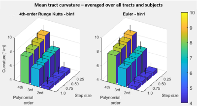 |
45 | Impact of diffusion tractography tracking parameters and smoothing polynomial order on muscle fiber measurements
Carly Anne Lockard1,2, Melissa T Hooijmans3, Xingyu Zhou2,4, and Bruce M Damon1,2
1Stephens Family Clinical Research Institute, Carle Health, Urbana, IL, United States, 2Carle Clinical Imaging Research Program, Carle Health, Urbana, IL, United States, 3Department of Radiology and Nuclear Medicine, Amsterdam Movement Sciences, Amsterdam University Medical Center, Amsterdam, Netherlands, 4Biomedical Engineering, Vanderbilt University, Nashville, TN, United States
Diffusion-tensor MRI tractography has been applied to characterize muscle diffusion properties and architecture. Tract propagation is performed using various propagation algorithms, settings, and step sizes. Smoothing, such as by polynomial fitting, has been shown to reduce tract path errors due to noise. Optimal settings for tracking algorithm, parameters, and order of subsequent smoothing polynomials have not been established. This work examines the effect of tracking algorithm settings, step size, and polynomial order on muscle architecture and diffusion estimates and polynomial fit residuals behavior. Results emphasize the high sensitivity of curvature estimates to smoothing polynomial order.
|
||
1141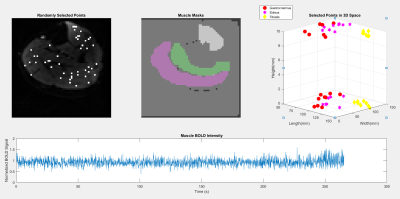 |
46 | Investigation into Graph Signal Processing Applications to Muscle BOLD
Thaejaesh Sooriyakumaran1, Joshua McGillivray1, and Michael D Noseworthy1
1School of Biomedical Engineering, McMaster University, Hamilton, ON, Canada
This investigation looks to use graph signal processing to generate a functional graph over the anatomical leg and assess the extracted functional information and its viability in modeling underlying physiological factors. The generated graph is constructed on the edge dimensions of node to node coherences and fractal dimension differences. The resultant graph structure is then analyzed to observe if extracted functional data shows alignment with underlying structures via a generalized linear mixed-effect mode
|
||
 Back to Meeting Home
 Back to the Program-at-a-Glance
The International Society for Magnetic Resonance in Medicine is accredited by the Accreditation Council for Continuing Medical Education to provide continuing medical education for physicians.
 Back to Meeting Home
 Back to the Program-at-a-Glance
 View the Presentation