Digital Poster
Cartilage
Joint Annual Meeting ISMRM-ESMRMB & ISMRT 31st Annual Meeting • 07-12 May 2022 • London, UK

| Computer # | ||||
|---|---|---|---|---|
2297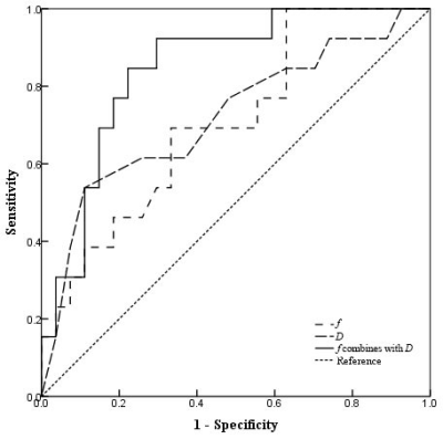 |
33 | Intravoxel Incoherent Motion Diffusion-Weighted MRI for Prediction of Prognosis in Intermediate Risk Acute Myeloid Leukemia
Jianting Li1, Jinliang Niu1, Wenjin Bian2, and Wenqi Wu1
1The Second Hospital of Shanxi Medical University, Taiyuan, China, 2Shanxi Medical University, Taiyuan, China
The mortality rate for patients with acute myeloid leukemia (AML) is still high. Cytogenetic features are the most important prognostic parameter, and the most appropriate treatment of intermediate risk AML (IR-AML), which accounts for about 60% of AML, is controversial. Our study suggested that the D value obtained from intravoxel incoherent motion (IVIM) could play a potential role in predicting treatment response of IR-AML patients. Moreover, f value and D* value in an IVIM model can independently predict OS of patients with IR-AML. These findings suggest that IVIM parameters can be useful imaging markers to predict prognosis for IR-AML.
|
||
2298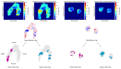 |
34 | Articular Cartilage T2 Relaxation Time Decreases in Regions of Loaded Contact in Cadaver Knees
Natasha M. Bzowey1, Marianne S. Black2, Lumeng Cui3, Chelsey S. Thorson1, Madeline M. Martel1, and Emily J. McWalter1
1Department of Mechanical Engineering, University of Saskatchewan, Saskatoon, SK, Canada, 2Department of Radiology, Stanford University, Stanford, CA, United States, 3Department of Biomedical Engineering, University of Saskatchewan, Saskatoon, SK, Canada
The aim of this work was to identify focal changes in T2 relaxation times inside and outside regions of tibiofemoral contact in loaded cadaver knee articular cartilage. We used a cluster analysis approach to identify focal regions of change in T2 relaxation time. We found clusters of increased and decreased T2 relaxation time when the cartilage was loaded; the majority of the clusters of decreased T2 relaxation time were observed within the tibial and femoral cartilage contact area.
|
||
2299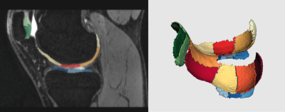 |
35 | Analysis of knee joint injury caused by training of freshmen based on 3T MRI and Automatic Cartilage Segmentation Technology :A prospective study Video Not Available
Lingling Liu1, Henan Liu2, Zhiming Zhen3, Yalan Zheng3, Xiaoyue Zhou4, Esther Raithel5, Jiang Du6, Yan Hu7, Wei Chen3, and Xiaofei Hu3
1Department of Radiology, First Affiliated Hospital of Army Medical University, chongqing, China, 2Department of nuclear medicine, First Affiliated Hospital of Army Medical University, chongqing, China, 3First Affiliated Hospital of Army Medical University, chongqing, China, 4Siemens Healthineers Ltd., shanghai, China, 5MR Application Predevelopment, Siemens Healthcare GmbH, Erlangen, Germany, 6Health Service Training Base of the Army Military Medical University., chongqing, China, 7Health Service Training Base of the Army Military Medical University, chongqing, China
Magnetic resonance imaging (MRI) is the preferred choice for evaluating knee joint injury, which commonly occurs during physical training. In this study, we compared the effects of long-walking and regular daily training on knee joint injuries and cartilage microstructure in 23 young freshmen by analyzing 3D-DESSWE and T2 mapping images using the automatic cartilage segmentation prototype software. Our results demonstrated that regular daily training strengthens the knee joints and protects against knee joint injury by increasing the volume of the knee cartilage. Moreover, knee joint injury caused by short-term long walking and high-intensity pull training was reversible.
|
||
2300 |
36 | Early Quantitative Diagnosis of Articular Cartilage Degeneration Using 3D Ultrashort Echo Time Cones Adiabatic T1ρ ( UTE Cones AdiabT1ρ) Imaging Video Not Available
Mei Wu1,2, Ya-jun Ma2, Zhouchonghao Wu2, Yanping Xue2, Yanting Zheng1, Zhao Wei2, Saeed Jerban2, Hyungseok Jang2, Douglas G Chang3, Liheng Ma4, Eric Y Chang2,5, and Jiang Du2
1Radiology, Guangzhou First People’s Hospital, School of Medicine, South China University of Technology, Guangzhou, China, 2Radiology, University of California San Diego, San Diego, CA, United States, 3Orthopaedic Surgery, University of California San Diego, San Diego, CA, United States, 4Imaging Department, The First Affiliated Hospital of Guangdong Pharmaceutical University, Guangzhou, China, 5Radiology Service, Veterans Affairs San Diego Healthcare System, San Diego, CA, United States This study aims to evaluate cartilage degeneration using 3D UTE-Cones-AdiabT1r imaging at 3T. In total 66 human subjects were recruited. KL and WORMS were evaluated by two MSK radiologists. The performance in evaluating cartilage degeneration was assessed via Spearman’s correlation coefficient and the area under the curve (AUC) calculated according to the receiver-operating characteristic (ROC) curve. Higher UTE-Cones-AdiabT1r values were observed in larger and deeper cartilage lesions. The diagnostic threshold of UTE-Cones-AdiabT1r for mild cartilage degeneration was 39.4 ms with 80.8% sensitivity and 63.5% specificity. The AUC value for mild cartilage degeneration (WORMS=1) was 0.8 according to the ROC curves. |
||
2301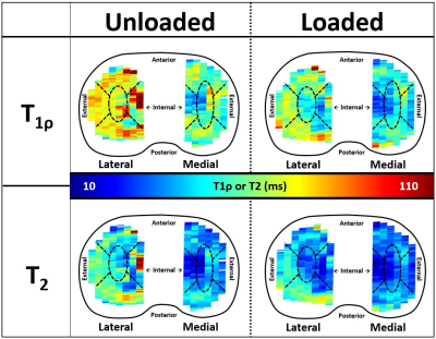 |
37 | Unloaded-to-loaded changes in regional tibial cartilage T1ρ and T2: Utilization of an MRI compatible loading device Video Permission Withheld
Erin C Argentieri1, Andrew C Zhu2, Sonia Bansal2, Amanda Wach2, Ashley Pekmezian2, Ryan E Breighner1, Hollis G Potter1, Suzanne A Maher2, and Matthew F Koff1
1Radiology and Imaging, Hospital for Special Surgery, New York, NY, United States, 2Biomechanics, Hospital for Special Surgery, New York, NY, United States
Unloaded and loaded tibial cartilage T1ρ and T2 values were evaluated regionally. Only T1ρ, significantly changed from the unloaded-to-loaded condition. Shortening of loaded T1ρ values may be attributed to matrix water loss due to PG damage and attendant loss of the cartilage FCD. Though not all regions exhibited significant changes in unloaded-to-loaded T1ρ values, previous work demonstrated that tibial cartilage tissue and fibril strains follow the pattern of changes in FCD distribution. Inter-regional T2 variation increased from unloaded-to-loaded conditions, suggesting that T2 values were affected by a decreased FCD of cartilage that allowed water to move more freely between regions.
|
||
2302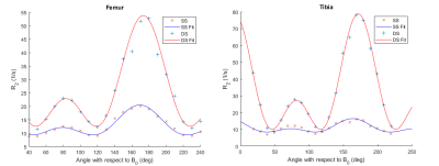 |
38 | Anisotropy of T2 and T1ρ Relaxation in Bovine Articular Cartilage at 3 T
Ville Kantola1, Jouni Karjalainen1, Tomi Jaakola1, Henri Leskinen2, Mikko Nissi1,2, Miika Nieminen1,3,4, and Victor Casula1,4
1Research Unit of Magnetic Imaging, Physics and Technology, University of Oulu, Oulu, Finland, 2University of Eastern Finland, Kuopio, Finland, 3Department of Diagnostic Radiology, Oulu University Hospital, Oulu, Finland, 4Medical Research Center, University of Oulu and Oulu University Hospital, Oulu, Finland
Multiple studies have investigated the orientation dependence of T2 and T1ρ relaxation in articular cartilage using ultra-high field magnets. This study aims to replicate these findings in a clinical measurement setting, performing a series of measurements at multiple orientations on a 3 T clinical scanner using large ex vivo joint samples. Function of a previously suggested relaxation model was confirmed in a low field-strength environment. Anisotropic components of T2 and T1ρ relaxation varied widely depending on the measured parameter, the spin-lock frequency, the magnetization preparation used for the spin-lock experiment (continuous wave/adiabatic), and the considered cartilage layer (superficial/deep half).
|
||
2303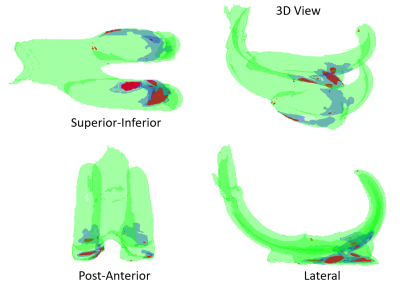 |
39 | Topographical Associations of Knee Cartilage MRI with Osteoarthritis Pain: Data from the Osteoarthritis Initiative.
Edward J Peake1,2, Tom D Turmezei3,4, and Dorothee P Auer1,2
1Radiological Sciences, Division of Clinical Neuroscience, University of Nottingham, Nottingham, United Kingdom, 2NIHR Nottingham Biomedical Research Centre, University of Nottingham, Nottingham, United Kingdom, 3Department of Radiology, Norfolk and Norwich University Hospitals NHS Foundation Trust, Norwich, United Kingdom, 4Norwich Medical School, University of East Anglia, Norwich, United Kingdom
Pain is a hallmark of knee osteoarthritis, a chronic condition with considerable health and socioeconomic burden. Osteoarthritic changes of the knee cartilage are often used as an outcome marker for clinical trials, but only weakly associated with pain. In this study, we aimed to examine the topographic correlation between intensity differences in knee cartilage MRI and knee pain. Voxel based morphometry (VBM) is used with linear effects modelling to show the spatial localization of knee OA contributing to pain. Local maxima and clusters with extents with P<0.05 are determined using random field theory.
|
||
2304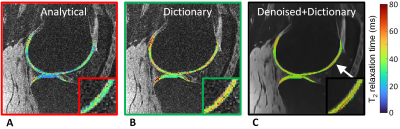 |
40 | Improved accuracy and precision in 3D T2 mapping of knee cartilage with dictionary fitting and patch-based denoising
Simon Kuhn1, Aurélien Bustin1,2,3, Aicha Lamri-Senouci1, Simone Rumac1, Jean-Baptiste Ledoux1,4, Roberto Colotti5, Jessica A. M. Bastiaansen1,6,7, Jérôme Yerly1,4, Julien Favre8, Patrick Omoumi1, and Ruud B. van Heeswijk1
1Department of Radiology, Lausanne University Hospital (CHUV) and University of Lausanne (UNIL), Lausanne, Switzerland, 2Department of Cardiovascular Imaging, Hôpital Cardiologique du Haut-Lévêque, CHU de Bordeaux, Bordeaux, France, 3IHU LIRYC Electrophysiology and Heart Modeling Institute, Université de Bordeaux, Bordeaux, France, 4CIBM Center for BioMedical Imaging, Lausanne, Switzerland, 5In Vivo Imaging Facility (IVIF), Department of Research and Training, Lausanne University Hospital (CHUV) and University of Lausanne (UNIL), Lausanne, Switzerland, 6Department of Diagnostic, Interventional and Pediatric Radiology (DIPR), Inselspital, Bern University Hospital, University of Bern, Bern, Switzerland, 7Translational Imaging Center, Sitem-Insel, Bern, Switzerland, 8Department of Musculoskeletal Medicine, Lausanne University Hospital (CHUV) and University of Lausanne (UNIL), Lausanne, Switzerland We implemented and compared three different reconstructions for 3D T2 mapping of the knee: I) a standard image reconstruction followed by an analytical fit, II) a standard image reconstruction followed by a dictionary fit, and III) a denoised image reconstruction followed by a dictionary fit. We optimized and compared these techniques in phantoms, five healthy volunteers, and five patients with mild osteoarthritis. The third reconstruction resulted in the highest accuracy and precision while retaining the spatial resolution, and allowed the load-bearing cartilage in the mild-OA patients to be differentiated from that in the healthy volunteers. |
||
2305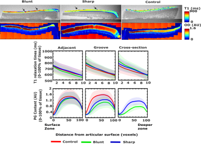 |
41 | 3-D T1 relaxation time measurements in an equine model of mild post-traumatic osteoarthritis using MB-SWIFT
Swetha Pala1, Olli Nykänen1,2, Nina Hänninen1,2, Ali Mohammadi1, Mohammadhossein Ebrahimi1,2, Nikae te Moller3, Harold Brommer3, P René van Weeren3, Janne T.A Mäkelä1, Rami K Korhonen1, Juha Töyräs1,4,5, Santtu Mikkonen1, and Mikko J Nissi1,2
1Department of Applied Physics, University of Eastern Finland, Kuopio, Finland, 2Research Unit of Medical Imaging, Physics and Technology, University of Oulu, Oulu, Finland, 3Department of Clinical Sciences, Faculty of Veterinary Medicine, Utrecht University, Utrecht, Netherlands, 4Science Service Center, University Hospital, Kuopio, Finland, 5School of Information Technology and Electrical Engineering, The University Queensland, Brisbane, Australia
In this study, we examined the variation in T1 relaxation times in a mild OA model of surgically grooved (sharp and blunt) articular cartilage of equine carpal joints. The study revealed that T1 relaxation time is significantly increased in bluntly-grooved cartilage with respect to control cartilage. No significant differences between sharply-grooved and control groups were seen for T1 relaxation. Moreover, strong variations in T1 relaxation times and proteoglycan (PG) contents were observed in the superficial half of the cartilage. T1 relaxation times correlated weakly (R≈0.33) with equilibrium mechanical modulus and with PG content (R≈0.23).
|
||
2306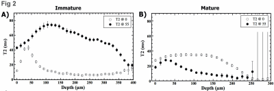 |
42 | Characteristics of maturation-related differences in rabbit cartilage by quantitative µMRI and PLM Video Permission Withheld
Yang Xia1, Hannah Mantebea1, Syeda Batool1, Amanveer Singh1, Mohammed Hammami1, and Farid Badar1
1Oakland Univ, Rochester Hills, MI, United States
Immature and mature articular cartilage from the humeral joints of rabbits were studied quantitatively and at microscopic resolutions by µMRI and polarized light microscopy (PLM). µMRI data revealed a number of differences between the immature and mature cartilage, including total thickness and T2 relaxation values. PLM data revealed in addition the cellular differences between the tissues. The mature cartilage has a clearly defined tidemark, which was absent in the immature tissue. The ability to differentiate specific maturation-related characteristics in cartilage could benefit translational studies of degenerative diseases such as osteoarthritis.
|
||
2307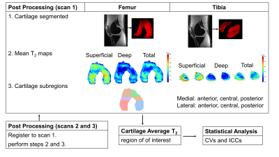 |
43 | Repeatability of Cartilage T2 relaxation times measures at 3T and 7T using quantitative double-echo in steady-state
Jessica Lauren Asay1, Anthony Andrea Gatti1, Arjun Divyang Desai1,2, Akshay Chaudhari1,3, Valentina Mazzoli1, Feliks Kogan1, and Garry Evan Gold1,4,5
1Radiology, Stanford University School of Medicine, Stanford, CA, United States, 2Electrical Engineering, Stanford University, Stanford, CA, United States, 3Biomedical Data Science, Stanford University School of Medicine, Stanford, CA, United States, 4Orthopaedic Surgery, Stanford University School of Medicine, Stanford, CA, United States, 5Bioengineering, Stanford University, Stanford, CA, United States
Quantitative MRI can be used to track subtle cartilage microstructure changes within a subject, which could be helpful in the early detection, treatment, and prevention of osteoarthritis. Increased field strength may offer higher resolution for detecting these changes. This study demonstrated the repeatability of mean T2 relaxation times measured at 3T and 7T. Full thickness cartilage T2 relaxation times at 3T and 7T had excellent ICCs, with CVs at 3T generally lower than 7T. This study suggests 3T and 7T measures are repeatable and can be used in longitudinal studies of cartilage changes.
|
||
2308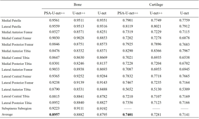 |
44 | Optimized U-net++: Novel Algorithm for Sub-regional Segmentation of Knee Cartilage and Bone Video Not Available
Lijie Zhong1, Jiaping Hu1, Shaolong Chen2, Yanjun Chen1, Zhongping Zhang3, Yingjie Mei3, Zhiyong Zhang2, and Xiaodong Zhang1
1Department of Medical Imaging, The Third Affiliated Hospital of Southern Medical University, Guangzhou, China, 2School of Electronics and Communication Engineering, Sun Yat-sen University, Shenzhen, China, 3China International Center, Philips Healthcare, Guangzhou, China
Cartilage degeneration and subchondral bone alterations play an important role in the pathogenesis and progression of knee osteoarthritis (OA). MRI can detect morphological or compositional change of cartilage and bone. Regional analysis of cartilage and bone lesions would significantly improve the diagnosis of OA and help to understand its role in OA. Automated segmentation of cartilage and bone on MRI is a necessary first step for quantitative measures. Therefore, we proposed an optimized U-net (PSA-U-net++) to solve the problem of sub-regional segmentation of bone and cartilage. The initiatory results showed that our model can accurately segment cartilage and bone.
|
||
2309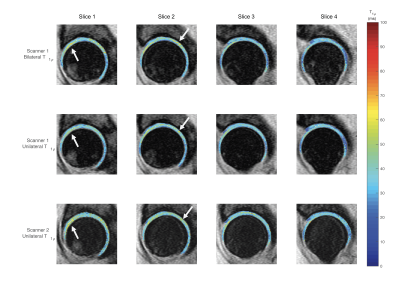 |
45 | Feasibility of Simultaneous Bilateral Hip Quantitative MRI
Koren Roach1, Misung Han1, Thomas Link1, Richard Souza1, Sharmila Majumdar1, and Valentina Pedoia1
1UCSF, San Francisco, CA, United States
Our group recently developed simultaneous bilateral hip MR acquisition methods to reduce total imaging time and employ during longitudinal evaluations of hip OA. This study evaluated the ability of simultaneous bilateral 3D FSE Cube and MAPSS acquisitions to maintain relevant morphological information compared to unilateral acquisitions. The mean T1ρ and T2 values of the simultaneous bilateral MAPSS acquisitions were not significantly different from unilateral acquisitions and relaxometry map patterns were preserved. Additionally, our simultaneous bilateral hip protocol promoted high resolution 3D Cube images for morphological abnormality detection and reduced scan time by 40%, to under 30 minutes.
|
||
2310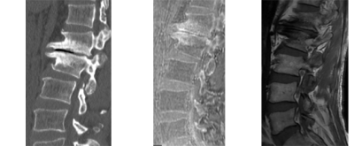 |
46 | Clinical assessment of the degenerative lumbar osseous morphology using zero echo time magnetic resonance imaging (ZTE-MRI)
Hou Bowen1, Chanyuan Liu1, Yitong Li1, Yan Xiong1, Jingyi Wang1, Weiyin Vivian Liu2, and Xiaoming Li1
1Radiology, Tongji Hospital, Tongji Medical College, Huazhong University of Science and Technology, Wuhan, Hubei Province, China, China, 2GE Healthcare, Beijing, China
Lumbar degenerative disease increases with age and is common in the elderly population. The symptoms caused by the osseous degeneration affect the quality of life. Zero echo time (ZTE) sequence could capture and reveal short-T2 materials, allowing to obtain osseous information without ionizing radiation. The ZTE performed similarly to multi-slice computed tomography (MSCT) on the ordinal variables and most measurements of osseous spine. ZTE-MRI could offer more cortical bone details than conventional MRI images and might be a valid alternative to CT for assessment of lumbar osseous morphology to some extent.
|
||
2311 |
47 | Highly Accelerated 4D Compressed Sensing Flow Sequence for Calf Muscle Strain and Strain Rate Tensor Mapping under Isometric Contractions
Usha Sinha1, Vadim Malis2, Brandon Cunnane1, Ning Jin3, and Shantanu Sinha2
1Physics, San Diego State University, San Diego, CA, United States, 2Radiology, UC San Diego, San Diego, CA, United States, 3Siemens Medical Solutions, Cleveland, OH, United States
The 4D CS-Flow sequence can be applied to image calf muscle motion during isometric contraction in order to extract 3D Strain tensors. The ability to extract the full 3D strain tensors should enable one to answer questions related to muscle deformation anisotropy in the transverse plane, volumetric strain and maximum shear strains.
|
||
 Back to Meeting Home
 Back to the Program-at-a-Glance
The International Society for Magnetic Resonance in Medicine is accredited by the Accreditation Council for Continuing Medical Education to provide continuing medical education for physicians.
 Back to Meeting Home
 Back to the Program-at-a-Glance
 View the Presentation