Digital Poster
Bone
Joint Annual Meeting ISMRM-ESMRMB & ISMRT 31st Annual Meeting • 07-12 May 2022 • London, UK

| Computer # | ||||
|---|---|---|---|---|
1596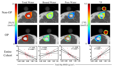 |
51 | Solid-state MR based quantitative assessment of bone water and 31P differentiates between postmenopausal women with and without osteoporosis
Brandon Clinton Jones1,2, Cheng-Chieh Cheng1,3, Xia Zhao1, Hyunyeol Lee1,4, Mona Al Mukaddam5, Peter J Snyder5, Chamith S Rajapakse1, Hee Kwon Song1, and Felix W Wehrli1
1Radiology, University of Pennsylvania, Philadelphia, PA, United States, 2Bioengineering, University of Pennsylvania, Philadelphia, PA, United States, 3Computer Science and Engineering, National Sun Yat-sen University, Kaohsiung, Taiwan, 4School of Electronics, Kyungpook National University, Daegu, Korea, Republic of, 5Department of Endocrinology, University of Pennsylvania, Philadelphia, PA, United States
Fifteen osteoporotic, treatment-naïve, and 19 non-osteoporotic postmenopausal women have been examined in an ongoing study to evaluate tibial cortical bone health via solid-state MRI. Proton dual-echo UTE and IR-prepared rapid-UTE sequences were used for quantification of pore and bound water concentrations, and a 31P PETRA-ZTE sequence for quantification of bone mineralization. Osteoporotics showed elevated pore and total water concentration, thinner cortices, decreased 31P content and degree of bone mineralization (DBM). Our preliminary results suggest that solid-state MR biomarkers of bone porosity and DBM may be useful in evaluating cortical bone health.
|
||
1597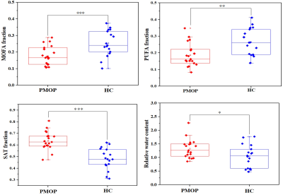 |
52 | 2D iDQC-MRS suggests that bone marrow fatty acids are more saturated for postmenopausal osteoporosis Video Not Available
Jianfeng Bao1, Xiao Wang1, Yong Zhang1, and Jingliang Cheng1
1Department of Magnetic Resonance Imaging, The First Affiliated Hospital of Zhengzhou University, Zhengzhou, China
Postmenopausal osteoporosis (PMOP) in the female elderly population has become one of serious health problems of the rapidly aging society in China. The aim of this work was to preliminary investigate the changes of bone marrow fatty acid composition in the presence of trabecular bone of PMOP and age-matched healthy controls (HC) using 2D intermolecular double-quantum coherence based magnetic resonance spectroscopy (iDQC-MRS). We found monounsaturated fatty acids (P < 0.001) and polyunsaturated fatty acids(p < 0.01) were significant higher lower in the PMOP group. The lower unsaturated fatty acids levels may play an important role in the pathogenesis of PMOP.
|
||
1598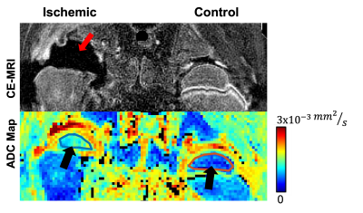 |
53 | IVIM Detects Bone Ischemia in a Piglet Model of Legg-Calvé-Perthes Disease
Erick Odoyo Buko1,2, Sampada Bhave1, Ferenc Tóth1, and Casey P Johnson1,2
1Veterinary Clinical Sciences, University of Minnesota, St. Paul, MN, United States, 2Center for Magnetic Resonance Research, University of Minnesota, Minneapolis, MN, United States
Intravoxel incoherent motion (IVIM) is a promising method to measure both tissue diffusion and perfusion using a single multi b-value diffusion-weighted imaging (DWI) acquisition. In this work, we investigated whether IVIM is sensitive in detecting surgically-induced femoral head ischemia in a piglet model of avascular necrosis. We found that the IVIM perfusion coefficient (Df) and fraction (f) decreased in the ischemic vs. contralateral-control femoral heads. Conversely, the IVIM diffusion coefficient (Ds) increased as a result of subsequent injury to the operated femoral head. These findings suggest that IVIM may provide a non-contrast-enhanced means to assess bone ischemia and perfusion.
|
||
1599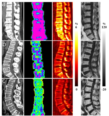 |
54 | Comprehensive Assessment of Osteoporosis in Lumbar Spine Using Compositional MR Imaging of Trabecular Bone Video Not Available
Jin Liu1, Jian-Wei Liao1, Jia-Xin Feng1, Wei Li1, Xiao-Jun Chen1, Lin Yao1, Pan-Hui Huang1, Long Qian2, Ya-Jun Ma3, and Shao-Lin Li1
1Department of Radiology, The Fifth Affiliated Hospital of Sun Yat-Sen University, Zhuhai, China, 2MR Research, GE Healthcare, Beijing, China, 3Department of Radiology, University of California San Diego, San Diego, CA, United States
Compositional MR Imaging, which is combined of a Short TR Adiabatic Inversion Recovery prepared Ultrashort Echo Time (STAIR-UTE) and the Iterative decomposition of water and fat with echo asymmetry and least squares estimation (IDEAL-IQ) techniques, is developed to measure collagen bound water proton fraction, free water proton fraction, total water proton fraction of trabecular bone in lumbar spine. These biomarkers are highly correlated with bone mineral density, T score and Fracture Risk Assessment Tool scores, which demonstrates that the compositional MR imaging has great potential in diagnosis of patient with osteoporosis.
|
||
1600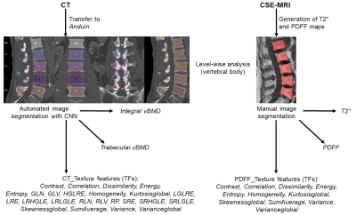 |
55 | Texture analysis using chemical shift imaging improves differentiation between patients with and without osteoporotic vertebral fractures Video Permission Withheld
Nico Sollmann1,2,3, Edoardo A. Becherucci2, Christof Boehm4, Malek El Husseini2, Stefan Ruschke4, Egon Burian2, Jan S. Kirschke2, Thomas M. Link3, Karupppasamy Subburaj5, Dimitrios C. Karampinos4, Roland Krug3, Thomas Baum2, and Michael Dieckmeyer2
1Department of Diagnostic and Interventional Radiology, University Hospital Ulm, Ulm, Germany, 2Department of Diagnostic and Interventional Neuroradiology, Technical University of Munich, Munich, Germany, 3Department of Radiology and Biomedical Imaging, University of California San Francisco, San Francisco, CA, United States, 4Department of Diagnostic and Interventional Radiology, Technical University of Munich, Munich, Germany, 5Engineering Product Development (EPD) Pillar, Singapore University of Technology and Design, Singapore, Singapore
Osteoporosis is characterized by increased skeletal fragility with vertebral fractures (VFs). Areal bone mineral density (BMD) from dual-energy X-ray absorptiometry (DXA) is the reference standard but has well-known limitations. Texture analysis (TA) can provide parameters of tissue microstructure using spine chemical shift encoding-based water-fat MRI (CSE-MRI) and computed tomography (CT), thus potentially improving fracture risk estimation. This study found that a model including volumetric BMD (vBMD) and several texture features (TFs) from CSE-MRI and CT predicts 81% of the variance regarding osteoporotic VF status, compared to 47% when based on vBMD and the proton density fat fraction (PDFF) only.
|
||
1601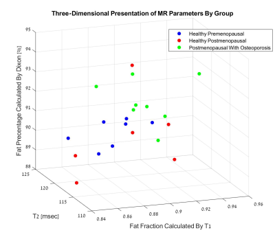 |
56 | Quantitative MR of the Distal Radius Bone Marrow as a marker of Osteoporosis Video Permission Withheld
Tamar K. De-Levie1,2, Yael S. Schiffenbauer1, Ido Druckmann3, Vanessa Rouach4, Naftali Stern2,4,5,6, Itzhak Binderman7, and Uri Nevo1,5
1Department of Biomedical Engineering, Tel Aviv University, Tel-Aviv, Israel, 2Sackler Faculty of Medicine, Tel Aviv University, Tel-Aviv, Israel, 3Skeletal Imaging Division, Tel Aviv-Sourasky Medical Center, Tel-Aviv, Israel, 4Institute of Endocrinology, Metabolism and Hypertension, Tel Aviv-Sourasky Medical Center, Tel-Aviv, Israel, 5The Sagol School of Neuroscience, Tel Aviv University, Tel-Aviv, Israel, 6The Sagol Center for Epigenetics, Tel Aviv-Sourasky Medical Center, Tel-Aviv, Israel, 7Department of Oral Biology, School of Dental Medicine, Tel Aviv University, Tal-Aviv, Israel
In-vivo detection of osteoporosis-related changes in the distal radius bone marrow, using MR protocols, could provide safe and accessible means for screening and monitoring. A clinical trial was performed, including 26 women assigned into three study groups: healthy premenopausal (n=7), healthy postmenopausal (n=10) and osteoporotic postmenopausal (n=9). Fat composition was evaluated using T2 maps, two-compartment model of T1 and Dixon sequence. The osteoporotic group exhibited higher fat content and lower T2 values compared to the healthy premenopausal group. This study provides proof of concept for the use of the distal radius bone marrow as probing site for osteoporosis.
|
||
1602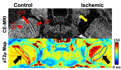 |
57 | MRI Relaxation Times Change in the Metaphysis Following Ischemic Injury to the Femoral Head: An In Vivo Piglet Model Study
Erick Buko1,2, Alexandra R Armstrong1, Ferenc Tóth1, and Casey P Johnson1,2
1Veterinary Clinical Sciences, University of Minnesota, ST. Paul, MN, United States, 2Center for Magnetic Resonance Research, University of Minnesota, Minneapolis, MN, United States
Quantitative mapping of T2, T1ρ, adiabatic T1ρ, and adiabatic T2ρ relaxation times may be useful to assess ischemic injury to the femoral head. In this work, we investigated whether these relaxation times are sensitive in detecting compositional changes to the primary spongiosa (the region of the metaphysis adjacent to the growth plate) following ischemic injury to the femoral head in a piglet model. We found that T1ρ and adiabatic T2ρ decreased in the primary spongiosa following ischemic injury to the femoral epiphysis, which suggests these methods may be useful to assess femoral head growth disturbances following ischemic injury.
|
||
 Back to Meeting Home
 Back to the Program-at-a-Glance
The International Society for Magnetic Resonance in Medicine is accredited by the Accreditation Council for Continuing Medical Education to provide continuing medical education for physicians.
 Back to Meeting Home
 Back to the Program-at-a-Glance
 View the Presentation