Digital Poster
Joints
Joint Annual Meeting ISMRM-ESMRMB & ISMRT 31st Annual Meeting • 07-12 May 2022 • London, UK

| Computer # | ||||
|---|---|---|---|---|
1403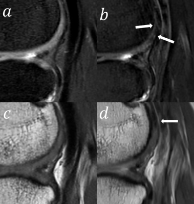 |
27 | The value of Modulated Flip Angle in Refocused Imaging with Extended Echo Trains with Compressed Sensing (MATRIX) in Knee MRI Imaging Video Not Available
He Sui1, Yu Gong2, Yunfei Zhang3, Yongming Dai3, and Zhanhao Mo1
1China-Japan Union Hospital of Jilin University, Changchun, China, 2Linyi People’s Hospital, Linyi, China, 3Central Research Institute, United Imaging Healthcare, Shanghai, China
Three-Dimensional (3D) sequence of magnetic resonance imaging (MRI) plays a critical role in musculoskeletal joints imaging; however, its long acquisition time limits the clinical application. In such condition, Compressed sensing (CS) is introduced to accelerate MRI during clinical practice. We aimed to investigate the feasibility of an isotropic 3D variable-flip-angle fast spin echo (FSE) sequence with CS technique (CS-MATRIX) compared to conventional 2D sequences on knee imaging.
|
||
1404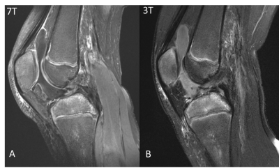 |
28 | Joint effusion contrast differences in 2D fat-saturated T2-weighted pulse sequences in 3T and 7T MRI
Sina Tafti1,2, Bruce Damon3, and Dean Hoffmeister4
1Siemens Medical Solutions USA, Inc., Urbana, IL, United States, 2Beckman Institute, University of Illinois at Urbana-Champaign, Urbana, IL, United States, 3Stevens Family Clinical Research Institute, Carle Health, Urbana, IL, United States, 4Radiology, Carle Health, Urbana, IL, United States
7T MRI provides excellent resolution of the structures in the knee. Fat-saturated T2-weighted imaging at 7T has been used clinically for assessing knee joint effusion, but may provide different contrast than 3T MRI in cases of acute injury involving suspected blood products in the fluid. Here, we show three cases in which joint effusion due to acute injury had dark contrast in T2-weighted images acquired at 7T. For one case, T2-weighted 3T MRI was acquired in which the effusion appeared bright. These differences illustrate the potential for high field MRI to elucidate pathological components not observable at lower field strength.
|
||
1405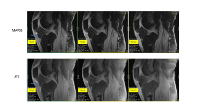 |
29 | Comparison of MAPSS- vs UTE-based T1rho Sequences in the Knee
Michael Carl1, Dina Moazamian2, Alecio F. Lombardi2, Amir Masoud Afsahi2, Yajun Ma2, and Jiang Du2
1GE Healthcare, San Diego, CA, United States, 2UCSD, San Diego, CA, United States
We compared two different T1rho sequence approaches and assessed their performance for quantitative musculoskeletal imaging. We found that using either a MAPSS or UTE acquisition after T1rho preparation resulted in similar T1rho decay values for tissues of moderate to long T2s (e.g., cartilage or meniscus). The UTE acquisition sequence, however, was also readily able to obtain T1rho decay values for tissues with short T2s (e.g., tendons), thereby facilitating whole knee assessment.
|
||
1406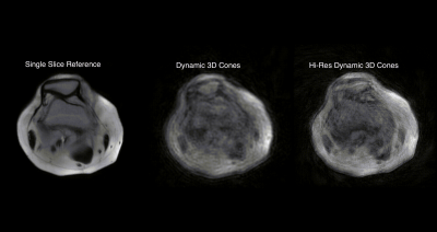 |
30 | Three-Dimensional real-time dynamic knee MRI using 3D cones with a multiscale low-rank reconstruction
Laurel Hales1, Chris Sandino1, Valentina Mazzoli2, and Feliks Kogan2
1Electrical Engineering, Stanford University, Stanford, CA, United States, 2Radiology, Stanford University, Stanford, CA, United States
3D real-time imaging of joints in motion can help us gain valuable insight into joint disorders. The combination of 3D golden-ratio re-ordered cones and multiscale low-rank reconstruction allow for continuous acquisition and high accelerations making 3D real time dynamic imaging of knees possible. We demonstrated the feasibility of this approach to capture patellar motion caused by a quadriceps flexion in 16 slices and with a temporal resolution of ~500ms. Although the resulting images are blurred, this method shows promise.
|
||
1407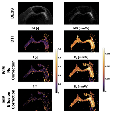 |
31 | Intravoxel Incoherent Motion (IVIM) to study synovitis in Osteoarthritis: a feasibility study
Valentina Mazzoli1 and Feliks Kogan1
1Radiology, Stanford University, Stanford, CA, United States
Synovial inflammation (synovitis) is present in the majority of people with Osteoarthritis(OA) and has been linked with pain, disease severity and progression of knee OA. However, synovitis is not widely studied due to challenges differentiating synovial inflammation from adjacent fluid on conventional MRI and contraindications and time, cost and complexity of gadolinium-based contrast-enhanced methods. In this work, we evaluate the feasibility of the intravoxel incoherent motion(IVIM) imaging model for characterization of synovitis activity. By characterizing the diffusion from synovial fluid effusions, we showed that the intravoxel signal from effusions can be removed allowing isolation of diffusion effects from the synovium.
|
||
1408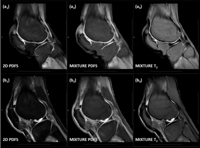 |
32 | Simultaneous morphologic and quantitative imaging of the knee joint using pseudo-3D MIXTURE sequences: from deliberation to implementation Video Permission Withheld
Teresa Nolte1, Shuo Zhang2, Nicola Pridöhl1, Malin Ciba1, Masami Yoneyama3, Christiane Kuhl1, and Sven Nebelung4
1Diagnostic and Interventional Radiology, University Hospital Aachen, Aachen, Germany, 2Philips GmbH Market DACH, Hamburg, Germany, 3Philips Japan, Tokyo, Japan, 4Clinic for Diagnostic and Interventional Radiology, Uniklinik RWTH Aachen University, Aachen, Germany
This work aimed to propose a clinical sequence platform for simultaneous morphologic and quantitative imaging of joints and identified TSE-based MIXTURE (Multi-Interleaved X-prepared Turbo-Spin Echo with Intuitive Relaxometry) sequences in terms of PDFS-T2 and T1-T1ρFS combinations for clinical implementation. Even though capable of isotropic resolution, MIXTURE sequences were acquired as pseudo-3D (thicker slices, higher in-plane resolution) to match standard-clinical 2D TSE sequences. After identification of clinical demands, MIXTURE sequences were systematically optimized, while maintaining clinically feasible acquisition times of 5:00 min (PDFS-T2 for T2-mapping) and 6:40 min (T1-T1ρFS for T1ρ-mapping), and evaluated in a cadaveric human knee cartilage defect model.
|
||
1409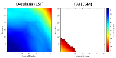 |
33 | Zero Echo Time (ZTE) MRI-Based 3D Collision Modeling of Femoroacetabular Impingement
Ryan E Breighner1, Emily Davidson1, and Hollis G Potter1
1Radiology and Imaging, Hospital for Special Surgery, New York, NY, United States
To simultaneously improve assessment of patholomorphologies of the hip and reduce the reliance on CT imaging, this study sought to utilize Zero Echo Time (ZTE) MRI to create patient-specific 3D models of the hip and perform in silico kinematic collision simulations to quantify hip range of motion in a cohort of patients with radiologic signs of FAI and hip dysplasia. Range of motion assessments indicate that patients' simulated ranges of motion comport with their radiologically diagnosed conditions.
|
||
1410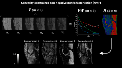 |
34 | Data-driven characterization of knee structures using non-negative matrix factorization of quantitative UTE Spiral VIBE MRI
Céline Smekens1, Pieter Van Dyck2, Thomas Janssens3, Jan Sijbers1, and Ben Jeurissen1
1imec-Vision Lab, Department of Physics, University of Antwerp, Wilrijk, Belgium, 2Department of Radiology, Antwerp University Hospital and University of Antwerp, Edegem, Belgium, 3Siemens Healthcare NV/SA, Beersel, Belgium
Model-driven methods for knee structure characterization, such as bi-exponential T2* mapping, may result in unreliable parameter estimation. In contrast, data-driven approaches, such as non-negative matrix factorization (NMF), allow to robustly identify multiple compartments. In this work, convexity-constrained NMF was used to decompose quantitative UTE MRI of three asymptomatic and one degenerative knees for structural characterization. Decomposition in four compartments led to minimal residual errors and low inter-subject differences in normalized mean compartment weights. Shifts in compartment weight distributions correlated to structural abnormalities, suggesting that the proposed approach may aid in the detection, grading and monitoring of internal knee derangements.
|
||
1411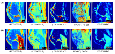 |
35 | Quantitative UTE Double Echo Steady State (qUTE-DESS) for Simultaneous Mapping of T1, T2, and Diffusivity of Short T2 Tissues – Ex Vivo Study
Hyungseok Jang1, Yajun Ma1, Amir Masoud-Afsahi1, Saeed Jerban1, Alecio F Lombardi1, Eric Y Chang1,2, Christine B Chung1,2, and Jiang Du1
1Radiology, University of California San Diego, San Diego, CA, United States, 2Radiology Service, Veterans Affairs San Diego Healthcare System, San Diego, CA, United States
Quantitative double echo steady state (DESS) imaging allows simultaneous estimation of T1, T2, and apparent diffusion coefficient (ADC) maps with high spatial resolution. Recently, ultrashort echo time (UTE) based DESS has been investigated targeting short T2 tissues. However, ADC mapping for tissues with short T2 values using DESS is challenging due to the two competing factors: sensitivity to diffusion that requires a long TR and sensitivity to short T2 component that requires a short TR. In this study, the feasibility of using quantitative UTE DESS (qUTE-DESS) to image connective tissues in the knee was investigated.
|
||
1412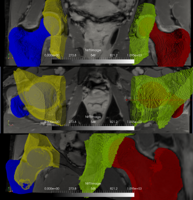 |
36 | Automatic segmentation of the hip bony structures on 3D Dixon MRI datasets using transfer learning from a neural network developed for the shoulder
Eros Montin1, Cem Murat Deniz1, Tatiane Cantarelli Rodrigues2, Soterios Gyftopoulos3, Richard Kijowski3, and Riccardo Lattanzi1,4
1Center for Advanced Imaging Innovation and Research (CAI2R) Department of Radiology, New York University Grossman School of Medicine, New York, NY, United States, 2Department of Radiology, Hospital do Coração (HCOR) and Teleimagem, São Paulo, Brazil, 3Department of radiology, New York University Grossman School of Medicine, New York, NY, United States, 4Vilcek Institute of Graduate Biomedical Sciences, New York University Grossman School of Medicine,, New York, NY, United States
We describe a network for automatic segmentation of acetabulum and femur on 3D-Dixon MRI data. Given the limited number of labeled 3D hip datasets publicly available, our network was trained using transfer learning from a network previously developed for the segmentation of the shoulder bony structures. Using only 5 hip datasets for training, our network achieved segmentation dice of 0.719 and 0.92 for acetabulum and femur, respectively. More training data is needed to improve results for the acetabulum. We show that transfer learning can enable automatic segmentation of the hip bones using a limited number of labeled training data.
|
||
1413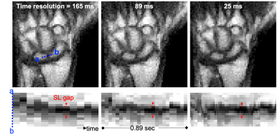 |
37 | Feasibility of real-time MRI of the actively moving wrist at 0.55 Tesla Video Not Available
Yongwan Lim1, Sophia X. Cui2, Robert M. Szabo3, Robert D. Boutin4, Abhijit J. Chaudhari5, and Krishna S. Nayak1
1Ming Hsieh Department of Electrical and Computer Engineering, University of Southern California, Los Angeles, CA, United States, 2Siemens Medical Solutions USA, Inc., Los Angeles, CA, United States, 3Orthopaedic Surgery, University of California Davis, Davis, CA, United States, 4Radiology, Stanford University School of Medicine, Stanford, CA, United States, 5Radiology, University of California Davis, Davis, CA, United States
In this work we show results from a first-in-human study utilizing a high-performance 0.55T system and balanced SSFP (bSSFP) real-time MRI acquisition to assess tissues of the actively moving wrist’s uninterrupted radial-ulnar deviation and clenched fist maneuvers. We show that at 0.55T, bSSFP is less sensitive to off-resonance and banding artifacts that frequently obscure critical wrist structures. We show that the high temporal resolution of 25 ms per frame at high-performance 0.55T enables improved characterization of wrist joint motion. These benefits could make it a promising technology to evaluate dynamic wrist instability.
|
||
1414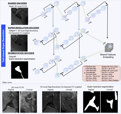 |
38 | Super Resolution Segmentation of the Scapula from Clinical MRI Video Not Available
Francesco Caliva1, Victoria Wong1, Favian Su1, Drew Lansdown1, and Valentina Pedoia1
1University of California San Francisco, San Francisco, CA, United States
We propose a deep learning approach to simultaneous super resolution and segmentation. We experiment our framework on the challenging task of generating 3D high-resolution axial shoulder MRI from 2D clinical MRI sequences, and demonstrate the ability to produce precise 3D scapula bone models. With an extensive experimental study, we show that with super-resolution, it is possible to produce high resolution bone models, which are invaluable for surgeons in the pre-operative planning of patients with various shoulder conditions. This method has the potential to reduce patients' need for undergoing CT scans, hence preventing exposure to radiation.
|
||
 Back to Meeting Home
 Back to the Program-at-a-Glance
The International Society for Magnetic Resonance in Medicine is accredited by the Accreditation Council for Continuing Medical Education to provide continuing medical education for physicians.
 Back to Meeting Home
 Back to the Program-at-a-Glance
 View the Presentation