Digital Poster
Meniscus, Ligament & Joint
Joint Annual Meeting ISMRM-ESMRMB & ISMRT 31st Annual Meeting • 07-12 May 2022 • London, UK

| Computer # | ||||
|---|---|---|---|---|
1486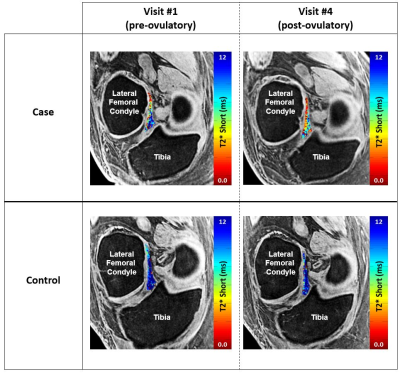 |
36 | Changes in ACL T2* metrics from pre- to post-ovulatory phases of the menstrual cycle: A new biomarker for ACL-injury risk in females? Video Permission Withheld
Erin C Argentieri1, Ryan E Breighner1, Matthew F Koff1, and Hollis G Potter1
1Radiology and Imaging, Hospital for Special Surgery, New York, NY, United States ACL T2* metrics were evaluated over the course of the menstrual cycle. During the pre-ovulatory phase, normally ovulating case subjects exhibited significantly shortened mean and median T2*S metrics compared to the post-ovulatory phase. Anovulatory control subjects displayed no significant changes in any ACL T2* metrics over time. Findings suggest that an increase in collagen bound water (T2*S) is present within the ACL during the pre-ovulatory phase, when both ACL-injury risk and anterior knee laxity are also increased. |
||
1487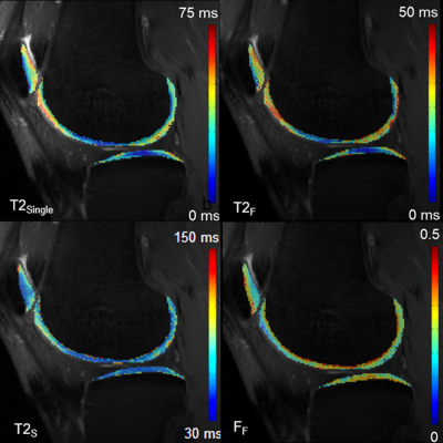 |
37 | Bi-Component Cartilage T2 Analysis in Subjects With Anterior Cruciate Ligament Reconstruction
Richard Kijowski1, Robert Moskwa2, and Fang Liu3
1Radiology, New York University School of Medicine, New York, NY, United States, 2Medical Physics, University of Wisconsin, Madison, WI, United States, 3Radiology, Massachusetts General Hospital, Boston, MA, United States
mcDESPOT was used to measure single-component T2 relaxation time (T2Single) and the bi-component T2 parameter fraction of the fast relaxing macromolecular bound water component (FF) of the cartilage of the knee at 3.0T in 10 subjects with ACL reconstruction and 10 control subjects. There were significant differences (p<0.05) between ACL reconstruction and control subjects on 5 of 6 articular surfaces of the knee for FF compared with 2 of 6 surfaces for T2Single with FF having higher absolute effect size than T2Single for all surfaces. The results demonstrate the superiority of bi-component over single-component T2 analysis for detecting cartilage degeneration.
|
||
1488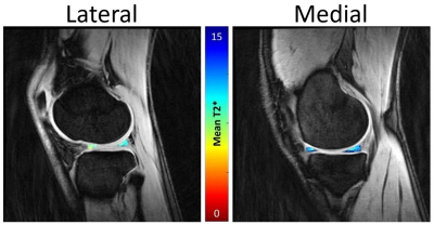 |
38 | Longitudinal comparison of medial and lateral meniscal T2* values within elite basketball players and swimmers Video Permission Withheld
Erin C Argentieri1, Garry Gold2, Sharmila Majumdar3, Hollis G Potter1, and Matthew F Koff1
1Radiology and Imaging, Hospital for Special Surgery, New York, NY, United States, 2Stanford University, Stanford, CA, United States, 3University of California, San Francisco, San Francisco, CA, United States
Basketball players represent a population with an inherently high risk of sustaining meniscal injuries. Evaluation of quantitative MRI (qMRI) metrics of the meniscus within these athletes may improve clinical insight. To date, no studies have longitudinally evaluated meniscal T2* values in high performance athletes. Therefore, the purpose of this study was to utilize ultra-short TE (UTE) MRI to longitudinally evaluate medial and lateral meniscal T2* values within elite weight-bearing (basketball) and non-weight bearing (swimmers) athletes. Significant differences of T2* values were found between the medial and lateral menisci. No significant difference of meniscal T2* values were found between basketball players and swimmers.
|
||
1489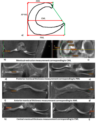 |
39 | Meniscal deformation MRI study using knee joint axial loading device and finite-element-model
Sandeep Panwar Jogi1, Rafeek Thaha1, Sriram Rajan2, Vidur Mahajan2, Vasantha Kumar Venugopal2, Amit Mehndiratta1,3, and Anup Singh1,3
1Center for Biomedical Engineering, Indian Institute of Technology, Delhi, India, 2Mahajan Imaging Center, Delhi, India, 3Center for Biomedical Engineering, All India Institute of Medical Sciences, Delhi, India The meniscal-extrusion may disturb the stability of the knee-joint, and depicts laxity in the meniscal-hoops. The degraded meniscal site usually show changes in the MR images (T1, T2, and Proton-Density-weighted images). However, the laxity of a specific meniscal segment and its effect on adjacent segment’s deformation is unknown. The current study attempt to address this problem using MRI data (with and without loading) and a finite-element-model. Each meniscal-hoop’s (lateral and medial) lengths, thickness, and meniscus-extrusion was measured to evaluate load-bearing changes. Medial meniscus hoop’s Anterior-meniscal-length(ML), Posterior-ML, Central-ML, and meniscus-extrusion show substantial change(>5%) during load. |
||
1490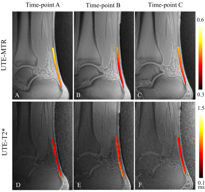 |
40 | Assessment of Achilles Tendon Changes After Long-Distance Running Using Ultrashort Echo Time Magnetization Transfer MR Imaging Video Permission Withheld
Yijie Fang1, Dantian Zhu1, Wenjun Yu1, Wenhao Wu1, Wei Li1, Long Qian2, Yajun Ma3, and Shaolin Li1
1The Fifth Affiliated Hospital of Sun Yat-sen University, Zhuhai, China, 2MR Research, GE Healthcare, Beijing, China, 3University of California, San Diego, CA, United States
Long-distance running is one of the common causes of Achilles tendon injury. UTE-MT is a magic angle-insensitive MRI technique and an excellent fit for the assessment of Achilles tendon which has a highly anisotropic collagen structure.This study aims to explore the feasibility of quantitative UTE-MT imaging in the assessment of Achilles tendon changes for subjects before and after long-distance running, then compare the technique’s performance with that of UTE-T2*.
|
||
1491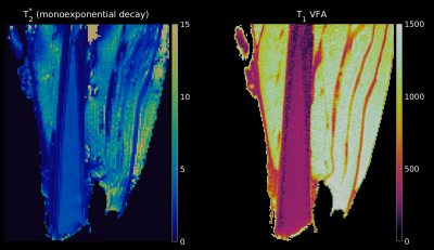 |
41 | T1 and T2* mapping of sheep forelimb aponeuroses with high-resolution ultra-short echo time (UTE) imaging
Marta B. Maggioni1, Martin Krämer1,2, Heiko Stark3, and Jürgen R. Reichenbach1,2,4,5,6
1Medical Physics Group, Institute of Diagnostic and Interventional Radiology, Jena University Hospital - Friedrich-Schiller-University, Jena, Germany, 2Institute of Diagnostic and Interventional Radiology, Jena University Hospital - Friedrich Schiller University, Jena, Germany, 3Institute of Zoology and Evolutionary Research with Phyletic Museum, Friedrich-Schiller-University, Jena, Germany, 4Michael Stifel Center for Data-driven and Simulation Science Jena, Friedrich Schiller University, Jena, Germany, 5Abbe School of Photonics, Friedrich Schiller University, Jena, Germany, 6Center of Medical Optics and Photonics, Friedrich Schiller University, Jena, Germany Aponeuroses play a crucial role in the transmission of mechanical load in the muscle-tendon unit. Their contribution is, however, still under debate and they are difficult to image with MRI due to their short T2* decay. In this work, we use high-resolution 3D ultra-short echo time imaging with 50 echoes to investigate the T2* decay of the aponeurosis and build a three-component model to quantify the T2* values. Additionally, we quantified T1 using variable flip angle imaging. To our knowledge, this work is the first to report T1 and T2* values of the aponeurosis. |
||
1492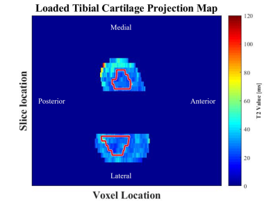 |
42 | Changes in Tibial Articular Cartilage T2 Relaxation Time with Load – An in vivo Repeatability Study
Katie Glynn Sofko1, Ibukunoluwa Elebute1, Lumeng Cui2, Natasha Bzowey1, and Emily J McWalter3
1Department of Mechanical Engineering, University of Saskatchewan, Saskatoon, SK, Canada, 2Department of Biomedical Engineering, University of Saskatchewan, Saskatoon, SK, Canada, 3Department of Mechanical and Biomedical Engineering, University of Saskatchewan, Saskatoon, SK, Canada
Tibial articular cartilage plays the important role of transmitting forces through the knee joint; however, we most often study it without any applied loads. The aim of this work was to assess the repeatability of T2 relaxation time maps in the unloaded and loaded knees of five healthy participants. Using qDESS, it is possible to obtain repeatable, in vivo T2 relaxation time maps of the entire tibial cartilage surface as well as inside and outside regions of tibiofemoral contact in both unloaded and loaded knees.
|
||
1493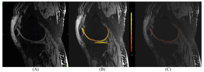 |
43 | Comparison of Two Methods for Measuring Knee Cartilage Thickness in Lower Extremities Using 7T Magnetic Resonance Imaging: A Cadaver Study
Dominik Vilimek1,2, Benedikt Hager3, Markus Schreiner4, Pavla Hanzlikova5,6, Radana Kahankova1, Veronika Janacova2, Didier Laurent7, Christoph Fuchssteiner8, Wolfgang Weninger8, Reinhard Windhager4, Pavol Szomolanyi2, Siegfried Trattnig2,3,9, and Vladimir Juras2
1Department of Cybernetics and Biomedical Engineering, VSB–Technical University of Ostrava, Ostrava, Czech Republic, 2High Field MR Centre, Department of Biomedical Imaging and Image-Guided Therapy, Medical University of Vienna, Vienna, Austria, 3Institute for Clinical Molecular MRI in the Musculoskeletal System, Karl Landsteiner Society, Vienna, Austria, 4Department of Orthopedics and Trauma Surgery, Medical University of Vienna, Vienna, Austria, 5Department of Imaging Method, Faculty of Medicine, University of Ostrava, Ostrava, Czech Republic, 6Department of Imaging Method, University Hospital Ostrava, Ostrava, Czech Republic, 7Novartis Institutes for Biomedical Research, Translational Medicine, Basel, Switzerland, 8Center for Anatomy and Cell Biology, Division of Anatomy, Medical University of Vienna, Vienna, Austria, 9CD laboratory for Clinical Molecular MR imaging (MOLIMA), Vienna, Austria Knee articular cartilage thickness may potentially serve as a marker for monitoring of the knee cartilage status. However, it is challenging to measure and quantify the articular cartilage thickness using in vivo MRI. In the present study, we evaluated 6 unpaired lower extremies of body donors using both a prototype segmentation software and a semi-automatic approach. Our results showed a low correlation (r = 0.45) between the two methods, indicating the challenge of determining cartilage thickness from MR images. |
||
1494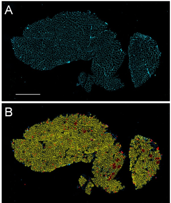 |
44 | Correlation of Fractional Anisotropy and Histological Measures of Muscle Architecture in Vastus Lateralus in Legs with Injured ACL
Peter Andrew Hardy1, Christopher Fry2, Anders Andersen3, Katherine Thompson4, Thorsten Feiweier5, and Brian Noehren6
1Radiology, University of Kentucky, Lexington, KY, United States, 2Athletic Training and Clinical Nutrition, University of Kentucky, Lexington, KY, United States, 3MRISC, University of Kentucky, Lexington, KY, United States, 4Statistics, University of Kentucky, Lexington, KY, United States, 5Diagnostic Imaging, Siemens Healthcare, GmbH, Erlangen, Germany, 6Physical Therapy, University of Kentucky, Lexington, KY, United States
We measured diffusion tensor properties of the vastus lateralus muscle in 30 subjects who had a torn ACL. DTI was acquired with a stimulated echo acquisition with a diffusion time of 185 ms. From biopsies of the muscle taken simultaneously we developed histological measures of muscle fiber cross sectional area and minimum Feret diameter. Linear regression analysis demonstrated strong association of the fractional anisotropy and minimum Feret diameter.
|
||
1495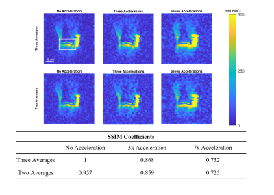 |
45 | Compressed sensing and the use of 3D UTE acquisition for high-resolution accelerated 23NA imaging at 3T
Cameron Villarreal1, Xin Shen1, Ali Calgar Ozen2, Serhat Ilbey2, Mark Chiew3, Ahmad Alhulial4, Evan Pogue5, Uzay Emir1,5, and Deva Chan1
1Weldon School of Biomedical Engineering, Purdue University, West Lafayette, IN, United States, 2Department of Radiology, Medical Physics, Medical Center, University of Freiburg, Freiburg, Germany, 3Wellcome Centre for Integrative Neuroimaging, University of Oxford, Oxford, United Kingdom, 4College of Applied Medical Sciences, Prince Sattam bin Abdulaziz University, Al Kharj, Saudi Arabia, 5School of Health Sciences, Purdue University, West Lafayette, IN, United States
There is a need for methods to detect early compositional changes during musculoskeletal diseases like osteoarthritis, but current diagnostic tools . Sodium (23Na) MRI enables quantification of sodium content which is correlated with cartilage health. However, several limitations preclude its widespread clinical use including long scan times and low resolution. We have developed a UTE 23Na MRI sequence with rosette acquisition capable of overcoming some of these limitations. In this study we demonstrate the feasibility of reducing our acquisition time through compressed sensing while conserving signal quality in a healthy human knee.
|
||
1496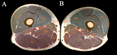 |
46 | The association of thigh muscle volume and fatty infiltration with knee joint structural damage
Amir M. Pirmoazen1, Upasana U. Bharadwaj 1, John A. Lynch2, Valentina Pedoia1, Michael C. Nevitt2, Charles E. McCulloch2, Gabby B. Joseph1, and Thomas M. Link1
1Radiology and Biomedical Imaging, University of California, San Francisco, San Francisco, CA, United States, 2Epidemiology and Biostatistics, University of California, San Francisco, San Francisco, CA, United States Knee osteoarthritis results from inflammatory, mechanical, and metabolic processes, leading to chronic degenerative changes. The purpose of this study was to investigate the cross-sectional associations between thigh muscle volume or intermuscular fat volume, and relative muscle sizes, from T1-weighted MR images of 100 thighs, and radiographic knee osteoarthritis (ROA) using Kellgren-Lawrence grades. The results showed that lower volumes of the quadriceps and the anterior muscle compartment of the thigh relative to total muscle volume, as well as relative higher hamstring muscle volume were associated with presence of ROA (P<0.05). |
||
 Back to Meeting Home
 Back to the Program-at-a-Glance
The International Society for Magnetic Resonance in Medicine is accredited by the Accreditation Council for Continuing Medical Education to provide continuing medical education for physicians.
 Back to Meeting Home
 Back to the Program-at-a-Glance
 View the Presentation