Digital Poster
Nerve, Spine & Tumors
Joint Annual Meeting ISMRM-ESMRMB & ISMRT 31st Annual Meeting • 07-12 May 2022 • London, UK

| Computer # | ||||
|---|---|---|---|---|
2885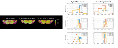 |
95 | Investigations of the effects of differently trained subject cohorts on T1 and T2 of the lumbar spine muscles
Marta B. Maggioni1, Martin Krämer1,2, Christoph Anders3, and Jürgen R. Reichenbach1,2,4,5,6
1Medical Physics Group, Institute of Diagnostic and Interventional Radiology, Jena University Hospital - Friedrich-Schiller-University, Jena, Germany, 2Institute of Diagnostic and Interventional Radiology, Jena University Hospital - Friedrich Schiller University, Jena, Germany, 3Institute for Pathophysiology and Pathobiochemistry, Motor Research Group, Friedrich-Schiller-University, Jena, Germany, 4Michael Stifel Center for Data-driven and Simulation Science Jena, Friedrich Schiller University, Jena, Germany, 5Abbe School of Photonics, Friedrich Schiller University, Jena, Germany, 6Center of Medical Optics and Photonics, Friedrich Schiller University, Jena, Germany The majority of studies investigating changes in skeletal muscle parameters with MRI have focused on analyzing the effects before and after exercise training. In this work, we analyze the effects of regular repetitive training on low back muscle by determining T1 and T2 parameters in three cohorts of subjects characterized by different levels of physical activity. |
||
2886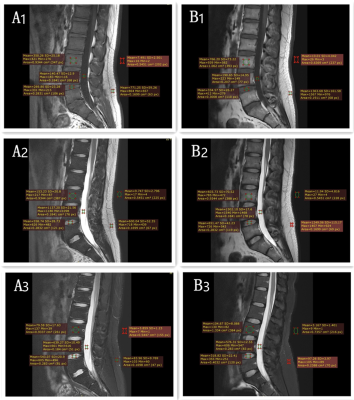 |
96 | Comparison of AI-assisted Compressed Sensing (ACS) and Conventional Two-Dimensional Sequences on Lumbar Spine Imaging Video Not Available
He Sui1, Yu Gong2, Yunfei Zhang3, Yongming Dai3, and Zhanhao Mo1
1China-Japan Union Hospital of Jilin University, Changchun, China, 2Linyi People’s Hospital, Linyi, China, 3Central Research Institute, United Imaging Healthcare, Shanghai, China
MRI is widely used in the diagnosis of spinal diseases, but due to the limitation of long scanning time, it is often difficult for patients to tolerate. This study aims at exploring whether the ACS (AI-assisted Compressed Sensing) accelerated sequences can assure similar image quality and diagnostic accuracy of lumbar spine diseases compared to 2D conventional sequences. Results suggested that 2D ACS sequence has become a well-anticipated technique for routine examination of lumbar spine and lumbar spine lesions diagnosis, while ensuring high image quality and reduced scan time effectively than traditional 2D sequences in MRI.
|
||
2887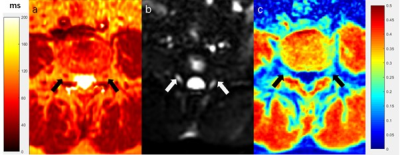 |
97 | Assessment of Lumbar Nerve Root Changes in Disc Herniation Using Ultrashort Echo Time Magnetization Transfer (UTE-MT) Imaging Video Not Available
Jin Liu1, Jia-Xin Feng1, Jian-Wei Liao1, Wei Li1, Xiao-Jun Chen1, Lin Yao1, Pan-Hui Huang1, Long Qian2, Ya-Jun Ma3, and Shao-Lin Li1
1Department of Radiology, The Fifth Affiliated Hospital of Sun Yat-Sen University, Zhuhai, China, 2MR Research, GE Healthcare, Beijing, China, 3Department of Radiology, University of California San Diego, San Diego, CA, United States
Ultrashort echo time magnetization transfer (UTE-MT) imaging can quantify macromolecular content changes in tissues. This study aims to investigate the feasibility of UTE-MT ratio (UTE-MTR) in assessment of lumbar nerve root changes due to disc herniation. T2 mapping and diffusion tensor imaging (DTI) were also employed for comparison. Our results demonstrated that the UTE-MTR is able to detect nerve root changes due to disc herniation and it has a better performance than T2 mapping and DTI imaging in differentiation of disc herniation and non-disc herniation.
|
||
2888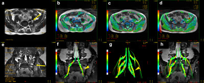 |
98 | Quantitative Evaluation Of Potential L5/S1 Spinal Nerve Degeneration In LSTV With Bertolotti's Syndrome By Diffusion Tensor Imaging
Shi Yin1, Weiqiang Dou2, and Hongyuan Ding1
1The First Affiliated Hospital of Nanjing Medical University, Nanjing, China, 2GE Healthcare, MR Research, Beijing, China
This study aims to investigate if quantitative MR diffusion tensor imaging (DTI) technology can be applied to quantitatively evaluate the potential L5/S1 spinal nerve degeneration in patients with lumbosacral transitional vertebrae. By quantitative evaluating 52 patients, we found the L5/S1 spinal nerves' DTI FA values showed lower in type Ⅱ-Ⅳ LSTV cases while the nerves run through the bony articulation between the vertebra and sacrum. Therefore , quantitative MR DTI has a good performance for quantitative diagnosis of LSTV with Bertolotti's syndrome.
|
||
2889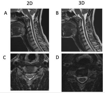 |
99 | Evaluation of Deep-Learning Reconstructed High-Resolution 3D Cervical Spine MRI to Improve Foraminal Stenosis Evaluation
Meghan Jardon1, Ek T. Tan1, J. Levi Chazen1, Meghan Sahr1, Yan Wen2, Alyssa M. Vanderbeek3, and Darryl B. Sneag1
1Department of Radiology and Imaging, Hospital for Special Surgery, New York, NY, United States, 2GE Healthcare, Chicago, IL, United States, 3Biostatistics Core, Research Administration, Hospital for Special Surgery, New York, NY, United States
Isotropic 3D MRI, with the addition of deep-learning based reconstruction algorithms (DLRecon), facilitates faster acquisition times and multiplanar reformatting. We compared interobserver agreement for cervical spine neural foraminal (NF) stenosis assessment of 3D T2-weighted fast spin echo (T2w-FSE) MR images with DLRecon to those of standard-of-care (SOC) 2D T2w-FSE images. We demonstrated that inter-observer agreement was high for both 2D and 3D sequences in the assessment of NF stenosis, but agreement was more consistent between readers at each level for the 3D acquisition. 3D DLRecon images were also more frequently free of motion, when compared to corresponding 2D sequences.
|
||
2890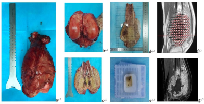 |
100 | Associations between DCE-MRI parameters and MVD, VEGF and HIF-1α in osteosarcoma based on MRI-histopathological comparison Video Permission Withheld
Huan Ma1, Yao Wang1, Kun Li1, Yingying Ding1, Jialu Li2, and Xiaoyong Zhang3
1Department of Radiology, The Third Affiliated Hospital of Kunming Medical University, Kunming, Yunnan Province, China, 2Department of Radiology, HangZhou Medical College Affiliated Lin'An People's Hospital, Hangzhou, Zhejiang Province, China, 3Clinical Science, Philips Healthcare, Chengdu, China, Chengdu, China
Tumor necrosis, angiogenesis and hypoxia are related to therapeutic effect, aggressiveness, metastasis and prognosis for osteosarcoma. In this study, DCE-MRI were matched ROI-to-ROI with histopathological tissues to explore the relationship between the perfusion parameters, including Ktrans, Kep, and Ve in each ROI and TNR, MVD, VEGF and HIF-1α in corresponding histopathological specimens, aiming to offer information about angiogenesis and hypoxia in the microenvironment of osteosarcoma timely and non-invasively. We found the perfusion parameters of DCE-MRI have a moderate sensitivity and specificity to evaluate MVD, VEGF and HIF-1α in the histopathological tissues of osteosarcoma, thereby helping microvessel status and hypoxia evaluating.
|
||
2891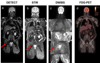 |
101 | Noninvasive Detection and Assessment of Treatment Response in Multiple Myeloma using Whole-body DETECT: Preliminary Findings
Sheng-Qing Lin1, Sebastian Fonseca1, Alberto Diaz De Leon1, Orhan Oz1,2, Durga Udayakumar1,3, Gurbakhash Kaur4, Larry D. Anderson, Jr.4, Ankit Kansagra4, and Ananth J. Madhuranthakam1,3
1Radiology, UT Southwestern Medical Center, Dallas, TX, United States, 2Nuclear Medicine, UT Southwestern Medical Center, Dallas, TX, United States, 3Advanced Imaging Research Center, UT Southwestern Medical Center, Dallas, TX, United States, 4Internal Medicine, UT Southwestern Medical Center, Dallas, TX, United States
We present the preliminary findings on the ability of our novel whole-body MRI using Dual-Echo T2-weighted acquisition for Enhanced Conspicuity of Tumors (WBMRI-DETECT) for detection and longitudinal assessment of therapy response in multiple myeloma (MM). Data from 4 MM patients undergoing induction therapy at 4 timepoints in an ongoing prospective research study are presented. Our results show that WBMRI-DETECT provides higher SNR, improved lesion conspicuity with shorter scan times compared to WBMRI-STIR and WBMRI-DWIBS, and minimal geometric distortions compared to WBMRI-DWIBS. In addition, quantitative FF measurements from WBMRI can serve as a prognostic imaging biomarker for the treatment response assessment.
|
||
2892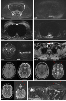 |
102 | Image quality in MY-RADS whole-body MRI protocols applied in a prospective multi-centre multiple myeloma study
Sam Keaveney1, Alina Dragan1, Mihaela Rata1, Matthew Blackledge1, Erica Scurr1, Jessica Winfield1, Dow-Mu Koh1, Nuria Porta1, Antonio Candito1, Alexander King2, Winston Rennie3, Suchi Gaba4, Priya Suresh5, Paul Malcolm6, Amy Davis7,8, Anjumara Nilak9, Aarti Shah10, Sanjay Gandhi11, Mauro Albrizio12, Arnold Drury13,
Sadie Roberts14, Matthew Jenner2, Sarah Brown14, Martin Kaiser1, and Christina Messiou1
1Royal Marsden Hospital & Institute of Cancer Research, Sutton, United Kingdom, 2Southampton General Hospital, Southampton, United Kingdom, 3Leicester Royal Infirmary, Leicester, United Kingdom, 4Royal Stoke University Hospital, Stoke-on-Trent, United Kingdom, 5Derriford Hospital, Plymouth, United Kingdom, 6Norfolk & Norwich University Hospital, Norwich, United Kingdom, 7Epsom & St. Helier University Hospital, Epsom, United Kingdom, 8Spire St. Anthony's Hospital, Sutton, United Kingdom, 9Worcestershire Royal Hospital, Worcester, United Kingdom, 10Basingstoke & North Hampshire Hospital, Basingstoke, United Kingdom, 11North Bristol NHS Trust, Bristol, United Kingdom, 12Nottingham University Hospitals, Nottingham, United Kingdom, 13Royal Bournemouth & Christchurch Hospitals, Bournemouth, United Kingdom, 14University of Leeds, Leeds, United Kingdom An assessment of image quality is presented from a multicentre WB-MRI study. A radiologist assessed image quality and the presence/severity of several common artefacts/image quality issues and metrics were also defined to measure these quantitatively. Image quality was consistently good or excellent, with only one DWI examination deemed non-diagnostic. In the case of most artefacts, the quantitative measurements were found to correlate with radiological assessment and a statistically significant ordinal regression model was found to predict DW image quality score using the quantitative measurements. These measurements could form part of an automated quality control pipeline for multi-centre WB-MRI studies. |
||
2893 |
103 | MR-guided high intensity focused ultrasound of extra-abdominal desmoid tumors: a retrospective study of 61 patients
Daniel Duex1, Vipul Sheth1, Ryan Brunsing1, Kristen Ganjoo2, Raffi Avedian3, Rachelle Bitton1, and Pejman Ghanouni1
1Radiology, Stanford University, Stanford, CA, United States, 2Medicine - Med/Oncology, Stanford University, Stanford, CA, United States, 3Orthopaedic Surgery, Stanford University, Stanford, CA, United States
We reviewed MR-guided high intensity focused ultrasound treatments (MRgFUS) of extra-abdominal desmoid tumors (DTs) in our institution from 02/2013 until 12/2020. The primary endpoints for assessing efficacy are post-treatment change in tumor volume, response based on RECIST and mRECIST; secondary endpoints are change in pain and quality of life. Complications are also reported. Our study demonstrates MRgFUS is an effective treatment option for first line and salvage therapy of desmoid tumors.
|
||
 Back to Meeting Home
 Back to the Program-at-a-Glance
The International Society for Magnetic Resonance in Medicine is accredited by the Accreditation Council for Continuing Medical Education to provide continuing medical education for physicians.
 Back to Meeting Home
 Back to the Program-at-a-Glance
 View the Presentation