Digital Poster
Quantitative & Technical Innovations for MSK
Joint Annual Meeting ISMRM-ESMRMB & ISMRT 31st Annual Meeting • 07-12 May 2022 • London, UK

Digital Poster
Quantitative & Technical Innovations for MSK
| Computer # | ||||
|---|---|---|---|---|
1686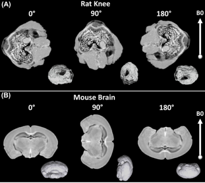 |
55 | Magic Angle effect in Diffusion Tensor Imaging
Nian Wang1,2, Qiuting Wen1, Surendra Maharjan1, and Charles Spritzer3
1Radiology and Imaging Sciences, Indiana University, Indianapolis, IN, United States, 2Stark Neurosciences Research Institute, Indiana University, Indianapolis, IN, United States, 3Radiology Department, Duke University, Durham, NC, United States
Magic Angle Effect (MAE) has been demonstrated to play an important role in both T2 and T1rho relaxation times in articular cartilage, its impact on the DTI measurements is still unknown. In this study we imaged rat knee, rat brain, and human brain at different orientation to explore the magic angle effect.
|
||
1687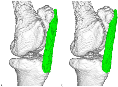 |
56 | Minimum number of scans for magic angle directional imaging based on a priori anatomical information
Harry Lanz1, Karyn Elizabeth Chappell2, and Mihailo Ristic3
1Mechanical Engineering, Imperial College London, London, United Kingdom, 2Surgery and Cancer, Imperial College London, London, United Kingdom, 3Mechanical Engineering Department, Imperial College London, London, United Kingdom
By exploiting the field-dependent anisotropy of collagen, valuable clinical information can be obtained, such as fibre tractography. We investigated the use of a priori anatomical knowledge such as scout scan, using both simulations and experimentally acquired images. We conclude that only 3 scanning directions may be sufficient for robust analysis, instead of 9 or more in the case when no such information is used. The method is also compatible with low SNR values associated with low-field MRI.
|
||
1688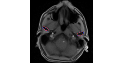 |
57 | Real-time MRI in axial plane allows quantitative evaluation of mandibular condyles motion symmetricity
Karyna Isaieva1, Justine Leclère1,2, Xavier Dubernard2, Jacques Felblinger1,3, and Pierre-André Vuissoz1
1IADI, Université de Lorraine, INSERM, Nancy, France, 2Oral Medicine Department, University Hospital of Reims, Reims, France, 3CIC-IT, CHRU de Nancy, INSERM, Nancy, France
Current diagnosis of temporomandibular disorders includes clinical examination and MRI; however, static MR images in only two key positions are not sufficient for detection of some cases of temporomandibular disk displacement. We acquired jaw opening and closure movements for 5 healthy volunteers with a real-time MRI in axial plane. The condyles were segmented with a convolutional neural network approach and motion curves of their mass centers were calculated. It was shown that the proposed protocol gives straighforward evaluation of condylar motion assymetry.
|
||
 |
1689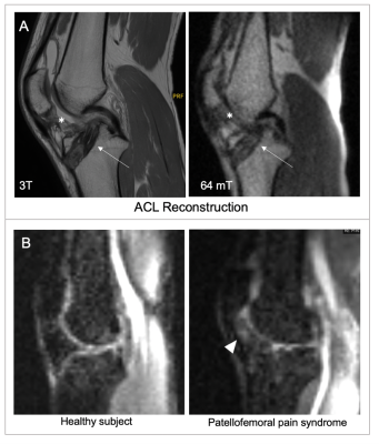 |
58 | Portable, Low-Field MRI for Evaluation of the Knee Joint
Jennifer Morgan Watchmaker1, Ding Xia2, Etan Dayan1, Idoia Corcuera-Solano1, Justin E Ngeow1, Samantha T By3, Gang Chen3, Elena Kaye3, John Pitts3, Rafael O'Halloran3, Zahi A Fayad1,2, Mingqian Huang1, and Li Feng2
1Department of Diagnostic, Molecular and Interventional Radiology, Icahn School of Medicine at Mount Sinai, New York, NY, United States, 2BioMedical Engineering and Imaging Institute, Icahn School of Medicine at Mount Sinai, New York, NY, United States, 3Hyperfine, Guilford, CT, United States
In this work, we present results of knee imaging using a portable, point-of-care MRI system in healthy subjects and subjects with knee pathology. To the best of our knowledge, this is the first reported results for knee imaging using the portable Swoop MRI system, a portable 0.064 T MRI scanner. The low-field images show good performance in visualizing the knee joint, and there was not a significant difference in the readers’ ability to evaluate the anatomic structures.
|
|
1690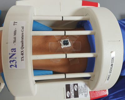 |
59 | 23Na in vivo MRI with 1mm isotropic resolution and real-time prospective motion correction implemented in Pulseq
Maxim Zaitsev1, Olgica Zaric2, and Sigfried Trattnig2
1High Field MR Center, Center for Medical Physics and Biomedical Engineering, Medical University of Vienna, Vienna, Austria, 2High Field MR Center, Department of Biomedical Imaging and Image- Guided Therapy, Medical University of Vienna, Vienna, Austria
Sodium imaging has a great potential for basic research and clinical MRI, but its applications are limited due to poor SNR achievable in typical in vivo scanning times. Image acquisition times can be substantially extended if involuntarily subject motion is compensated for, e.g. by using an external optical motion tracking system. In this proof-of-concept study we demonstrate 1mm isotropic 23Na MRI with the achieved image quality sufficient for the anatomical orientation based on the native sodium images. The used ultra-short echo time 3D imaging sequence was implemented in Pulseq, an open-source platform-independent development environment.
|
||
1691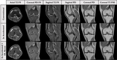 |
60 | Comparison of New MR Approaches for Accelerated Knee Imaging
Ananya Goyal1, Christopher Beaulieu1, Maggie Fung2, Trevor Kolupar3, Akshay Chaudhari1, Kathryn Stevens1, and Feliks Kogan1
1Radiology, Stanford University, Stanford, CA, United States, 2GE Healthcare, New York, NY, United States, 3GE Healthcare, Milwaukee, WI, United States
Conventional knee MRI protocols use numerous 2D-FSE sequences with multiple contrasts, which have long scan times and don’t fully use modern parallel imaging and improved SNR technologies.In this abstract, we outline four approaches that reduce protocol times to <6 minutes and potentially add diagnostic value through quantitative data or oblique reformats.These include an accelerated conventional 2D protocol, SNR-efficient 3D qDESS and CUBE approaches, and a thin-Slice protocol with high slice resolution.We propose a study that will evaluate the performance of each of these knee MRI protocols, and determine diagnostic utility and confidence with respect to fluid sensitivity and tissue pathologies.
|
||
1692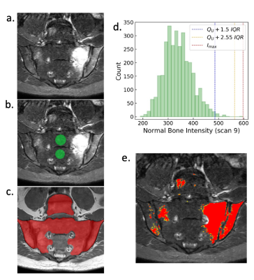 |
61 | Volume of hyperintense inflammation (VHI): a deep learning-enabled quantitative imaging biomarker of inflammation load
Timothy JP Bray1,2, Carolyna JP Hepburn1, Alexis Jones3, Alan Bainbridge4, Hui Zhang5, and Margaret A Hall-Craggs1,2
1Centre for Medical Imaging, University College London, London, United Kingdom, 2Department of Imaging, University College London Hospital, London, United Kingdom, 3Rheumatology, University College London Hospital, London, United Kingdom, 4Medical Physics, University College London Hospital, London, United Kingdom, 5Centre for Medical Image Computing, University College London, London, United Kingdom
Short inversion time inversion recovery (STIR) MRI is widely used in clinical practice to identify and quantify inflammation in patients with axial spondyloarthritis. However, assessment of STIR images is limited by the qualitative nature of image interpretation, which depends on observer expertise, and can be biased by the clinical setting. To address this, we propose the volume of hyperintense inflammation (VHI) as a quantitative imaging biomarker of inflammation load, underpinned by a recently-described segmentation method incorporating deep learning and intensity-based segmentation.
|
||
1693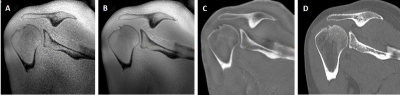 |
62 | Comparison of CT versus Zero Echo Time MRI techniques in the management of patients with shoulder instability
Laura Carretero1,2, Pablo García-Polo1, Alejandro Congo3, Michael Carl4, Graeme C McKinnon5, Maggie Fung6, and Mario Padrón3
1GE Healthcare, Madrid, Spain, 2Rey Juan Carlos University, Madrid, Spain, 3Clínica Cemtro, Madrid, Spain, 4GE Healthcare, San Diego, CA, United States, 5GE Healthcare, Waukesha, WI, United States, 6GE Healthcare, New York, NY, United States In this study we evaluate the viability of using ZTE, a novel MRI sequence for bone imaging, with deep-learning (DL) reconstruction to assess glenohumeral shoulder instability, aiming to improve signal-to-noise ratio (SNR) and get comparable images to CT, the gold standard technique for surgical planning. Bone loss measurements were performed on both techniques achieving almost perfect inter-modality agreement on 20 patients. This approach could prevent the patient from receiving ionizing radiation concomitant to CT examination and could be combined in a single routine shoulder examination with other MR sequences for a complete study and optimized patient workflow. |
||
1694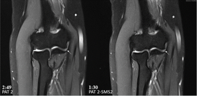 |
63 | 6-Minute Turbo Spin Echo MRI of the Elbow Using Combined Simultaneous Multi-Slice and Parallel Imaging Acceleration
Erol Akkoc1, Jan Fritz1, and Hoi Cheung Zhang1
1NYU Grossman School of Medicine, New York, NY, United States Elbow MRI contributes essential information in acute and chronic elbow injuries. Elbow MRI is ideally obtained in the superman position (prone position with elbow extended, arm elevated above the head, and hand pronated); however, this position can be challenging for patients due to injury-related pain and positioning-related discomfort. To shorten the exam time and reduce patient discomfort, we developed a 4-fold accelerated 5-sequence 6-minute clinical MRI protocol using combined simultaneous multi-slice and parallel imaging acceleration of turbo spin echo sequences. In this study, we compared the image quality of our conventional and novel accelerated elbow MRI protocols. |
||
1695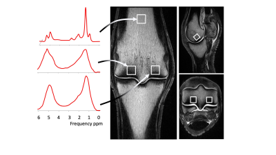 |
64 | Proton magnetic resonance spectroscopy of the distal metacarpus/tarsus in Thoroughbred racehorses with and without catastrophic fractures.
Charlotte L Hewitt-Dedman1, Sarah E Taylor1, Tobias Schwarz1, Carola R Daniel1, Maria Chiara Pressanto1, and Lucy E Kershaw2
1Royal Dick School of Veterinary Studies and Roslin Institute, Royal Dick School of Veterinary Studies and Roslin Institute, University of Edinburgh, Edinburgh, United Kingdom, 2BHF Centre for Cardiovascular Science and Edinburgh Imaging, BHF Centre for Cardiovascular Science and Edinburgh Imaging, University of Edinburgh, Edinburgh, United Kingdom
Fractures of the third metacarpal/tarsal bone are common in racehorses. Racehorse cadaver limbs underwent single voxel magnetic resonance spectroscopy (MRS) and computed tomography. The percentage fat content (FC) and the mean bone mineral density (BMD) was calculated at 3 locations within the bone. A significant negative correlation was identified for mean BMD and percentage FC for all condyles and in bone marrow of the third metacarpal/tarsal distal diaphysis. The median percentage FC was lower in horses with fractures compared to controls. These findings suggest that fat and bone are capable of mutual regulation in Thoroughbred racehorses.
|
||
 Back to Meeting Home
 Back to the Program-at-a-Glance
The International Society for Magnetic Resonance in Medicine is accredited by the Accreditation Council for Continuing Medical Education to provide continuing medical education for physicians.
 Back to Meeting Home
 Back to the Program-at-a-Glance
 View the Presentation