Digital Poster
Imaging OA & Arthroplasties
Joint Annual Meeting ISMRM-ESMRMB & ISMRT 31st Annual Meeting • 07-12 May 2022 • London, UK

| Computer # | ||||
|---|---|---|---|---|
2403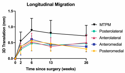 |
32 | Measuring Fixation in Cementless Total Knee Replacement Tibial Baseplates with RSA and MRI
Jordan Broberg1,2,3, Matthew Koff4, James Howard5, Brent Lanting5, Hollis Potter4, and Matthew Teeter1,2,3,5
1Medical Biophysics, Western University, London, ON, Canada, 2Robarts Research Institute, London, ON, Canada, 3Lawson Health Research Institute, London, ON, Canada, 4Radiology and Imaging, Hospital for Special Surgery, New York, NY, United States, 5Surgery, Western University, London, ON, Canada
This prospective evaluation of cementless total knee replacement tibial baseplate fixation using RSA and MRI at paired time points identified a stabilization of migration and mostly normal integration of the bone-implant interface by 3 months post-operation. A potential relationship between RSA migration magnitude and the evaluated bone-implant interface supports the use of MRI as a tool for evaluating implant fixation.
|
||
2404 |
33 | MRI of the Post-Operative Hip Following Arthroscopic Repair: What are the Imaging Characteristics of Labrum Re-Tear and Capsular Complications?
Douglas L Handley1, Joseph Y Tang1, Andrea M Spiker2, and Donna G Blankenbaker1
1Radiology, University of Wisconsin School of Medicine and Public Health, Madison, WI, United States, 2Orthopedic Surgery, University of Wisconsin School of Medicine and Public Health, Madison, WI, United States
After acetabular labrum repair an intact repair, labrum re-tear, adhesions, and capsular disruption can have variable appearance on MRI. Some signs have been described as evidence of labral re-tear and adhesions. We retrospectively evaluated 25 hip MRIs in patients who later underwent revision arthroscopy utilizing these signs. Results were compared to arthroscopic findings. Fluid signal within the labrum demonstrated a high specificity for re-tear. Signs of adhesion were often identified accurately but could be imitated by synovitis. Capsular disruption was also identified with high sensitivity.
|
||
2405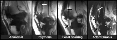 |
34 | Using MRI to Differentiate Synovial Reponses in Patients with Total Knee Arthroplasty
Matthew F. Koff1, Elexis Padgett2, David Landy2, Peter Sculco3, Thomas Sculco3, Timothy Wright2, and Hollis G. Potter1
1Department of Radiology and Imaging, Hospital for Special Surgery, New York, NY, United States, 2Department of Biomechanics, Hospital for Special Surgery, New York, NY, United States, 3Department of Orthopaedic Surgery, Hospital for Special Surgery, New York, NY, United States
Many patients are satisfied following total knee arthroplasty (TKA) surgery, but post-operative challenges occur. This study evaluated synovial reactions in patients with TKA and related pre-operative MRI findings to direct evaluation of retrieved tibial polyethylene liners from revision surgery. Patients with synovial response of 'arthrofibrosis' had less damage to the tibial insert than patients with a synovial response of 'polymeric'. MRI was able to distinguish specific synovial responses directly to clinically confirmed arthrofibrosis and aseptic loosening.
|
||
2406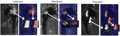 |
35 | Radiomic Variance within Soft Tissue Pathologies Near Total Hip Arthroplasty: A Cluster-based Analysis
Kevin Koch1, Robin Ausman1, John Neri2, Hollis Potter2, and Matthew Koff2
1Radiology, Medical College of Wisconsin, Milwaukee, WI, United States, 2Radiology and Imaging, Hospital for Special Surgery, New York City, NY, United States
In this exploratory study, radiomic measures were computed across radiologist-segmented image patches within 3D-MSI STIR images collected on symptomatic total hip replacements (THR). Utilizing radiologist classifications of normal, infection, polymeric wear, or metallosis debris for each image patch, principle-component based cluster analysis was deployed to visualize and estimate radiomic distinctions between different soft-tissue pathology classifications. The study results offer insights to future studies seeking to utilize quantitative MR to characterize and classify soft-tissue complications near THR.
|
||
2407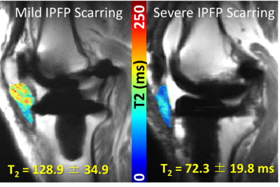 |
36 | Infrapatellar Fat Pad T2 values Associated with Fat Pad Scarring Severity in Individuals with Total Knee Arthroplasty
Sara E. Sacher1, John P. Neri1, Madeleine A. Gao1, Hollis G. Potter1, and Matthew F. Koff1
1Department of Radiology, Hospital for Special Surgery, New York, NY, United States
The infrapatellar fat pad (IPFP) has been implicated as a source of pain after total knee arthroplasty (TKA), but regions close to metallic implants are susceptible to metal artifacts in MRI. Multi-acquisition variable-resonance image combination (MAVRIC) based T2 mapping technique can mitigate these artifacts. The goal of this study was to evaluate how T2 values of the IPFP vary with the degree of IPFP scarring. T2 values were shorter and displayed a wider range for individuals with more severe scarring. MAVRIC T2 mapping may be used as a quantitative biomarker of IPFP scarring.
|
||
2408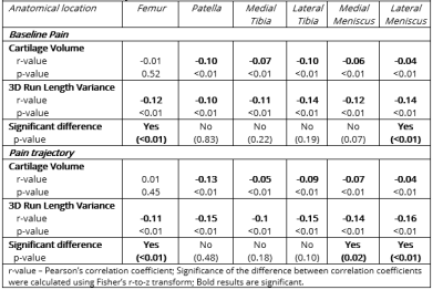 |
37 | Knee cartilage radiomics biomarkers are associated with osteoarthritis pain phenotypes
Edward J Peake1,2, Stefan Kluzek3,4, Dorothee Auer1,2, and Maja Radojčić3,5
1NIHR Nottingham Biomedical Research Centre, University of Nottingham, Nottingham, United Kingdom, 2Radiological Sciences, University of Nottingham, Nottingham, United Kingdom, 3Nuffield Department of Orthopaedics, Rheumatology and Musculoskeletal Sciences, University of Oxford, Oxford, United Kingdom, 4Department of Sports Medicine, University of Nottingham, Nottingham, United Kingdom, 5Centre for Sport, Exercise and Osteoarthritis Research Versus Arthritis, University of Oxford, Oxford, United Kingdom
Pain is a hallmark of knee osteoarthritis, a chronic condition with considerable health and socio-economic burden. Phenotyping this heterogeneous disease is a step towards improved and personalised treatment, and long-term pain phenotypes based on latent class trajectory analysis and knee MRI may facilitate it. In this study, we aimed to examine the correlation between baseline knee cartilage MRI features with established 9-year pain trajectories. Using data from the Osteoarthritis Initiative (n = 9385, 9-year follow-up), we demonstrated a weak correlation between knee cartilage, pain scores and pain trajectories and significant differences in cartilage radiomics between the knee pain trajectories.
|
||
2409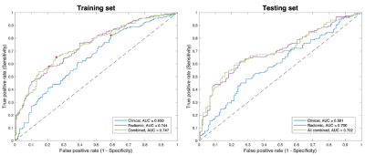 |
38 | Infrapatellar fat pad is predictive of incident knee osteoarthritis one year prior to diagnosis: data from the osteoarthritis initiative
Jia Ying1, Keyan Yu2, Tianyun Zhao1, Xiaodong Zhang2, and Chuan Huang1,3
1Biomedical Engineering, Stony Brook University, Stony Brook, NY, United States, 2Medical Imaging, The Third Affiliated Hospital of Southern Medical University – Guangdong Academy of Orthopedics, Guangzhou, Guangdong, China, 3Radiology, Stony Brook Medicine, Stony Brook, NY, United States
The infrapatellar fat pad (IPFP) plays an important role in the incidence of knee osteoarthritis (OA). However, whether the IPFP can serve as an independent biomarker for OA development is yet unknown. Radiomics is a powerful tool that can extract high-dimensional quantitative features for clinical outcomes. In this work, we proposed a prediction model for incident radiographic knee OA (iROA), using radiomic features of the IPFP, one year prior to diagnosis. The prediction performance was assessed, and our results from 604 knees demonstrated that MR-based radiomic features from the IPFP are predictive of iROA.
|
||
2410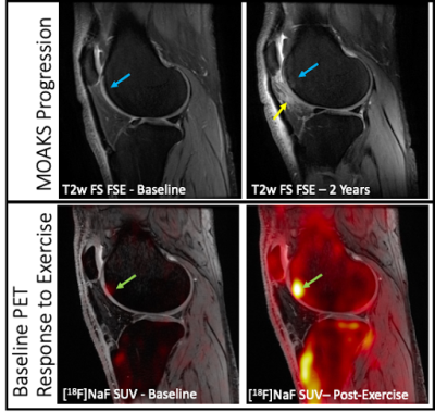 |
39 | Metabolic bone response to exercise loading is predictive of joint degeneration after 2 years in OA knees
Janelle Baker1, James MacKay2, Feliks Kogan3, and Lauren Watkins3,4
1Biomechanical Engineering, Stanford University, Stanford, CA, United States, 2Radiology, Cambridge University, Cambridge, United Kingdom, 3Radiology, Stanford University, Stanford, CA, United States, 4Steadman Philippon Research Institute, Vail, CO, United States
Hybrid [18F]sodium fluoride PET-MRI was used to evaluate the relationship between the bone metabolic response to exercise and joint changes over two years in osteoarthritic (OA) knees, including changes in cartilage T2 relaxation times as well as structural changes in bone and cartilage. Regions in OA knees that experienced structural changes over two years had increased PET metabolic bone response to exercise compared to regions that did not. Bone response to exercise may be a useful metric to predict joint changes in OA.
|
||
 Back to Meeting Home
 Back to the Program-at-a-Glance
The International Society for Magnetic Resonance in Medicine is accredited by the Accreditation Council for Continuing Medical Education to provide continuing medical education for physicians.
 Back to Meeting Home
 Back to the Program-at-a-Glance
 View the Presentation