Digital Poster
Quantitative Neuroimaging: Data Acquisition & Analysis
Joint Annual Meeting ISMRM-ESMRMB & ISMRT 31st Annual Meeting • 07-12 May 2022 • London, UK

Digital Poster
Quantitative Neuroimaging: Data Acquisition & Analysis
| Computer # | ||||
|---|---|---|---|---|
1906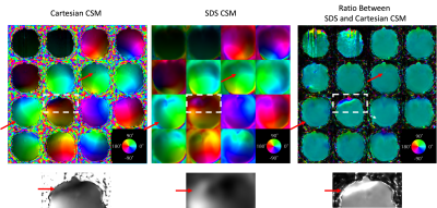 |
1 | Using a Spherically Distributed Spirals Trajectory to Collect Coil Sensitivity Maps for Iterative SENSE Reconstruction
Le Zhang1, Tzucheng Chao1, Dinghui Wang1, Guruprasad Krishnamoorthy2, and James Pipe1
1Radiology, Mayo Clinic, Rochester, MN, United States, 2MR R&D, Philips Healthcare, Rochester, MN, United States Conventionally, Cartesian pre-scans are collected over several seconds to create accurate coil sensitivity maps (CSM) for SENSE reconstruction of under-sampled MRI data, with body-coil images used for signal normalization. Since body coils have a larger sensitivity region than phased-array coils, these pre-scans typically employ a very large field-of-view (FOV) to avoid aliasing, leading to low-resolution CSM and potential small errors in subsequent reconstructions. This abstract proposes a new CSM pre-scan sequence using a spherically-distributed-spirals trajectory. The acquisition efficiency and incoherent aliasing offered by this trajectory allow for smaller FOV and higher resolution while maintaining similar scanning time as conventional pre-scans. |
||
1907 |
2 | Machine learning for detecting intracranial dural arteriovenous fistula on susceptibility weighted image using a convolutional neural network Video Permission Withheld
Bejoy Thomas1, Jithin S S1, Ajimi mol Anzar1, and Santhosh Kannath2
1Imaging Sciences and Interventional Radiology, Sree Chitra Tirunal Institute for Medical Sciences and Technology, Thiruvananthapuram, India, 2Sree Chitra Tirunal Institute for Medical Sciences and Technology, Thiruvananthapuram, India
Artificial intelligence techniques are widely used in medical imaging and diagnostics. In this retrospective study, a convolutional neural network (CNN) architecture was developed to classify intracranial dural arteriovenous fistula (DAVF) on Susceptibility Weighted Images (SWI). The dataset used was a total of 3965 SWI image slices of DAVF patients and 4380 images of controls. The proposed classifier showed significant accuracy in the diagnosis of DAVF and it could be developed as a computer assisted diagnosis tool to identify unsuspected DAVF in routine MR imaging.
|
||
1908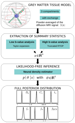 |
3 | Full posterior estimation of Gray Matter cytoarchitecture using a three-compartment model with exchange: a simulation-based study
Thomas Meunier1, Chengran Fang2, Maëliss Jallais1, and Demian Wassermann1
1INRIA Saclay, Paris, France, 2INRIA Saclay, Palaiseau, France
We extract cytoarchitectural characteristics of brain gray matter from diffusion MRI signals including soma size, neurite signal fraction and water exchange. Our model improves on state-of-the-art in that 1) we extract an invertible system leading to stable parameters estimation, 2) our simulation-based inference approach allows to obtain the full posterior distribution of the parameters given a signal. Our solution is a two-step model. First, a new forward model relates summary statistics of the dMRI signal to different tissue parameters. Then, a likelihood-free inference-based algorithm is applied to invert the model, and returns a full posterior distribution over the parameter space.
|
||
1909 |
4 | Analyzing Task-based fMRI Time Series using Machine Learning
Elaine Yuen Fong Kuan1,2, Viktor Vegh1,2, Kieran O'Brien3, Amanda Hammond3, Javier Urriola Yaksic1, and David Reutens1,2
1Centre of Advanced Imaging, The University of Queensland, Brisbane, Australia, 2ARC Training Centre for Innovation in Biomedical Imaging Technology, The University of Queensland, Brisbane, Australia, 3Siemens Healthcare Pty Ltd, Brisbane, Australia
Common approaches for analyzing task-based fMRI data rely upon the use of regressors, which in some experimental paradigms are difficult to define. A machine learning method is proposed to overcome this challenge. Three machine learning methods with established utility for time series classification were used to classify areas of activation and non-activation in a language fMRI study. Machine learning methods were able to identify the activation regions identified by analyses using the General Linear Model (GLM). Machine learning may be useful for fMRI time series analysis, particularly when regressors required for GLM-based analyses are difficult to define.
|
||
1910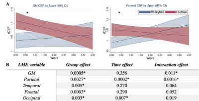 |
5 | Longitudinal decrement of cerebral blood flow in high-impact sports
Mahta Karimpoor1, Moss Zhao1, Brian Mills1, Marios Georgiadis1, Dean Tran1, Maged Goubran2, Nicole Mouchawar1, Sohrab Sami3, Max Wintermark1, Gerald Grant4, David Camarillo5, Greg Zaharchuk1, and Michael Zeineh1
1Radiology, Stanford University, Stanford, CA, United States, 2Medical Biophysics, University of Toronto, Toronto, ON, Canada, 3Stanford Center for Clinical Research, Stanford, CA, United States, 4Neurosurgery, Stanford University, Stanford, CA, United States, 5Bioengineering, Stanford University, Stanford, CA, United States
Longitudinal changes (over four years) of cerebral blood flow (CBF) using arterial spin labeling MRI were investigated in a population of high-contact sport football college athletes and were compared to low-contact cohort of volleyball athletes. A linear-mixed-effects model was applied to assess CBF (normalized to the cerebellum) by sport (football vs. volleyball), time from baseline MRI, and the interaction between sport and time. Longitudinal analysis showed a prospective decline in perfusion in football compared to volleyball. Fourteen football players experienced an in-study concussion; in contrast to the longitudinal findings, football players exhibited acutely a mild increase in occipital lobe CBF.
|
||
1911 |
6 | Imaging grey and white matter microstructure simultaneously on a clinical scanner is now possible
Simona Schiavi1, Marco Palombo2,3,4, Domenico Zacà5, Francesco Tazza1, Caterina Lapucci6, Lucio Castellan7, Mauro Costagli1, and Matilde Inglese1,8
1Department of Neuroscience, Rehabilitation, Ophthalmology, Genetics, Maternal and Child Health (DINOGMI), University of Genoa, Genoa, Italy, 2Centre for Medical Image Computing (CMIC), Department of Computer Science, University College London, London, United Kingdom, 3Cardiff University Brain Research Imaging Centre (CUBRIC), School of Psychology, Cardiff University, Cardiff, United Kingdom, 4School of Computer Science and Informatics, Cardiff University, Cardiff, United Kingdom, 5Siemens Healthcare s.r.l, Milan, Italy, Milan, Italy, 6HNSR, IRRCS Ospedale Policlinico San Martino, Genoa, Italy, 7Department of Neuroradiology, IRCCS Ospedale Policlinico San Martino, Genoa, Italy, 8IRCCS Ospedale Policlinico San Martino, Genoa, Italy Diffusion MRI is a powerful technique that, thanks to advanced signal modelling like the Soma And Neurite Density Image (SANDI) can probe microstrucutral information of both grey and white matter. However, this model requires multishell acquisitions including b-values that are at least 6 times higher than those used in clinical practice. Here we propose a 10-minute acquisition protocol that enables to acquire such images on a clinical 3T scanner. We show the reproducibility of our approach on five healthy subjects as well as potential clinical impact on two subjects affected by multiple sclerosis. |
||
1912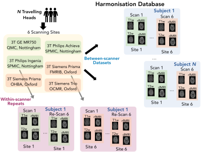 |
7 | A resource for development and comparison of harmonisation methods for multi-modal brain MRI data
Asante Ntata1, Olivier Mougin2, Matteo Bastiani1, Fidel Alfaro Almagro3, Jon Campbell3, Paul S Morgan2, Mark Jenkinson3,4, and Stamatios N Sotiropoulos1,3
1Sir Peter Mansfield Imaging Centre, School of Medicine, University of Nottingham, Nottingham, United Kingdom, 2Sir Peter Mansfield Imaging Centre, School of Physics, University of Nottingham, Nottingham, United Kingdom, 3Wellcome Centre for Integrative Neuroimaging (WIN - FMRIB), University of Oxford, Oxford, United Kingdom, 4Australian Institute for Machine Learning, University of Adelaide, Adelaide, Australia
A key challenge in robustly extracting quantitative information from MRI data is the dependence of derived features on nuisance factors, such as the scanning protocol, hardware and software, which are different between vendors and vary with site. While there exist several harmonisation approaches, what’s missing is objective ways and datasets to compare them. Here we present a novel multi-modal neuroimaging data resource for evaluating and comparing harmonisation approaches based on a “travelling heads” paradigm. We further demonstrate how such a resource can be used to a) map the need for harmonisation for different imaging-derived features, b) evaluate existing harmonisation approaches.
|
||
1913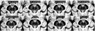 |
8 | CNN denoising of FGATIR MRI improves direct visualization of subcortical anatomy
Benjamin Ades-Aron1,2, Mohammed Elsayed1, Michael Hoch3, Gregory Lemberskiy1, Yao Wang2, Dmitry S. Novikov1, Els Fieremans1, and Timothy M. Shepherd1
1Center for Advanced Imaging Innovation and Research (CAI2R), Department of Radiology, New York University, school of medicine, New York, NY, United States, 2Electrical and computer engineering, New York University, Tandon school of engineering, Brooklyn, NY, United States, 3Radiology, University of Pennsylviania, Philadelphia, PA, United States
The basal ganglia, thalamus and brainstem are affected by movement disorders and contain key targets for functional neurosurgery. Targeting however is based on indirect coordinates originally derived from pneumoencephalograms! 3D Fast Gray Matter Acquisition T1 Inversion Recovery (FGATIR) can directly visualize potential targeted structures (e.g. dentatorubrothalamic tract), but is signal-starved in clinically-feasible acquisitions. We developed a convolutional neural network to improve FGATIR quality. Expert rater assessment suggested this CNN improved contrast resolution of individual structures and overall clinical image quality of 1-average data to the level of 4-averages. This could further enable investigations of functional neurosurgery for movement disorders.
|
||
1914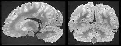 |
9 | Exploring Ultra-High Resolution Imaging of the Ex Vivo Whole Brain: Initial Results with Balanced Steady State Free Precession Sequences at 3T
Matthias Weigel1,2,3, Peter Dechent4, Riccardo Galbusera1,2, Erik Bahn5, Ludwig Kappos1,2, Wolfgang Brück5, Christine Stadelmann5, and Cristina Granziera1,2
1Translational Imaging in Neurology (ThINk) Basel, Department of Biomedical Engineering, Faculty of Medicine, University Hospital Basel and University of Basel, Basel, Switzerland, 2Neurological Clinic and Policlinic, MS Center and Research Center for Clinical Neuroimmunology and Neuroscience Basel (RC2NB), University Hospital Basel and University of Basel, Basel, Switzerland, 3Dept. of Radiology, Division of Radiological Physics, University Hospital Basel, Basel, Switzerland, 4Department of Cognitive Neurology, MR-Research in Neurology and Psychiatry, University Medical Center Göttingen, Göttingen, Germany, 5Institute of Neuropathology, University Medical Center Göttingen, Göttingen, Germany
Balanced steady state free precession (bSSFP) sequences provide the highest signal intensity per unti time, which makes them virtually predestined for ultra-high resolution MR imaging at 3T. Their sensitivity to susceptibility effects and demand for high performance, however, represent two major drawbacks. It will be shown that a carefully chosen protocol, which also includes common phase cycling techniques, will enable artifact-free 200-microns isotropic bSSFP acquisitions of the entire human fixed brain within approximately 27h.
|
||
1915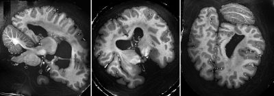 |
10 | High Resolution Postmortem Brain Imaging at 7 Tesla using Parallel Transmission and a Simple Set-Up Video Permission Withheld
Eberhard Daniel Pracht1, Markus Cremer2, Daniel Loewen1, Franck Mauconduit3, Aurelien Massire4, Sebastian Bludau2, Nicolas Boulant3, Katrin Amunts2,5, and Tony Stoecker1,6
1German Center for Neurodegenerative Diseases (DZNE), Bonn, Germany, 2Institute for Neuroscience and Medicine, INM-1, Structural and Functional Organisation of the Brain, Forschungszentrum Juelich GmbH, Juelich, Germany, 3University of Paris-Saclay, CEA, CNRS, BAOBAB, NeuroSpin, Gif sur Yvette, Paris-Saclay, France, 4Siemens Healthcare SAS, Saint-Denis, France, 5Cecile and Oskar Vogt Institute for Brain Research, Heinrich Heine University Duesseldorf, University Hospital, Duesseldorf, Germany, 6Department of Physics and Astronomy, University of Bonn, Bonn, Germany
In this work we present a simple set-up for high resolution postmortem imaging at ultra high field strength. The processed images have high, as well as homogeneous gray/white matter contrast over the whole brain. Additionally, during post-processing, the background signal of the fixation liquid is almost completely removed, allowing for an optimal image registration process after histological sectioning.
|
||
1916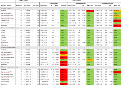 |
11 | Comparison of ComBat harmonization methods for longitudinal magnetic resonance imaging data in a travelling subject cohort
Sophie Richter1, Stefan Winzeck1,2, Marta M Correia3, Evgenios N Kornaropoulos4, Anne Manktelow1, Joanne Outtrim1, Doris Chatfield1, Jussi Posti5, Olli Tenovuo5, Guy B Williams6, David K Menon1, and Virginia F J Newcombe1
1Division of Anaesthesia, University of Cambridge, Cambridge, United Kingdom, 2BioMedIA Group, Department of Computing, Imperial College, London, United Kingdom, 3MRC Cognition and Brain Sciences Unit, University of Cambridge, Cambridge, United Kingdom, 4Diagnostic Radiology, Lund University, Lund, Sweden, 5Department of Neurosurgery and Turku Brain Injury Centre, Turku University Hospital and University of Turku, Turku, Finland, 6Wolfson Brain Imaging Centre, Department of Clinical Neurosciences, University of Cambridge, Cambridge, United Kingdom The trend in neuroimaging towards multi-site studies requires validated harmonization approaches to eradicate scanner differences which mask the biological effect of interest. Here, the harmonization algorithm ComBat and its modification for longitudinal data (LongComBat) were compared on a large travelling subject sample (n=23 for structural MRI and n=31 for diffusion tensor MRI).
In structural data scanner difference are not apparent in unharmonized data but can be created by harmonization. For DTI data, scanner differences in unharmonized data are large and, both ComBat and LongComBat successfully diminished those in most regions of interest, with LongComBat achieving slightly lower false positive rates. |
||
1917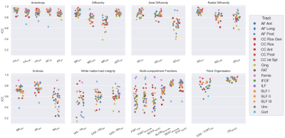 |
12 | Reliability and test-retest reproducibility of advanced diffusion metrics in healthy older adults
Rachel L. C. Barrett1, Robert Dallyn1, Marie-Stephanie Cahart1, Richard Stones2, Maarten Timmers3, Steven Williams1, and Flavio Dell'Acqua2
1Department of Neuroimaging, King's College London, London, United Kingdom, 2Department of Forensics and Neurodevelopmental Sciences, King's College London, London, United Kingdom, 3Janssen Research and Development, Janssen Pharmaceutica NV, Beerse, Belgium
Reliability and test-retest reproducibility of simple and advanced diffusion metrics were evaluated in healthy older adults. Advanced models such as Diffusion Kurtosis Imaging, White Matter Tract Integrity, Neurite Orientation Dispersion and Density Imaging and other multi-compartment methods have greater specificity but lower reliability and reproducibility than simple models such as DTI and spherical harmonic metrics. All models nevertheless had good or acceptable reproducibilty and reliability in certain cases. Particular care may be given when planning clinical research applications with advanced metrics, as greater sample sizes and/or improved data quality may be needed.
|
||
1918 |
13 | The Feasibility of Meningeal Imaging using multi-echo gradient echo sequence mFFE with optimized scan parameters
Han Dou1, Xiaoming Wang1, Yang Zheng1, and Zhiwei Shen2
1Department of Radiology, Shengjing Hospital of China Medical University, Shenyang, China, 2Philips healthcare,Beijing,China, Beijing, China At present, the detection of meningeal diseases is mainly based on MRI enhanced scan sequences, however, the poor image quality of affected the degree of meninges visualization due to the limited concentration of contrast agent staying in the meningeal after administration. In this study, the feasibility of meningeal imaging with multi-echo gradient echo MR sequence (Mffe) was explored, and optimized the main scan parameters. The optimal imaging parameters were obtained by analyzing the meninge-skull signal intensity ratio (SIR) and contrast-noise ratio (CNR) of healthy volunteers. |
||
1919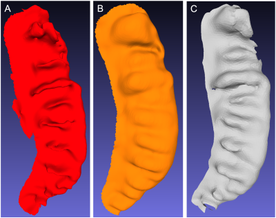 |
14 | A proposed method to objectively quantify hippocamapal dentation through midlayer AUC analysis
Lawrence Ver Hoef1,2 and Anandh Kilpattu Ramaniharan1
1Neurology, University of Alabama at Birmingham, Birmingham, AL, United States, 2Neurology, Birmingham VA Medical Center, Birmingham, AL, United States
We present a novel method to objectively quantify a morphological feature known as hippocampal dentation from ultra high resolution 3D T2w images.
|
||
1920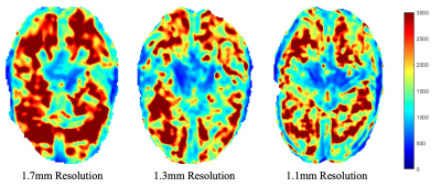 |
15 | Parameter Optimization for High-Resolution MR Elastography of the Human Brain at 7T
Emily Rose Triolo1, Oleksandr Khegai2, Jelle Veraart3, Akbar Alipour2, Trey Hedden4, Mehmet Kurt1,2, and Priti Balchandani2
1Mechanical Engineering, Stevens Institute of Technology, Hoboken, NJ, United States, 2Biomedical Engineering and Imaging Institute, Ichan School of Medicine at Mount Sinai, New York City, NY, United States, 3New York University, New York City, NY, United States, 4Ichan School of Medicine at Mount Sinai, New York City, NY, United States
MRE, a technique used to characterize the viscoelasticity of tissues, is typically performed at 3T for the human brain. MRE at 7T has the promise to deliver higher resolution, however, is subject to several artifacts and limitations. Here, we developed and tested an MRE setup and sequence at 7T on one subject at three different resolutions to investigate how both changing resolution and using MP-PCA denoising affects the estimated shear modulus (|G*|). Our pilot study has shown an increase in OSS-SNR after using MP-PCA denoising, and has demonstrated a relationship between resolution, OSS-SNR, and |G*| that requires further investigation.
|
||
1921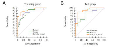 |
16 | A radiomics approach to assess high risk carotid plaques: a non-invasive imaging biomarker, retrospective study Video Not Available
Sihan celestine Chen1, Yunfei Zha2, Changsheng Liu2, Xixiang chen3, Ling ma4, and Weiyin vivian Liu5
1Radiology, Renmin Hospital of Wuhan University and Hubei General Hospital, Wuhan, China, 2Radiology, enmin Hospital of Wuhan University and Hubei General Hospital, Wuhan, China, 3Renmin Hospital of Wuhan University and Hubei General Hospital, Wuhan, China, 4He Kang Corporate Management (SH) Co.Ltd, Shanghai, China, Wuhan, China, 5Advanced Application specialist, GE Healthcare, Beijing, China, Beijing, China
This study aimed to construct a radiomics-based T2-weighted imaging (T2WI) approach from high-resolution multi-contrast magnetic resonance imaging (hrMRI) in combination with clinical high-risk factors for non-invasive assessment, to differentiate symptomatic carotid atherosclerotic plaques from asymptomatic ones. Radscore showed a better diagnostic performance. The combination model of texture features and clinical data had the best performance in assessment of lesion vulnerability. This study demonstrated that hrMRI radiomics features provided incremental value for assessment of the carotid atherosclerotic vulnerability.
|
||
1922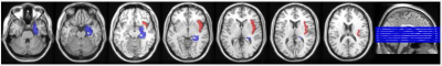 |
17 | Frailty assessment using synthetic MRI
Di Wang1, Chunmei Li2, Yuhui Chen2, Pu-Yeh Wu3, and Min Chen1
1Beijing Hospital, National Center of Gerontology; Institute of Geriatric Me, Beijing, China, 2Beijing Hospital, Beijing, China, 3GE Healthcare, Beijing, China, Beijing, China
Synthetic MRI is a novel method which can provides quantitative relaxation mapping and synthetic contrast-weighted images at the same time. In this study, we adopted this technique to assessed the frailty. We found T2 relaxation of right insular lobe and right limbic subcortical nuclei had significant difference between frailty and prefrailty patients. T1 and T2 relaxation of right limbic subcortical nuclei and right basal ganglia significantly correlated with the short physical performance battery total scores. Overall, our findings suggested the synthetic MR can be considered an effective tool for assessing frailty.
|
||
 Back to Meeting Home
 Back to the Program-at-a-Glance
The International Society for Magnetic Resonance in Medicine is accredited by the Accreditation Council for Continuing Medical Education to provide continuing medical education for physicians.
 Back to Meeting Home
 Back to the Program-at-a-Glance
 View the Presentation