Digital Poster
Imaging of the Spinal Cord
Joint Annual Meeting ISMRM-ESMRMB & ISMRT 31st Annual Meeting • 07-12 May 2022 • London, UK

| Computer # | ||||
|---|---|---|---|---|
1451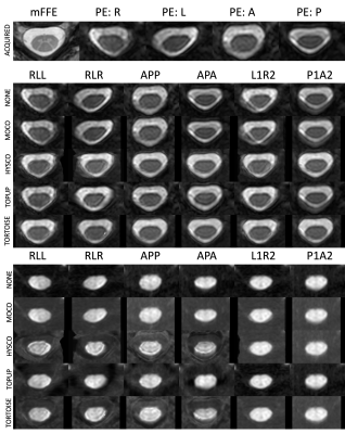 |
1 | Evaluating acquisition and preprocessing methods for diffusion MRI in the cervical spinal cord
Kurt Schilling1, Anna J.E. Combes2, Logan Prock2, Kristin P. O'Grady1, Bennett A Landman2, and Seth A Smith1
1Vanderbilt University Medical Center, Nashville, TN, United States, 2Vanderbilt University, Nashville, TN, United States
The aim of this study was to evaluate acquisition and preprocessing strategies for diffusion MRI in the cervical spinal cord. We tested 6 acquisition and 5 preprocessing strategies, and quantified evaluation criteria, and found that pipelines that include distortion correction significantly improve data quality. However, motion and lack of cardiac triggering significantly impact quality measures. Standard diffusion processing packages need to be adapted and modified for spinal cord microstructure to ensure accurate and robust diffusion quantification.
|
||
1452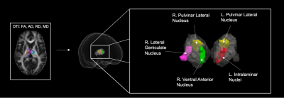 |
2 | Thalamic Involvement in the Pediatric Chronic Spinal Cord Injured Population with Respect to the Severity of Injury
KiChang Kang1, Kristen Fleming1, Anish V. Sathe1, Jennifer Muller1, Devon Middleton1, Feroze Mohamed1, Laura Krisa1, and Mahdi Alizadeh1
1Thomas Jefferson University, Philadelphia, PA, United States
Little is known about microstructural alterations in the thalamus following spinal cord injury (SCI), especially in the pediatric population. In this study, we used diffusion tensor imaging (DTI) derived metrics to characterize microstructural changes in thalamic nuclei of pediatric subjects with chronic SCIs with respect to the severity of injury based on the American Spinal Injury Association Impairment Scale (AIS). We report significant differences in mean diffusion metrics in thalamic nuclei that are suggestive of microstructural alterations between different AIS classifications.
|
||
1453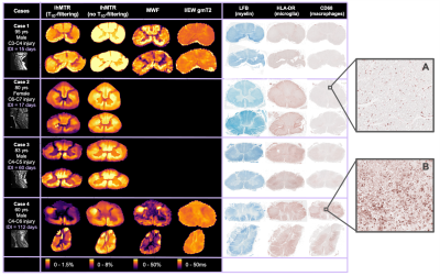 |
3 | Myelin-sensitive MRI metrics correlate with inflammation in human traumatic spinal cord injury
Sarah Rosemary Morris1,2,3, Taylor Swift-LaPointe2, Andrew Yung1,3,4, Valentin Prevost1,3,4, Shana George1, Andrew Bauman4, Piotr Kozlowski1,2,3,4, Farah Samadi1,5, Caron Fournier1,5, Lisa Parker6, Kevin Dong1, Femke Streijger1, Veronica Hirsch-Reinshagen1,5,6, G.R. Wayne Wayne Moore1,5,6, Brian K Kwon1,7, and Cornelia Laule1,2,3,5
1International Collaboration on Repair Discoveries (ICORD), Vancouver, BC, Canada, 2Physics & Astronomy, University of British Columbia, Vancouver, BC, Canada, 3Radiology, University of British Columbia, Vancouver, BC, Canada, 4Faculty of Medicine, UBC MRI Research Centre, Vancouver, BC, Canada, 5Pathology & Laboratory Medicine, University of British Columbia, Vancouver, BC, Canada, 6Vancouver General Hospital, Vancouver, BC, Canada, 7Orthopaedics, University of British Columbia, Vancouver, BC, Canada We investigated the correlation of inhomogeneous magnetisation transfer ratio (ihMTR), myelin water fraction (MWF) and intra/extra-cellular water geometric mean T2 (I/EW gmT2) with the distribution of inflammatory cells as measured by HLA-DR and CD68 histological staining for macrophages and activated microglia in human traumatic spinal cord injury. We found that ihMTR and MWF decreases were strongly correlated with the presence of HLA/CD68+ immune cells in white matter and both ihMTR and MWF were strongly correlated with LFB, a stain for myelin lipids. Our results demonstrate a strong link between ihMTR and MWF and inflammation. |
||
1454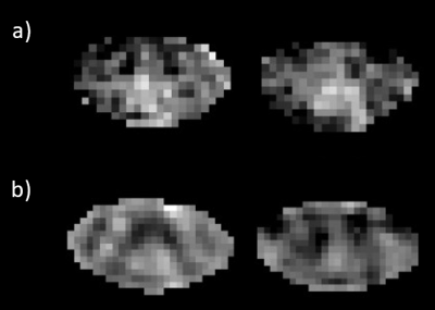 |
4 | Making myelin water imaging “normal”: Robust acquisition for cervical cord with low peripheral nerve stimulation
Sharada Balaji1, Adam Dvorak1, Neale Wiley1, Laura Barlow2, Irene M. Vavasour3,4, Alex MacKay1,3, and Shannon H. Kolind1,3,4,5
1Physics and Astronomy, University of British Columbia, Vancouver, BC, Canada, 2MRI Research Centre, University of British Columbia, Vancouver, BC, Canada, 3Radiology, University of British Columbia, Vancouver, BC, Canada, 4International Collaboration on Repair Discoveries, Vancouver, BC, Canada, 5Medicine, University of British Columbia, Vancouver, BC, Canada
The standard myelin water imaging (MWI) sequence for cervical spinal cord, 3D gradient and spin echo (GRASE), was modified to allow scanning in “normal mode” (limiting imaging parameters that may cause physiologic stress) for patients who may not tolerate peripheral nerve stimulation or tissue heating due to MR-conditional implants or medical issues. Traditional 32-echo GRASE was replaced with 48-echo GRASE in normal mode. Myelin water fraction maps from the new sequence had better repeatability than the standard sequence. 48-echo GRASE is the recommended sequence for MWI in spinal cord, particularly for subjects who cannot be scanned outside of normal mode.
|
||
1455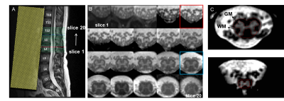 |
5 | Lumbosacral spinal cord changes after acute spinal cord injury: preliminary results of an MRI study
Silvan Büeler1, Patrick Freund2, Thomas M. Kessler1, Martina D. Liechti1, and Gergely David1,2
1Department of Neuro-Urology, Balgrist University Hospital, University of Zürich, Zürich, Switzerland, 2Spinal Cord Injury Center, Balgrist University Hospital, University of Zürich, Zürich, Switzerland
Imaging of the lumbosacral enlargement (LSE) has previously demonstrated remote degeneration below a cervical spinal cord injury leading to progressive atrophy of grey and white matter. Here, we investigate, for the first time, neurodegeneration in the conus medullaris (including the LSE) after acute spinal cord injury using an optimized multi-echo gradient-echo sequence. At 1-month after injury, preliminary results show lower grey matter volume in the conus medullaris. This is the first MRI investigation indicating remote tissue-specific volumetric changes in the conus medullaris in spinal cord injury.
|
||
1456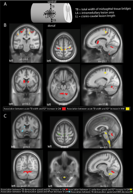 |
6 | Traumatic spinal cord injury – transition from a focal lesion to widespread neurodegeneration: Lessons from 60-month observational data
Tim Max Emmenegger1, Dario Pfyffer1, Simon Schading1, Alan Thompson2, Gabriel Ziegler3,4, John Ashburner5, Karl Friston5, Nikolaus Weiskopf6, Armin Curt1, and Patrick Freund1,5,6,7
1Spinal Cord Injury Center, Balgrist University Hospital Zurich, University of Zurich, Zurich, Switzerland, 2Queen Square Multiple Sclerosis Centre, Institute of Neurology, University College London, London, United Kingdom, 3Institute of Cognitive Neurology and Dementia Research, Otto-von-Guericke-University Magdeburg, Magdeburg, Germany, 4German Center for Neurodegenerative Diseases (DZNE), Bonn, Germany, 5Wellcome Centre for Human Neuroimaging, Queen Square Institute of Neurology, University College London, London, United Kingdom, 6Department of Neurophysics, Max Planck Institute for Human Cognitive and Brain Sciences, Leipzig, Germany, 7Department of Brain Repair and Rehabilitation,, Queen Square Institute of Neurology, University College London, London, United Kingdom
Traumatic spinal cord injury (SCI) triggers a cascade of neurodegenerative events across the neuroaxis. The trajectories of lesion characteristics and brain and spinal cord macro-and microstructural changes were analysed over five years in 23 SCI patients and 21 healthy controls. Initially, SCI patients showed higher volume and iron content in the spinal cord which decreased over time. They showed lower myelin-sensitive MTsat values in the dorsal column and cortex which also decreased over time and were associated with acute lesion characteristics. These observations illustrate the widespread and progressive neuroplastic processes after SCI, its magnitude being predicted by acute lesion characteristics.
|
||
1457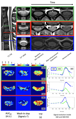 |
7 | Dynamic Contrast Enhanced MR perfusion of the Spinal Cord with radial streaking artefacts reduction at 3T: Preliminary results and applications
Guillaume Frébourg1,2,3, Kaissar Farah4, Thomas Troalen5, Lauriane Pini1,3, Aurélien Destruel1,2,3, Stéphane Fuentes4, Maxime Guye1,3, and Virginie Callot1,2,3
1Aix-Marseille University, CNRS, CRMBM, Marseille, France, 2iLab-Spine International Associated Laboratory, Marseille-Montréal, France, 3AP-HM, Hôpital de la Timone, CEMEREM, Marseille, France, 4AP-HM, Hôpital Timone, Neurosurgery Dept., Marseille, France, 5Siemens Healthcare SAS, Saint-Denis, France
3T T1-GRASP acquisition combined with an optimized dynamic radial streaking artefacts reduction have been set up to analyze Dynamic Contrast Enhanced MRI (DCE) over time and spinal cord levels. Acquisition with Gd-injection using the best temporal resolution parameters have been applied on two healthy volunteers and one patient showing perfusion dysfunction with compression. Semi-quantitative parameters (SQP) have been calculated in different regions of interest along the cervical levels and voxel-wise to provide SQP axial mapping of the spinal cord.
|
||
1458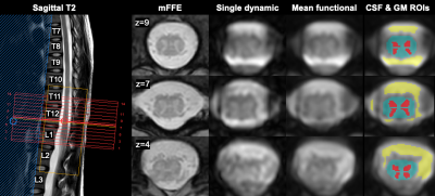 |
8 | Detection of resting-state functional connectivity networks in the human lumbar spinal cord at 3T
Anna JE Combes1,2, Anirban Sengupta1,2, Baxter P Rogers1,2, Delaney Houston1, Logan Prock1, Francesca Bagnato3, Colin D McKnight3, John C Gore1,2,4, Seth A Smith1,2,4, and Kristin P O'Grady1,2
1Vanderbilt University Institute of Imaging Science, Vanderbilt University Medical Center, Nashville, TN, United States, 2Radiology and Radiological Sciences, Vanderbilt University Medical Center, Nashville, TN, United States, 3Neurology, Vanderbilt University Medical Center, Nashville, TN, United States, 4Biomedical Engineering, Vanderbilt University, Nashville, TN, United States
3D multishot GRE resting-state BOLD-sensitive images of the lumbar spinal cord were acquired in 10 healthy participants as part of a functional MRI (fMRI) protocol. Temporal SNR in the gray matter (GM) was 18.2±4.3 after post-processing. Average correlations at rest were 0.49±0.17 between ventral horns, and 0.55±0.13 between dorsal horns, and could be detected at the single-subject level using seed-based analysis. Independent Component Analysis in 6 subjects, each scanned on two different occasions, identified network nodes within GM that were consistent across sessions. FMRI of the lumbar spine is feasible and may reflect functional integrity of the lumbar cord.
|
||
1459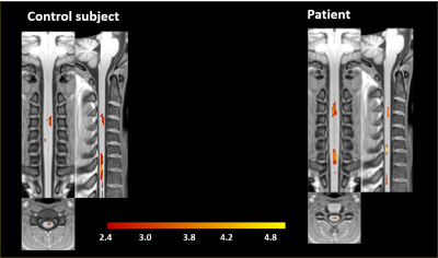 |
9 | Characterization of the spinal cord fMRI signal in the healthy subjects and in multiple sclerosis patients
Michela Fratini1, Laura Maugeri2, Maria Guidi3, Mauro Di Nuzzo4, Marta Moraschi5, Fabio Mangini6, Irene Egidi3, Daniele Mascali4, Valerio Pisani6, Ugo Nocentini6, and Federico Giove4
1CNR- Nanotec, Roma, Italy, 2CNR- Nanotec, Lecce, Italy, 3Enrico Fermi Reserch Centre, Rome, Italy, 4CREF-Centro Fermi, Roma, Italy, 5campus biomedico, Roma, Italy, 6fondazione Santa Lucia, Roma, Italy
Functional Magnetic Resonance Imaging (fMRI) has become one of the most powerful tools in neuroscience research, with promising applications in clinical practice. Particularly, fMRI based on blood oxygenation level dependent (BOLD) contrast has gained a primary role in the study of human brain and spinal cord, for the characterization of normal and pathological brain/spinal cord (SC) activity. In this framework, we have studied and report the characterization of the functional response of the SC to a multilevel motor task, designed to assess the linearity of the hemodynamic response as a function of the intensity of a graded task.
|
||
1460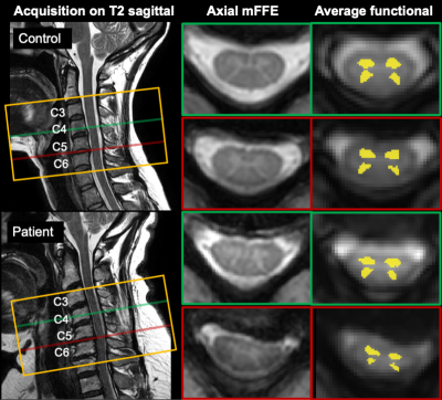 |
10 | Cord compression from stenosis reduces resting-state functional connectivity in the cervical spinal cord
Anna JE Combes1,2, Kristin P O'Grady1,2, Baxter P Rogers1,2, Wuraola Adesinasi3, Logan Prock1, Delaney Houston1, Hamid Shah3, David Edwards4, Silky Chotai3, Byron F Stephens5, Li Min Chen1,2, Seth A Smith1,2,6, and John C Gore1,2,6
1Vanderbilt University Institute of Imaging Science, Vanderbilt University Medical Center, Nashville, TN, United States, 2Radiology and Radiological Sciences, Vanderbilt University Medical Center, Nashville, TN, United States, 3Neurological Surgery, Vanderbilt University Medical Center, Nashville, TN, United States, 4Anesthesiology, Vanderbilt University Medical Center, Nashville, TN, United States, 5Orthopaedic Surgery, Vanderbilt University Medical Center, Nashville, TN, United States, 6Biomedical Engineering, Vanderbilt University, Nashville, TN, United States Spinal cord stenosis presents with highly variable clinical manifestations and prognoses. Resting-state functional MRI of the cord could provide complementary information in addition to structural measures. We sought to determine whether functional connectivity (FC) measures were altered in patients with compression of the lower cervical spinal cord, and whether those were related to morphometric measures at and above the level of pathology. Compression in the lower segments was significantly associated with lower FC of the ventral and dorsal networks in patients. This finding is a first step in exploring the FC features of the cord in compressive pathology. |
||
1461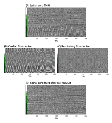 |
11 | Spinal cord fMRI heatmaps reveal a structured cardiac artifact “traveling” along the length of the cord
Kimberly J. Hemmerling1,2, Mark A. Hoggarth2, Todd Parrish3, and Molly G. Bright1,2
1Biomedical Engineering, Northwestern University, Evanston, IL, United States, 2Physical Therapy & Human Movement Sciences, Northwestern University, Chicago, IL, United States, 3Department of Radiology, Northwestern University, Chicago, IL, United States
Denoising of spinal cord fMRI data is important to address physiological noise confounds. Heatmaps used to visualize structured variance in spinal cord fMRI data reveal a unique artifact of aliased cardiac signals from pulsatile flow adjacent to the cord, which appears to travel along the longitudinal cord axis. Cardiac-related RETROICOR noise maps indicate that this artifact is successfully modeled and removed. The artifact velocity along the length of the spinal cord was calculated and is moderately correlated to the heart rate. This artifact may also provide insight into CSF flow in the spinal canal.
|
||
1462 |
12 | Comparison of Physiological Noise Models for Thermal Stimulus fMRI in the Cervical Spinal Cord at 7T
Alan C Seifert1,2,3 and S Johanna Vannesjo4
1Biomedical Engineering and Imaging Institute, Icahn School of Medicine at Mount Sinai, New York, NY, United States, 2Department of Radiology, Icahn School of Medicine at Mount Sinai, New York, NY, United States, 3Graduate School of Biomedical Sciences, Icahn School of Medicine at Mount Sinai, New York, NY, United States, 4Department of Physics, Norwegian University of Science and Technology (NTNU), Trondheim, Norway
A physiological noise model (PNM) is crucial for maximizing sensitivity and specificity to activation in 7T spinal cord fMRI. The PNM must balance exhaustive modeling of non-BOLD signal, preservation of degrees of freedom, and avoidance of nuisance regressors that may be correlated with the task. We compared four candidate PNMs. A 37-term PNM and a 35-term PNM excluding heart rate and respiratory volume per unit time, which may be correlated with the task, performed approximately equally well. A 32-term PNM containing only regressors derived from physiological recordings, and a 3-term PNM containing only image-derived regressors, were also evaluated.
|
||
1463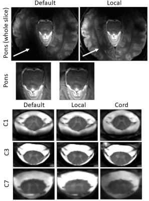 |
13 | B1 shimming for cervical spine 7T parallel transmission MRI: preliminary in vivo imaging and preparation of virtual observation points
Aurelien Destruel1,2,3, Pierre Jomin4, Tobias Wichmann5, Maxime Guye1,2, Redha Abdeddaim 4, and Virginie Callot1,2,3
1Aix-Marseille Université, CNRS, CRMBM, Marseille, France, 2AP-HM, Hôpital de la Timone, Pôle d'imagerie médicale, CEMEREM, Marseille, France, 3iLab-Spine - Laboratoire international associé - Imagerie et Biomécanique du rachis, Canada/, France, 4Aix Marseille Univ, CNRS, Centrale Marseille, Institut Fresnel, Marseille, France, 5RAPID Biomedical GmbH, Rimpar, Germany
Parallel transmission for 7T MRI of the spinal cord is a promising area of research, as high specific absorption rate and coil inefficiency in some levels may limit certain applications. In the absence of virtual observation points (VOP) provided by the coil manufacturer, preliminary testing and first in vivo applications on a healthy volunteer were done in a conservative SAR-restricted, showing potential in regions that suffered from signal drops with the default shim parameters. The workflow to ensure radiofrequency safety with virtual observation points, based on simulations of the specific absorption rate and their validation, is also described.
|
||
1464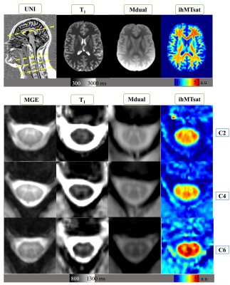 |
14 | Early aging, iron accumulation and demyelination in cervical spinal cord and brain as long-term effects of rugby practice?
Arash Forodighasemabadi1,2,3,4, Guillaume Baucher1,2,5, Lucas Soustelle1,2, Thomas Troalen6, Olivier Girard1,2, Guillaume Duhamel1,2, Jean-Philippe Ranjeva1,2, Maxime Guye1,2, Jean-Baptiste Grisoli7, and Virginie Callot1,2,4
1Aix-Marseille Univ, CNRS, CRMBM, Marseille, France, 2APHM, Hopital Universitaire Timone, CEMEREM, Marseille, France, 3Aix-Marseille Univ, Université Gustave Eiffel, LBA, Marseille, France, 4iLab-Spine International Associated Laboratory, Marseille-Montreal, Marseille, France, 5APHM, Hopital Universitaire Nord, Neurosurgery Dept, Marseille, France, 6Siemens Healthcare SAS, Saint-Denis, France, 7APHM, Hopital Universitaire Timone, Pole MPR, Marseille, France Brain alterations due to cumulative effects of impacts have been reported in rugby players. However, literature on long-term effects is scarce, and no study has been conducted to characterize potential spinal cord impairments. In this study using a multiparametric MR protocol dedicated to both brain and cord, and combining state-of-the-art T1 relaxometry and ihMT imaging, we observed diffuse T1 increase in the cervical spinal cord, together with increased T1 and decreased ihMTsat in specific brain WM tracts in retired players. These preliminary results also suggest early aging, tissue degeneration, iron accumulation, and different aging processes in retired players. |
||
1465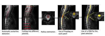 |
15 | Automatic spinal cord segmentation in pediatric MR images
Colline Blanc1,2, Shiva Shahrampour3, Feroze Mohamed3, and Benjamin De Leener1,2,4
1NeuroPoly Lab, Institute of Biomedical Engineering, Polytechnique Montréal, Montreal, QC, Canada, Montreal, QC, Canada, 2Research Center, Ste-Justine Hospital University Centre, Montreal, QC, Canada, Montreal, QC, Canada, 3Thomas Jefferson University, Philadelphia, PA, United States, Philadelphia, PA, United States, 4Department of Computer and Software Engineering, Polytechnique Montréal, Montréal, QC, Canada, Montreal, QC, Canada
Segmentation of the spinal cord is an essential process for the accurate delineation of spinal cord structures. However, it is a long process and automatic segmentation tools are not adapted to segment the pediatric spinal cord. We therefore developed a tool mixing a neural network and a deterministic method to overcome the limitations. We succeeded in obtaining a segmentation with a dice coefficient of 0.86 on a patient with a spinal cord injury (SCI).
|
||
 Back to Meeting Home
 Back to the Program-at-a-Glance
The International Society for Magnetic Resonance in Medicine is accredited by the Accreditation Council for Continuing Medical Education to provide continuing medical education for physicians.
 Back to Meeting Home
 Back to the Program-at-a-Glance
 View the Presentation