Digital Poster
Quantitative Neuroimaging: Techniques in Preclinical Models & Humans
Joint Annual Meeting ISMRM-ESMRMB & ISMRT 31st Annual Meeting • 07-12 May 2022 • London, UK

Digital Poster
Quantitative Neuroimaging: Techniques in Preclinical Models & Humans
| Computer # | ||||
|---|---|---|---|---|
2011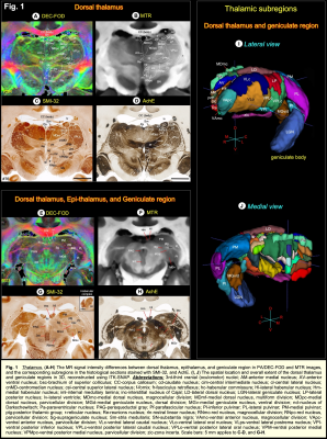 |
1 | Multimodal high-resolution mapping of subcortical regions in the macaque monkey revealed by combined MAP-MRI and histology
Kadharbatcha S Saleem1, Alexandru V Avram1, Daniel Glen2, Michal Komlosh1, and Peter J Basser3
1Neuroscience, Center for Neuroscience and Regenerative Medicine (CNRM), Henry M. Jackson Foundation (HJF) for the Advancement of Military Medicine, Bethesda, MD 20817; Section on Quantitative Imaging and Tissue Sciences (SQITS), Eunice Kennedy Shriver National Institute of Child Health and Human Development (NICHD), NIH, Bethesda, MD 20892, Bethesda, MD, United States, 2Scientific and Statistical Computing Core, Scientific and Statistical Computing Core, National Institute of Mental Health (NIMH), Bethesda, MD 20892, Bethesda, MD, United States, 3Section on Quantitative Imaging and Tissue Sciences (SQITS), Center for Neuroscience and Regenerative Medicine (CNRM), Henry M. Jackson Foundation (HJF) for the Advancement of Military Medicine, Bethesda, MD 20817; Section on Quantitative Imaging and Tissue Sciences (SQITS), Eunice Kennedy Shriver National Institute of Child Health and Human Development (NICHD), NIH, Bethesda, MD 20892, Bethesda, MD, United States
Despite its essential use as a model for neurological disorders in humans, the rhesus macaque lacks a comprehensive MRI-histology-based segmentation of subcortical regions. Here, we mapped the subcortical regions in the macaque monkey using MAP-MRI, and postmortem histology of the same brain. Our results demonstrate that, at high spatial resolution, MAP-MRI can distinguish a large number of gray and white matter structures in deep brain areas. The ability to delineate and validate these in a given subject is useful for neurosurgical planning and navigation of implantable devices to potential targets for deep brain stimulation in the macaque model of neurological or psychiatric disorders.
|
||
2012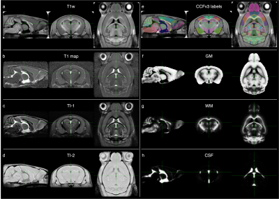 |
2 | Evaluation of a new, openly accessible in vivo MP2RAGE mouse brain template for registration and morphometric analysis
Eugene Kim1, Eilidh MacNicol1, Jemeen Sreedharan2, and Diana Cash1
1Neuroimaging, King's College London, London, United Kingdom, 2Basic & Clinical Neuroscience, King's College London, London, United Kingdom
The most popular mouse brain templates are created from ex vivo scans while in vivo templates are not widely available. We have released openly accessible in vivo multi-contrast mouse brain templates and associated tissue probability maps for analyses of in vivo mouse neuroimaging data. Deformation-based morphometry was performed on a test dataset using a study-specific template, our new “MouseIn” template, or the ex vivo DSURQE template. The results using the MouseIn template better matched those using the “gold-standard” study-specific template compared to those using the ex vivo template.
|
||
2013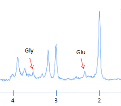 |
3 | Assessing the feasibility of measuring glycine with a TE-Averaged MRS sequence at 3 Tesla
Reggie Taylor1
1Institute of Mental Health Research, The Royal Ottawa Mental Health Centre, Ottawa, ON, Canada
A TE-Averaged sequence was created on a Siemens Biograph mMR 3 Tesla scanner for the purpose of detecting glycine at a clinical field strength in a healthy population. The sequence demonstrated that it can detect glycine and glutamate with moderate reproducibility, potentially providing a method to indirectly measure the health of the NMDA receptor
|
||
2014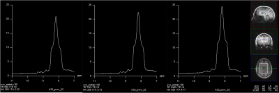 |
4 | Fast scan downfield MR spectra of human brain in vivo at 7.0 Tesla
Ravi Prakash Reddy Nanga1, Mark Elliott2, Neil Wilson2, Sophia Swago3, Walter Witschey2, and Ravinder Reddy2
1Perelman School of Medicine at The University of Pennsylvania, Philadelphia, PA, United States, 2Radiology, Center for Advance Metabolic Imaging in Precision Medicine, Perelman School of Medicine at The University of Pennsylvania, Philadelphia, PA, United States, 3Bioengineering, University of Pennsylvania, Philadelphia, PA, United States
In this study, we investigated the optimal acquisition parameters in terms of voxel size, SNR and total scan time to obtain high quality NAD+ 1H downfield (>4.7 ppm) spectra from human brain at 7 Tesla. Using a spectrally selective excitation spatially selective localization pulse sequence and without water suppression, large voxel size and shorter TR, we were able to acquire NAD+ spectra with an SNR of ~30 in about 5 min. Preliminary results of high SNR and resolution enhanced NAD+ spectra from human brain of healthy volunteers are presented.
|
||
2015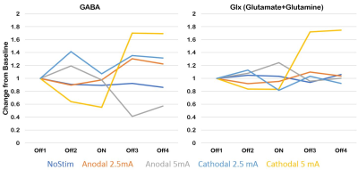 |
5 | A concurrent tDCS-MEGA PRESS MRS study to investigate polarity dependent effects on neurometabolites
Rajakumar Nagarajan1, Anant Shinde2,3, Muhammed Enes Gunduz4, and Gottfried Schlaug1,2,3
1Human Magnetic Resonance Center, Institute for Applied Life Sciences, University of Massachusetts Amherst, Amherst, MA, United States, 2Biomedical Engineering, University of Massachusetts Amherst, Amherst, MA, United States, 3University of Massachusetts Medical School - Baystate Medical Center, Springfield, MA, United States, 4University of Massachusetts Medical School, Worcester, MA, United States
Up- or downregulation of neurotransmitter/receptor concentration have been seen when transcranial direct current stimulation (tDCS) and proton Magnetic Resonance Spectroscopy (MRS) were combined in concurrent experiments; however, results have been mixed and not reliable across studies. In this study, we investigated dose and polarity effects of tDCS in an MRS voxel centered over the anterior cingulate/prefrontal region using three dose levels of tDCS with stimulation electrode placed over left a supraorbital area and return electrode over right mastoid bone. Dose and polarity dependent modulation of GABA and Glx was observed in a single subject study.
|
||
2016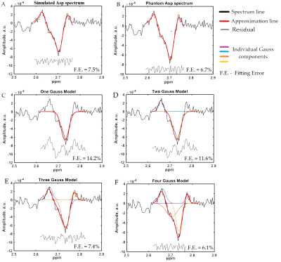 |
6 | Different approaches of aspartate approximation in MEGA-PRESS 1H MRS spectra
Petr Bulanov1, Petr Menshchikov2, Andrei Manzhurtsev3, Alexey Yakovlev3, Tolib Akhadov3, Richard Edden4, and Natalia Semenova3,5
1Lomonosov Moscow State Univesity, Moscow, Russian Federation, 2Emanuel Institute of Biochemical Physics of RAS, Moscow, Russian Federation, 3Clinical and Research Institute of Emergency Pediatric Surgery and Traumatology, Moscow, Russian Federation, 4Johns Hopkins University, Baltimore, MD, United States, 5Semenov Institute of Chemical Physics of the Russian Academy of Sciences, Moscow, Russian Federation
Changes in Aspartate (Asp) concentrations are a potential biomarker of malate-aspartate shuttle dysfunction, as well as disturbances in the synthesis of NAA. J-difference editing allows the detection of a resolved Asp signal at δAsp ≈ 2.71ppm. This resonance has a complex shape and therefore approximation of the Asp signal is a challenging task. In our study we compare the performance of six Asp-approximation models applied to data from three brain regions (anterior cingulate gyrus (ACC), dorsolateral pre-frontal area (DLPFA) and visual cortex (VC)). A model consisting of four Gauss signals shows the best performance for all brain regions studied.
|
||
2017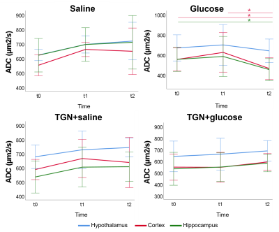 |
7 | Integrative assessment of the aquaporin-4 role to a glucose stimulus by magnetic resonance Video Permission Withheld
Balbino Yagüe1, Irene Guadilla1, Sebastián Cerdán1, Blanca Lizarbe1, and Pilar López1
1Instituto de Investigaciones Biomédicas Alberto Sols, Madrid, Spain Aquaporin-4 (AQP4) is a transmembrane water channel highly expressed in central nervous system, regulating fluid exchange by water transport between two sides of plasmatic membrane, and depending on concentration gradients of solutes, like glucose. On these grounds, we studied AQP4’s role in glucose uptake. AQP4 inhibitor TGN-020 was administrated in adult mice before vehicle and glucose stimulus. Diffusion and T2* images were acquired and apparent diffusion coefficient and T2* were measured. Ex vivo 1H high-resolution magic angle spinning spectra were acquired. Results showed that TGN avoid physiological cerebral response linked to glucose uptake, noting the AQP4 involvement in the process. |
||
2018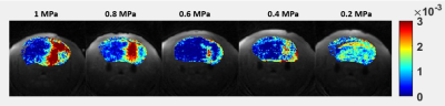 |
8 | Assessment of subtle BBB disruption using Focused Ultrasound: comparison between the contrast agent exchange rate and the water exchange rate
Jonghyun Bae1,2,3, Jin Zhang3, Mihaela Stavarache3, Ayesha Das3, Sawwal Qayyum3, Karl Kiser3, and Sungheon Gene Kim3
1Vilcek Institute of Biomedical Science, New York University School of Medicine, New York, NY, United States, 2Radiology, Center for Advanced Imaging Innovation and Research, New York, NY, United States, 3Radiology, Weill Cornell Medical College, New York, NY, United States
This study explores two different approachs of measuring subtle BBB disruption. To induce different levels of BBB disruption, we used a focused ultrasound (FUS) sonication with intravenously injected microbubbles with an animal model. Each animal underwent FUS procedure with different acoustic power levels. We compared the changes measured using DCE-MRI with Gadolinium-based contrast agent for volume transfer rate constant and Ferumoxytol-based ACE-MRI to measure transendotheliel water exchange rate. Our results suggest that both the water exchange rate and the contrast exchange rate show sensitive detection of subtle BBB disruption, which could shed light on understanding different permeability changes in BBB.
|
||
2019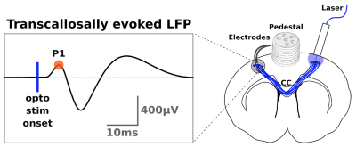 |
9 | Conduction velocity mapping a transcallosal pathway through a correlative multimodal structure-function relationship Video Permission Withheld
Christian Stald Skoven1, Mariam Andersson1, Marco Pizzolato1,2,3, Bente Pakkenberg4,5, Hartwig Roman Siebner1,5,6, and Tim Bjørn Dyrby1,3
1Danish Research Centre for Magnetic Resonance (DRCMR), Copenhagen Universital Hospital Amager and Hvidovre, Copenhagen, Hvidovre, Denmark, 2Signal Processing Lab (LTS5), École Polytechnique Fédérale de Lausanne, Lausanne, Switzerland, 3Department of Applied Mathematics and Computer Science, Technical University of Denmark (DTU), Kgs. Lyngby, Denmark, 4Research Laboratory for Stereology and Neuroscience, Copenhagen University Hospital Bispebjerg, Copenhagen, Denmark, 5Department of Clinical Medicine, Faculty of Medical and Health Sciences, University of Copenhagen, Copenhagen, Denmark, 6Department of Neurology, Copenhagen University Hospital Bispebjerg, Copenhagen, Denmark
The structure-function relationship of axons within the intact brain proves challenging to delineate. We recorded transcallosal conduction time in the rat brain with optogenetics and electrophysiology, estimated pathway length with tractography, - and axon diameters (AD) with dw-MRI and transmission electron microscopy. The modalities exhibit different sensitivity profiles. Recorded latencies correspond only to smaller axons below the mode of the distribution of ADs from histology. dw-MRI is only sensitive to larger axons, not accounted for by electrophysiology and only sparsely with histology. Future correlative studies should choose modalities with a sufficient sensitivity profile for the structural or functional metric of interest.
|
||
2020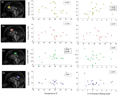 |
10 | A Neurochemical Atlas of the Cingulate Cortex in Relation to Pain.
Jessica Archibald1, Erin L. MacMillan2,3, Niklaus Zölch4, and John L.K. Kramer5
1Experimental Medicine, University of British Columbia, Vancouver, BC, Canada, 2Radiology, University of British Comulbia, Vancouver, BC, Canada, 3Philips, Vancouver, BC, Canada, 4Wissenschaftlicher Mitarbeiter Forensische Medizin und Bildgebung, Universität Zürich, Zürich, Switzerland, 5Anesthesiology, Pharmacology and Therapeutics, University of British Columbia, Vancouver, BC, Canada
1H-MRS provides a window into brain chemistry without the need to directly sample tissue. The growing prevalence of neurological conditions highlight the need for evidence based treatments. Specifically, preclinical and clinical evidence suggest a link between glutamate and pain. This study shows spatial neurochemical differences from different voxels across the cingulate cortex using sLASER at 3 T. A significant correlation between pain ratings and the anterior cingulate cortex is seen. As MRS moves towards becoming a standardized clinical technique, understanding the spatial distributions and variability will provide an important basis for the understanding of the healthy human brain’s biochemical environment.
|
||
2021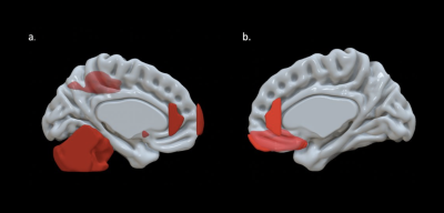 |
11 | Investigating Early Brain Development and Executive Function in Young Children
Colleen Pletcher1, Lauren Heinrich1, Marissa DiPiero1,2, Andrew Alexander1,3,4, Steven Kecskemeti 1, Elizabeth Planalp1, and Douglas Dean III1,3,5
1Waisman Center, University of Wisconsin-Madison, Madison, WI, United States, 2Neuroscience Training Program, University of Wisconsin-Madison, Madison, WI, United States, 3Department of Medical Physics, University of Wisconsin-Madison School of Medicine and Public Health, Madison, WI, United States, 4Department of Psychiatry, University of Wisconsin-Madison School of Medicine and Public Health, Madison, WI, United States, 5Department of Pediatrics, University of Wisconsin-Madison School of Medicine and Public Health, Madison, WI, United States
The emergence of executive function (EF) in children is an important developmental process that impacts later cognitive and behavioral outcomes; however, much remains unknown about the neural processes underlying the development of EF. In this study, we quantified volumetric measures from structural MRI to investigate associations between EF and brain structure in children aged 3 to 10 years old. Results indicated that there were correlations between EF and structures in subcortical brain regions and regions of the frontal and parietal lobes.
|
||
2022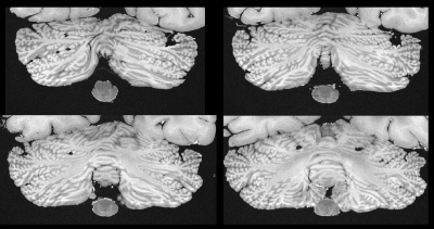 |
12 | T1 Weighted Postmortem MR Imaging of the Cerebellum at 3T: Preliminary Results between Feasibility and Desire
Matthias Weigel1,2,3, Riccardo Galbusera1,2, Peter Dechent4, Erik Bahn5, Govind Nair6, Ludwig Kappos1,2, Wolfgang Brück5, Christine Stadelmann5, and Cristina Granziera1,2
1Translational Imaging in Neurology (ThINk) Basel, Department of Biomedical Engineering, Faculty of Medicine, University Hospital Basel and University of Basel, Basel, Switzerland, 2Neurological Clinic and Policlinic, MS Center and Research Center for Clinical Neuroimmunology and Neuroscience Basel (RC2NB), University Hospital Basel and University of Basel, Basel, Switzerland, 3Dept. of Radiology, Division of Radiological Physics, University Hospital Basel, Basel, Switzerland, 4Department of Cognitive Neurology, MR-Research in Neurology and Psychiatry, University Medical Center Göttingen, Göttingen, Germany, 5Institute of Neuropathology, University Medical Center Göttingen, Göttingen, Germany, 6Translational Neuroradiology Section, Division of Neuroimmunology and Neurovirology, National Institute of Neurological Disorders and Stroke, National Institutes of Health, Bethesda, MD, United States
MRI of the fixed human brain is highly interesting, since it basically allows very long scan times for unprecedented MRI resolutions on clinical scanners. Recent work utilized T2* weighted RF spoiled gradient echo sequences to achieve isotropic resolutions up to 160-microns at 3T. This work establishes a T1 weighted MR imaging protocol based on RF spoiled gradient echo sequences that currently enables isotropic resolutions of 240-microns at 3T and depicts the complex fine structure of the fixed cerebellum very well.
|
||
2023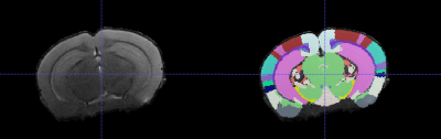 |
13 | Phenotyping a Mouse Model of TRAPPC9 Associated Intellectual Disability with High Resolution MRI and Diffusion Tensor Imaging
Mark David Platt1, Antonius Plagge2, and Harish Poptani1
1Centre for Preclinical Imaging, University of Liverpool, Liverpool, United Kingdom, 2Cellular and Molecular Physiology, University of Liverpool, Liverpool, United Kingdom
Microcephaly and intellectual disability is associated with mutation of the TRAPPC9 gene. A knockout mouse model of TRAPPC9-associated intellectual disability and microcephaly was characterised using high resolution T1 and T2 weighted Magnetic Resonance Imaging (MRI) and Diffusion Tensor Imaging (DTI) alongside behavioural assays. Behavioural differences suggested learning impairment in the model, MRI elucidated the developmental timeline of the disorder and highlighted several brain regions with reduced volume. DTI revealed reduced structural integrity in the corpus callosum.
|
||
 Back to Meeting Home
 Back to the Program-at-a-Glance
The International Society for Magnetic Resonance in Medicine is accredited by the Accreditation Council for Continuing Medical Education to provide continuing medical education for physicians.
 Back to Meeting Home
 Back to the Program-at-a-Glance
 View the Presentation