Digital Poster
Tractography Studies in the Brain
Joint Annual Meeting ISMRM-ESMRMB & ISMRT 31st Annual Meeting • 07-12 May 2022 • London, UK

Digital Poster
Tractography Studies in the Brain
| Computer # | ||||
|---|---|---|---|---|
2099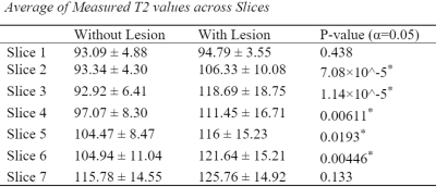 |
1 | Increases in Quantitative T2 Values in Spinal Cord of Chiari Patients with Syrinx
Maia J Baumbach1, Jason W. Allen2, Daniel L. Barrow2, David A. Reiter2, and John N. Oshinski2
1Georgia Institute of Technology, Atlanta, GA, United States, 2Emory University, Atlanta, GA, United States Here we use quantitative T2 mapping of the spinal cord in Chiari malformation patients to evaluate the ability to discriminate between spinal cord tissue in Chiari patients with syrinx and spinal cord tissue without. Spinal cord tissue adjacent to syrinx lesions show significantly elevated T2 values suggesting potential utility of T2 mapping for clinical management of Chiari patients. |
||
2100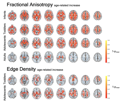 |
2 | Age-associated White Matter Microstructure and Connectome Abnormalities in Autism Spectrum Disorder
Clara F Weber1, Stefan P Haider1, Pratik Mukherjee2, Evelyn MR Lake1, Dustin Scheinost1, Nigel S Bamford3, Laura Ment3, Todd Constable1, and Sam Payabvash1
1Radiology and Biomedical Imaging, Yale School of Medicine, New Haven, CT, United States, 2University of California, San Francisco, San Francisco, CA, United States, 3Pediatrics, Neurology, Cellular and Molecular Physiology, Yale School of Medicine, New Haven, CT, United States
Analyzing four study cohorts spanning from infancy to adulthood, we compared DTI-derived diffusion metrics as well as connectome Edge Density between subjects with Autism Spectrum Disorders (ASD) and neurotypical controls. Additionally, we explored the performance of several machine learning algorithms applied to tract-based values for prediction of ASD. We found age- and ASD-related alterations in white matter microstructure and connectome in both voxel-wise and tract-based analyses that show how ASD-associated abnormalities emerge and change over time. Our machine learning analysis evaluated several different approaches and identified a model that achieved 0.75 AUC in the prediction of an ASD diagnosis.
|
||
2101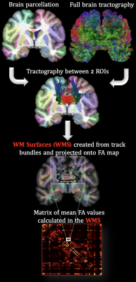 |
3 | Chronic anemia: the effects on the connectivity of white matter
Clio Gonzalez Zacarias1,2, Soyoung Choi1,2, Richard M. Leahy2,3, and John C. Wood4,5
1Neuroscience Graduate Program, University of Southern California, Los Angeles, CA, United States, 2Signal and Image Processing Institute, University of Southern California, Los Angeles, CA, United States, 3Ming Hsieh Department of Electrical and Computer Engineering, Viterbi School of Engineering, University of Southern California, Los Angeles, CA, United States, 4Department of Pediatrics and Radiology, Children's Hospital Los Angeles, Los Angeles, CA, United States, 5Biomedical Engineering, University of Southern California, Los Angeles, CA, United States
Chronic anemia is commonly observed in patients with hemoglobinopathies, mainly represented by disorders in hemoglobin structure (e.g., sickle cell anemia, SCA) and hemoglobin synthesis (e.g., thalassemia syndrome). These hemoglobin disorders have been associated with white matter alterations. In this study, changes in white matter connectivity on chronic anemic patients were characterized by quantifying the volumetric mean of fractional anisotropy along the pathway of tracks connecting two ROIs (defined by BrainSuite’s BCI-DNI atlas) and comparing it with healthy individuals. SCA patients showed interhemispheric FA derangements but not to the same extent as changes observed in the watershed areas of non-SCA patients.
|
||
2102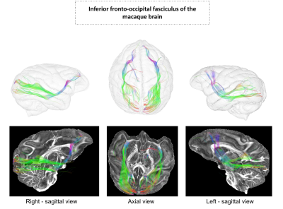 |
4 | Investigation of the inferior fronto-occipital fasciculus in the macaque fascicularis brain using 11.7 T ultra-high field diffusion data
Maëlig Chauvel1, Ivy Uszynski1, Clara Fischer1, Fabrice Poupon1, Christophe Destrieux2, Igor Maldonado2, and Cyril Poupon1
1BAOBAB, NeuroSpin, Université Paris-Saclay, CNRS, CEA, Gif-sur-Yvette, France, 2Unité Imagerie & Cerveau (iBrain), UMRS INSERM 1253, Université de Tours, Tours, France
In this study, we investigated the controversial presence of the inferior fronto-occipital fasciculus (IFOF) in the macaque species (Macaca fascicularis) using ultra-high field MRI (11.7 T) diffusion data. Thanks to a fiber clustering approach, we were able to reconstruct the IFOF in both hemispheres. We observed thin frontal connections and a more developed fasciculus on the right hemisphere compared to the left. The presence of this special fasciculus, known in Humans as being part of the ventral pathways for multi-modal language processing, is a new step forward concerning the comprehension of the macaque brain, and the primate brain in general.
|
||
2103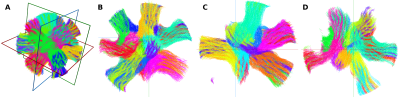 |
5 | Structural connectivity estimates are accurate: the outcome of diffusion-simulated connectivity (DiSCo) challenge
Gabriel Girard1,2,3, Jonathan Rafael-Patino2,3, Raphaël Truffet4, Dogu Baran Aydogan5,6,7, Nagesh Adluru8,9, Veena A. Nair9, Vivek Prabhakaran9, Barbara B. Bendlin10, Andrew L. Alexander8,11,12, Sara Bosticardo13,14, Ilaria Gabusi13,15, Mario Ocampo-Pineda13, Matteo Battocchio13,16, Zuzana Piskorova13,17, Pietro Bontempil13,18, Simona Schiavi19, Alessandro Daducci13, Aleksandra Stafiej20, Dominika Ciupek20,
Fabian Bogusz20, Tomasz Pieciak21, Matteo Frigo22, Sara Sedlar22, Samuel Deslauriers-Gauthier22, Ivana Kojcic22, Mauro Zucchelli22, Hiba Laghrissi22, Yang Ji22, Rachid Deriche22, Kurt G Schilling23, Bennett A Landman23,24, Alberto Cacciola25, Gianpaolo Antonio Basile25, Salvatore Bertino25, Nancy Newlin24, Praitayini Kanakaraj24, Francois Rheault24, Patryk Filipiak26, Timothy Shepherd26,
Ying-Chia Lin26, Dimitris G Placantonakis27, Fernando E Boada26, Steven H Baete26, Erick Hernández-Gutiérrez16, Alonso Ramírez-Manzanares28, Ricardo Coronado-Leija29, Pablo Stack-Sánchez28, Luis Concha30, Maxime Descoteaux16, Sina Mansour L31,32, Caio Seguin32,33, Andrew Zalesky31,32, Kenji Marshall3,34, Erick J Canales-Rodríguez3, Ye Wu35, Sahar Ahmad35, Pew-Thian Yap35, Antoine Théberge16, Florence Gagnon16,
Frédéric Massi16, Juan Luis Villarreal Haro3, Marco Pizzolato3,36, Emmanuel Caruyer4, and Jean-Philippe Thiran1,2,3
1CIBM Center for Biomedical Imaging, Lausanne, Switzerland, 2Radiology Department, Centre Hospitalier Universitaire Vaudois and University of Lausanne, Lausanne, Switzerland, 3Signal Processing Laboratory (LTS5), Ecole Polytechnique Fédérale de Lausanne (EPFL), Lausanne, Switzerland, 4Univ Rennes, Inria, CNRS, Inserm, IRISA UMR 6074, Empenn ERL U-1228, Rennes, France, 5A.I. Virtanen Institute for Molecular Sciences, University of Eastern Finland, Kuopio, Finland, 6Department of Neuroscience and Biomedical Engineering, Aalto University, Espoo, Finland, 7Department of Psychiatry, Helsinki University Hospital, Helsinki, Finland, 8Waisman Center, University of Wisconsin-Madison, Madison, WI, United States, 9Department of Radiology, University of Wisconsin-Madison, Madison, WI, United States, 10Department of Medicine, University of Wisconsin-Madison, Madison, WI, United States, 11Department of Medical Physics, University of Wisconsin-Madison, Madison, WI, United States, 12Department of Psychiatry, University of Wisconsin-Madison, Madison, WI, United States, 13Diffusion Imaging and Connectivity Estimation (DICE) Lab, Department of Computer Science, University of Verona, Verona, Italy, 14Translational Imaging in Neurology (ThINk), Department of Biomedical Engineering, University Hospital Basel and University of Basel, Basel, Switzerland, 15Department of Advanced Biomedical Sciences, University of Naples “Federico II”, Naples, Italy, 16Sherbrooke Connectivity Imaging Laboratory (SCIL), Department of Computer Science, University of Sherbrooke, Sherbrooke, QC, Canada, 17Brno Faculty of Electrical Engineering and Communication, Department of mathematics, University of Technology, Brno, Czech Republic, 18Department of neurosciences, biomedicine and movement sciences, University of Verona, Verona, Italy, 19Department of Neuroscience, Rehabilitation, Ophthalmology, Genetics, Maternal and Child Health (DINOGMI), University of Genoa, Genoa, Italy, 20AGH University of Science and Technology, Kraków, Poland, 21Laboratorio de Procesado de Imagen (LPI), Universidad de Valladolid, Valladolid, Spain, 22Athena Project Team, Centre Inria d'Université Côte d'Azur, Sophia Antipolis, France, 23Department of Radiology and Radiological Sciences, Vanderbilt University Medical Center, Nashville, TN, United States, 24Department of Electrical and Computer Engineering, Vanderbilt University, Nashville, TN, United States, 25Brain Mapping Lab, Department of Biomedical, Dental Sciences and Morphological and Functional Images, University of Messina, Messina, Italy, 26Center for Advanced Imaging Innovation and Research (CAI2R), Department of Radiology, NYU Langone Health, New York, NY, United States, 27Department of Neurosurgery, Perlmutter Cancer Center, Neuroscience Institute, Kimmel Center for Stem Cell Biology, NYU Langone Health, New York, NY, United States, 28Computer Science Department. Centro de Investigación en Matemáticas A.C., Guanajuato, Mexico, 29Center for Biomedical Imaging, Department of Radiology, NYU School of Medicine, New York, NY, United States, 30Instituto de Neurobiología, Universidad Nacional Autónoma de México, Juriquilla, Mexico, 31Department of Biomedical Engineering, The University of Melbourne, Parkville, Australia, 32Melbourne Neuropsychiatry Centre, Department of Psychiatry, The University of Melbourne and Melbourne Health, Parkville, Australia, 33The University of Sydney, School of Biomedical Engineering, Sydney, Australia, 34McGill University, Montréal, QC, Canada, 35Department of Radiology and Biomedical Research Imaging Center (BRIC), The University of North Carolina at Chapel Hill, Chapel Hill, NC, United States, 36Department of Applied Mathematics and Computer Science, Technical University of Denmark, Kgs. Lyngby, Denmark
The estimation of structural connectivity from DW-MRI is a challenging task, in part due to false-positive connections. Building on previous efforts, the MICCAI-CDMRI Diffusion-Simulated Connectivity (DiSCo) challenge was organized with the aim of evaluating connectivity methods using large-scale synthetic datasets obtained from DW-MRI Monte-Carlo simulations. The outcome of the challenge suggests that methods selected by the 14 teams participating in the challenge provide both high correlations between estimated and ground-truth connectivity weights and high accuracy in binary connectivity identification. Furthermore, the challenge provided unique data with realistic connectivity and microstructure properties to foster the development of connectivity estimation methods.
|
||
2104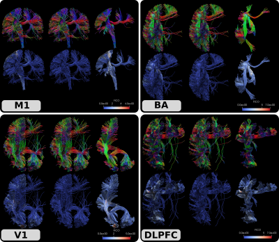 |
6 | Fiber coupling (FICO) measure using anisotropic smoothing of track orientation density images for tractogram filtering
Dogu Baran Aydogan1
1A.I. Virtanen Institute for Molecular Sciences, University of Eastern Finland, Kuopio, Finland
Tractography is a powerful tool to study structural connectivity of the brain. However, its accuracy is known to be limited and tractograms are contaminated with large numbers of false positive streamlines. In this work, we propose a novel tractogram filtering approach. Our method leverages the topographic regularity of connections with which nearby streamline tend to follow similar trajectories. With this observation, we introduce an anisotropic smoothing approach for track orientation density images. These images are back projected onto the streamlines, which provides information about fiber-to-bundle coupling (FICO). Streamlines are then filtered by thresholding their FICO value.
|
||
2105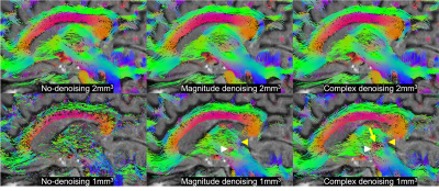 |
7 | MP-PCA denoising of complex imaging data for direct visualization of the dentatorubrothalamic tract using diffusion MRI at 3T
Benjamin Ades-Aron1, Santiago Coelho1, Gregory Lemberskiy1, Alon Mogilner2, Dmitry S. Novikov1, Timothy M. Shepherd1, and Els Fieremans1
1Center for Advanced Imaging Innovation and Research (CAI2R), Department of Radiology, New York University, school of medicine, New York, NY, United States, 2Neurosurgery, New York University, school of medicine, New York, NY, United States
Direct visualization of anisotropic structures in the brainstem, basal ganglia and thalamus could improve functional neurosurgery planning and characterize changes associated with pathology, treatment response or side effects. Postmortem MRI microscopy demonstrates the potential for high resolution diffusion MRI to detect such functional organization. However, in vivo signal-to-noise limitations have precluded accurate quantitative diffusion measurements at sufficient spatial resolution for these structures. We demonstrate that MP-PCA denoising of complex dMRI at 1-mm isotropic resolution and b-values up to 2000 s/mm2 enables direct visualization of the dentatorubrothalamic tract, the key white matter pathway for functional neurosurgical treatment of tremor.
|
||
2106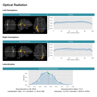 |
8 | Q-FiberMapper - A framework for tractography and tractometry of clinical data
Tommy Boshkovski1, Guillermo Gallardo1, Oscar Peña-Nogales1, Marc Ramos1, Kire Trivodaliev1, Paulo Rodrigues1, and Vesna Prchkovska1
1QMENTA Inc., Barcelona, Spain
Despite recent advances in tractography, automatically reconstructing brain bundles in clinical settings remains a challenge. Here we present Q-FiberMapper (QFM), a framework that automatically preprocesses clinical data and reconstructs 33 major brain bundles with minimal user intervention. Furthermore, it derives insightful biomarkers and summarizes the findings in a human readable report. We validate QFM using a large cohort of 600 subjects, and show that it correctly reconstructs the shape and lateralization of major white-matter bundles. By simplifying and speeding the process of clinical reconstruction, QFM could help clinicians in the diagnosis and monitoring of brain pathologies.
|
||
2107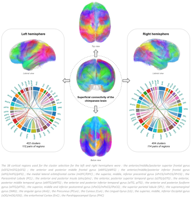 |
9 | Investigation of the singularity of the chimpanzee brain superficial white matter bundles using diffusion MRI and clustering-based approaches
Maëlig Chauvel1, Ivy Uszynski1, Alexandros Popov1, William Hopkins2, Jean-François Mangin1, and Cyril Poupon1
1BAOBAB, NeuroSpin, Université Paris-Saclay, CNRS, CEA, Gif-sur-Yvette, France, 2Keeling Center for Comparative Medicine and Research, The University of Texas MD Anderson Cancer Center, Bastrop, TX, United States
A way to better appreciate the complex human brain evolution relies on comparative investigations with homologous species. We present here the superficial connectivity organization of the Chimpanzee brain using a combination of image processing and DBSCAN clustering analyzes obtained from 39 chimpanzees DTI scans. The results revealed the presence of U-fiber, V-fiber and Curved-fiber shapes already identified in Human superficial fiber connectivity studies. This non-negligeable short fibers organisation resemblance between Chimpanzees and Humans, brings out new perspectives on hominid brain singularity knowledge.
|
||
2108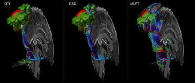 |
10 | Multi-level fiber tractography in brain tumor patients using functional mapping for seeding
Andrey Zhylka1, Josien Pluim1, Florian Kofler2,3,4, Ahmed Radwan5,6, Alberto De Luca7,8, Axel Schroeder9, Benedikt Wiestler2,4, Jan S. Kirschke2,10, Bjoern Menze3,11, Stefan Sunaert5,6,12, Alexander Leemans7, Sandro M. Krieg9,13, and Nico Sollmann2,13,14
1Biomedical Engineering, Eindhoven University of Technology, Eindhoven, Netherlands, 2Department of Diagnostic and Interventional Neuroradiology, School of Medicine, Klinikum rechts der Isar, Technical University of Munich, Munich, Germany, 3Department of Informatics, Technical University of Munich, Munich, Germany, 4TranslaTUM - Central Institute for Translational Cancer Research, Technical University of Munich, Munich, Germany, 5Department of Imaging and Pathology, Translational MRI, KU Leuven, Leuven, Belgium, 6Department of Neuroscience, Leuven Brain Institute (LBI), KU Leuven, Leuven, Belgium, 7Image Sciences Institute, University Medical Center Utrecht, Utrecht, Netherlands, 8Neurology Department, UMC Utrecht Brain Center, University Medical Center Utrecht, Utrecht, Netherlands, 9Department of Neurosurgery, School of Medicine, Klinikum rechts der Isar, Technical University of Munich, Munich, Germany, 10TUM-Neuroimaging Center, Klinikum rechts der Isar, Technical University of Munic, Munich, Germany, 11Department of Quantitative Biomedicine, University of Zurich, Zurich, Switzerland, 12Department of Radiology, UZ Leuven, Leuven, Belgium, 13TUM-Neuroimaging Center, Klinikum rechts der Isar, Technical University of Munich, Munich, Germany, 14Department of Diagnostic and Interventional Radiology, University Hospital Ulm, Ulm, Germany Navigated transcranial magnetic stimulation (nTMS) motor mapping allows locating function in the individual patient pre-operatively and, to a certain extent, decreases the false-positive rate for fiber tractography. However, the false-negative rate is still high. In this work we evaluate novel multi-level fiber tractography (MLFT) along with conventional deterministic algorithms in patients with motor-eloquent high-grade glioma, combined with nTMS mapping-based region of interest (ROI) placement. The results were compared based on the coverage of nTMS motor maps, revealing reconstruction of the corticospinal tract (CST) with significantly higher volume and better coverage of the eloquent motor cortex for MLFT. |
||
2109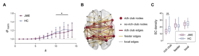 |
11 | Altered rich club organization and dynamic structure-function relationship in treatment-naïve new-onset juvenile myoclonic epilepsy Video Not Available
Guangyao Liu1, Hong Liu1, Pengfei Zhang1, Jing Zhang1, and Zhe Zhang2
1Department of Magnetic Resonance, Lanzhou University Second Hospital, Lanzhou, China, 2Department of Biomedical Engineering, Zhejiang University, Zhejiang, China
Neuroimaging studies have shown that juvenile myoclonic epilepsy (JME) is characterized by impaired brain networks. However, few studies have investigated the potential disruptions in the rich club organization. Our study described the alterations of anatomical rich club organization and abnormalities of dynamic SC-FC coupling in treatment-naïve newly diagnosed JME. The results showed that the anatomical rich club organization was disrupted in the patient group, along with decreased connectivity strength among rich club hub nodes. The aberrant dynamic SC-FC coupling of rich club organization suggests a selective influence of interconnected network core in JME at the early phase of the disease.
|
||
2110 |
12 | Cerebellum structural and functional connectivity alteration in relation to freezing of gait in Multiple system atrophy Video Not Available
Huaguang Yang1, Weiyin Vivian Liu2, Liang Li1, Zhi Wen1, and Yunfei Zha1
1Renmin Hospital of Wuhan University, Wuhan, China, 2GE Healthcare, Beijing, China
Cerebellum is responsible for posture and gait control. 67% of patients with multiple system atrophy experienced FOG. Patients with the damaged cerebellar locomotor region had gait-freezing-like symptoms, suggesting that the cerebellum may be involved in the occurrence and development of FOG symptoms. This study suggested that the cerebellum volume atrophy may be involved in FOG development in MSA patients and subsequently induce functional abnormality in the cerebellum-cerebral circuit. This study provided neuroimaging evidence for clinical understanding of cerebellum role in MSA patients with FOG injury.
|
||
 Back to Meeting Home
 Back to the Program-at-a-Glance
The International Society for Magnetic Resonance in Medicine is accredited by the Accreditation Council for Continuing Medical Education to provide continuing medical education for physicians.
 Back to Meeting Home
 Back to the Program-at-a-Glance
 View the Presentation