Digital Poster
Quantitative Neuroimaging: Clinical & Translational Studies I
Joint Annual Meeting ISMRM-ESMRMB & ISMRT 31st Annual Meeting • 07-12 May 2022 • London, UK

Digital Poster
Quantitative Neuroimaging: Clinical & Translational Studies I
| Computer # | ||||
|---|---|---|---|---|
2637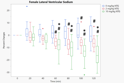 |
1 | On the basis of sex: Preclinical 23Na MRI at 21.1 T Delineates Sodium Distribution Differences in Females Following Central Sensitization
Samuel W. Holder1,2, Dayna L. Richter1,2, David C. Hike1,2, Michael G. Harrington3, and Samuel C. Grant1,2
1National High Magnetic Field Laboratory, Florida State University, Tallahassee, FL, United States, 2Chemical & Biomedical Engineering, FAMU-FSU College of Engineering, Tallahassee, FL, United States, 3Neurology, University of Southern California, Los Angeles, CA, United States
23Na FID-based 3D CSI was implemented in female rats at 21.1 T to examine sex differences in sodium response to NTG-triggered central sensitization. The female brainstem was resilient to NTG-triggered increases in sodium seen with males, despite sodium increases in the female cisterna magna and fourth ventricle. Lateral ventricles and thalamus sodium in females decreased with NTG trigger. Female resilience in brainstem and thalamus, both connected to trigeminal first order neuron, holds interesting implications for migraine.
|
||
2638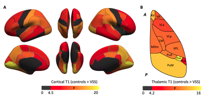 |
2 | Microstructural changes in the grey matter of patients with visual snow syndrome: an ultra-high field morphological and quantitative MRI study
Myrte Strik1, Meaghan Clough2, Emma J Solly2, Owen B White2, Scott C Kolbe2, and Joanne Fielding2
1Department of Radiology, University of Melbourne, Melbourne, Australia, 2Department of Neuroscience, Monash University, Melbourne, Australia
Visual snow syndrome (VSS) is a neurological disorder characterized by continuous visual disturbances, and accompanied by a range of non-visual symptoms, including tinnitus and migraine. Little is known about the pathological mechanisms underlying VSS. In this study we assessed brain morphometry and microstructure in VSS patients using high-resolution structural (MP2RAGE, 0.75 mm iso) and quantitative (T1 mapping) 7T MRI. In VSS patients, we observed similar morphometry, but widespread changes in the grey matter microstructure (lower T1 values), which followed a caudal-rostral pattern affecting the occipital cortices most profoundly. Migraine did not appear to independently affect these changes.
|
||
2639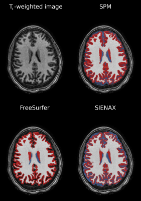 |
3 | BRAIN VOLUMES IN SICKLE CELL DISEASE ACROSS THE LIFESPAN: ALGORITHM PERFORMANCE AND CORRESPONDENCE WITH BLOOD OXYGEN CONTENT
Randall Sky Jones1, Manus Donahue2, Spencer Waddle3, Larry Taylor Davis3, Sumit Pruthi3, Chelsea Lee1, Niral Patel1, Michael DeBaun1, Adetola Kassim4, Mark Rodeghier5, and Lori Jordan1
1Pediatric Neurology, Vanderbilt University Medical Center, Nashville, TN, United States, 2Neurology, Vanderbilt University Medical Center, Nashville, TN, United States, 3Radiology, Vanderbilt University Medical Center, Nashville, TN, United States, 4Medicine; Hematology and Oncology, Vanderbilt University Medical Center, Nashville, TN, United States, 5Rodeghier Consulting, Chicago, IL, United States
Brain tissue volumes were calculated in adults and children with sickle cell disease (SCD) across the lifespan for different segmentation algorithms and clinical indicators of disease. Tissue volume reductions in persons with SCD without prior stroke were detected when using FreeSurfer, but not other software, highlighting algorithm bias. Arterial oxygen content was directly related to tissue volumes when using FreeSurfer and SIENAX. Brain tissue volume was not associated with silent cerebral infarcts (SCIs). Findings highlight the subtlety of tissue volume changes in SCD, provide age-specific reference-standards, and motivate an integrated approach of anatomical and functional assessments for informing SCD care.
|
||
2640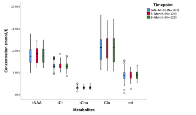 |
4 | Advancing Concussion Assessment in Pediatrics (A-CAP): Longitudinal changes in brain metabolites in pediatric concussion
Parker L La1, Ashley D Harris1, Robyn Walker1, Julie M Joyce1, Tiffany Bell1, William Craig2, Quynh Doan3, Miriam H Beauchamp4, Roger Zemek5, Pediatric Emergency Research Canada (PERC)6, Keith O Yeates7, and Ashley D Harris1
1Radiology, University of Calgary, Calgary, AB, Canada, 2Pediatrics, University of Alberta and Stollery Children's Hospital, Edmonton, AB, Canada, 3Pediatrics, University of British Columbia, Vancouver, BC, Canada, 4Psychology, University of Montreal and St Justine Hospital, Montreal, QC, Canada, 5Pediatrics and Emergency Medicine, University of Ottawa and Children's Hospital of Eastern Ontario, Ottawa, ON, Canada, 6University of Calgary, Calgary, AB, Canada, 7Psychology, University of Calgary, Calgary, AB, Canada
Disruptions in brain metabolites following concussion are commonly reported in the literature. However, studies have typically included only one timepoint, studied adults and/or had limited sample size. In the largest MRS dataset in pediatric concussion to date, we show that metabolites do not differ significantly over time points with longitudinal data collected sub-acutely (<14 days post-injury) and at 3- or 6-months follow-up. This lack of change may indicate that metabolites are relatively stable, that changes are regionally specific or occur more acutely, or there are subgroups that need to be specifically considered.
|
||
2641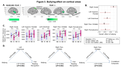 |
5 | Bullying is associated with reduced cognitive scores and altered brain morphometry over time in preadolescent children.
Miriam S Menken1, Pedro Rodriguez Rivera1, Amal Isaiah2,3, Thomas Ernst1, Christine C Cloak1, and Linda Chang1,4,5
1Diagnostic Radiology and Nuclear Medicine, University of Maryland School of Medicine, Baltimore, MD, United States, 2Otorhinolaryngology—Head and Neck Surgery, University of Maryland School of Medicine, Baltimore, MD, United States, 3Pediatrics, University of Maryland School of Medicine, Baltimore, MD, United States, 4Neurology, University of Maryland School of Medicine, Baltimore, MD, United States, 5Neurology, Johns Hopkins University School of Medicine, Baltimore, MD, United States Few studies identified brain morphometric changes associated with peer-victimization (“being bullied”), and no study examined how these changes can mediate the relationship between bullying and cognition. Using T1-weighted MRI scans and cognitive test scores from the Adolescent Brain Cognitive Development longitudinal dataset, we found that bullying is associated with reduced cognitive performance over time. Bullied children had smaller putamen volumes and right insula surface areas, thinner left precentral and banks of superior temporal sulcus cortices, but larger left entorhinal and right pars orbitalis surface areas. Importantly, these altered brain measures partially mediated the relationship between bullying and cognitive scores. |
||
2642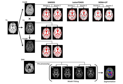 |
6 | Brain-wide Quantitative Imaging Measures in progressive Multiple Sclerosis: a Multi-modal Framework and Longitudinal Validation Study
Yukai Zou1,2, Aravinthan Varatharaj1,3, Charlotte Stuart1, Finn Lennartsson4, Angela Darekar2, Claudia AM Gandini Wheeler-Kingshott5, and Ian Galea1,3
1Clinical Neurosciences, University of Southampton, Southampton, United Kingdom, 2Medical Physics Department, University Hospital Southampton NHS Foundation Trust, Southampton, United Kingdom, 3Wessex Neurological Centre, University Hospital Southampton NHS Foundation Trust, Southampton, United Kingdom, 4Department of Clinical Sciences, Diagnostic Radiology, Lund University, Lund, Sweden, 5NMR Research Unit, Department of Neuroinflammation, Queen Square MS Centre, UCL Queen Square Institute of Neurology, London, United Kingdom
This study aims to develop quantitative MRI measures that are more statistically straightforward to correlate with clinical disability of progressive MS individuals, whose brains often show atrophy and tissue damage that vary amongst regions, which is hard to interpret clinically. The impacts of different software packages and MR contrasts on estimating brain atrophy were investigated. Individual DTI profiles characterised specific progression within each brain region, preserving the heterogeneity amongst individuals. Some of the measures were shown to capture the overall progression and correlate with clinical disability. In summary, this framework shows promise to integrate quantitative MRI into routine clinical care.
|
||
2643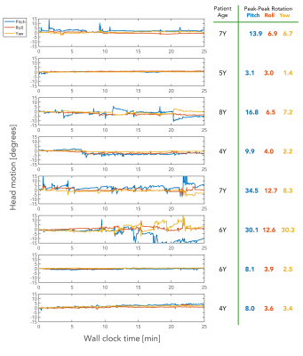 |
7 | A very motion-robust pediatric brain MRI protocol for clinical use
Stefan Skare1,2, Adam van Niekerk1,2, Tim Sprenger2,3, Aleksandra Ramolli1, Yords Österman1, Jan Svoboda1, Ola Norbeck1,2, Henric Rydén1,2, Enrico Avventi1,2, and Johan Berglund2
1Karolinska University Hospital, Stockholm, Sweden, 2Karolinska Institutet, Stockholm, Sweden, 3GE Healthcare, Stockholm, Sweden
Selected pediatric patients (3-9Y) scheduled for a clinical brain MRI under general anesthesia are enrolled in this ongoing study to be scanned awake, while watching a movie for entertainment, using a new motion-robust brain MRI protocol. The protocol consists solely of custom-made, inherently motion-robust, sequences, with all necessary MR contrasts for clinical use. All but one sequence uses real-time correction of head motion with an external tracking device. All eight unsedated pediatric patients scanned so far resulted in diagnostic images, despite some patients moving their heads in excess of 30 degrees (peak-peak) during the exam.
|
||
2644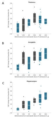 |
8 | Dose-dependent response of cerebral blood flow in healthy volunteers following administration of β2-adrenergic receptor agonist clenbuterol
Courtney A. Bishop1, Thomas Lodeweyckx2, Jan de Hoon2, Koen Van Laere3,4, Michel Koole4, Wim Vandenberghe5, Gaia Rizzo1, Eugenii Rabiner1,6, Renee Martin7, Anthony Ford7, and Gabriel Vargas7
1Invicro, London, United Kingdom, 2Center for Clinical Pharmacology, Department of Pharmaceutical and Pharmacological Sciences, KU Leuven, Leuven, Belgium, 3Division of Nuclear Medicine, University Hospital Leuven, Leuven, Belgium, 4Nuclear Medicine and Molecular Imaging, KU Leuven, Leuven, Belgium, 5Department of Neurology, University Hospital Leuven, Leuven, Belgium, 6Centre for Neuroimaging Sciences, Institute of Psychiatry, Psychology and Neuroscience, King’s College, London, United Kingdom, 7CuraSen Therapeutics, San Carlos, CA, United States
Initial evidence is provided for central effects of the β2-AR agonist clenbuterol on CBF in healthy volunteers. Regions showing particular effect (the hippocampus and thalamus) are associated with cognition and neurodegeneration in Parkinson’s and Alzheimer’s Disease, so similar targeting of β2-AR may prove beneficial in these conditions. Further work is required to determine the extent of central mediation of the effect of β2-AR agonism with clenbuterol in healthy volunteers.
|
||
2645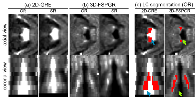 |
9 | A clinical protocol for 3D imaging of the locus coeruleus with super-resolution at 3 Tesla
Catarina Rua1, Mark R Symms2, Ali Ghayoor3, Brian Avants3, Courtney Bishop1, and Lino Becerra3
1Invicro LLC, A Konica Minolta Company, London, United Kingdom, 2GE Healthcare, London, United Kingdom, 3Invicro LLC, A Konica Minolta Company, Needham, MA, United States The locus coeruleus (LC) is the principal source of noradrenaline production in humans. Histology studies have shown that severe loss of neurons in the LC is associated with many neurodegenerative disorders. Damage is thought to be non-uniform and occurring in stages, hence there is a growing interest in imaging the LC in vivo in these patient populations. In this work we propose a protocol for imaging the LC at clinical field strengths using a 3D magnetization-transfer prepared imaging sequence strategy and the application of super-resolution techniques to increase the LC features within the brainstem region. |
||
2646 |
10 | Structural brain imaging with high-resolution 3D MRI on the Next-Generation 7T brain scanner
Alexander JS Beckett1,2, Jingjia Chen3, Shajan Gunamony4,5, An T Vu6,7, Salvatore Torrisi1,2, Chunlei Liu1,3, Jason Stockmann8,9, and David A Feinberg1,2
1Helen Wills Neuroscience Institute, University of California, Berkeley, CA, United States, 2Advanced MRI Technologies, Sebastopol, CA, United States, 3Department of Electrical Engineering and Computer Sciences, University of California, Berkeley, CA, United States, 4Imaging Centre of Excellence, University of Glasgow, Glasgow, United Kingdom, 5MR CoilTech Limited, Glasgow, United Kingdom, 6Radiology, University of California, San Francisco, CA, United States, 7San Francisco Veteran Affairs Health Care System, San Francisco, CA, United States, 8Athinoula A. Martinos Center for Biomedical Imaging, Massachusetts General Hospital, Charlestown, MA, United States, 9Harvard-MIT Health Sciences and Technology, MIT, Cambridge, MA, United States
The 128-channel receive, 16-channel parallel transmit capabilities of the new NexGen 7T system, in conjunction with the high amplitude and slew rate of the novel “Impulse” head gradient coil, provide many potential benefits for high-resolution 3D neuroimaging. High channel array coils allow acceleration on two phase encoded axes for very high total acceleration factors, and faster gradients can reduce image readout time for additional benefits. We demonstrate these benefits in different 3D imaging applications (MP2RAGE, FLAIR, FLASH, SWI) at 7T.
|
||
2647 |
11 | Assessment of Brain Activity during a Lower Extremity Sensorimotor Task using CEST MRI at 3T Video Permission Withheld
Rongwen Tain1,2, Benjamin M. Ellingson3,4, Craig Stark1,2,5, Catalina Raymond Guzman3,4, Kelli Sharp6,7, and Joanne Armour Smith8
1Campus Center for Neuroimaging, University of California, Irvine, CA, United States, 2Facility for Imaging and Brain Research, University of California, Irvine, CA, United States, 3Brain Tumor Imaging Laboratory, University of California, Los Angeles, CA, United States, 4Department of Radiology and Psychiatry, University of California, Los Angeles, CA, United States, 5Department of Neurobiology and Behavior, University of California, Irvine, CA, United States, 6Department of Physical Medicine and Rehabilitation, University of California, Irvine, CA, United States, 7Department of Dance, University of California, Irvine, CA, United States, 8Department of Physical Therapy, Chapman University, Orange, CA, United States A recent animal study demonstrates that CEST MRI is a potential tool to detect neuronal activation. In this work, we study CEST effects in the secondary somatosensory cortex during rest and during a goal-directed lower extremity sensorimotor task (small amplitude left leg lifting) in 10 healthy participants. Significantly higher CEST effects were observed in participants performing the leg task in the secondary somatosensory cortex, but not in a prefrontal cortex control region. This indicates that CEST MRI is a potential tool to detect brain metabolism associated with neuronal activity in human subjects at 3T. |
||
 Back to Meeting Home
 Back to the Program-at-a-Glance
The International Society for Magnetic Resonance in Medicine is accredited by the Accreditation Council for Continuing Medical Education to provide continuing medical education for physicians.
 Back to Meeting Home
 Back to the Program-at-a-Glance
 View the Presentation