Digital Poster
Quantitative Neuroimaging: Clinical & Translational Studies II
Joint Annual Meeting ISMRM-ESMRMB & ISMRT 31st Annual Meeting • 07-12 May 2022 • London, UK

Digital Poster
Quantitative Neuroimaging: Clinical & Translational Studies II
| Computer # | ||||
|---|---|---|---|---|
2733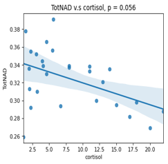 |
1 | Effect of circadian rhythm on brain NAD: an MRS study at 7 T
Zhiwei Huang1, Bernard Cuenoud2, Mickael Hartweg3, Daniel Wenz1, and Lijing Xin1
1Center for Biomedical Imaging, EPFL, Ecublens, Switzerland, 2Translation Research, Nestlé Health Science, Lausanne, Switzerland, 3Clinical Research Unit, Nestlé Research and Development, Lausanne, Switzerland
Our understanding of circadian rhythm has expanded to provide molecular insights into physiology and disease since the discovery of circadian clock.Pre-clinical experiments have shown that circadian rhythm is linked with the Nicotinamid Adenine Dinucleotide (NAD) levels and the redox ratio. However, clinical data is still lacking. In this study, we aim to study the effect of circadian rhythm on NAD levels in the human brain. By measuring the NAD levels in the occipital lobe with 31P-MRS, preliminary interim results suggest a negative correlation trend between cortisol level and total NAD level.
|
||
2734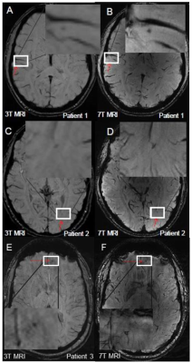 |
2 | Evaluation of brain volumetric changes and alterations in T1 relaxation times in American football players using 7 Tesla MRI Video Permission Withheld
Oliver Kraff1, Cornelius Deuschl2, Richard Dodel3, Janis Evers3,4, Anika Nietert1,5, Annika Verheyen1,6, and Harald H Quick1,7
1Erwin L. Hahn Institute for MRI, University Duisburg-Essen, Essen, Germany, 2Dept. of Diagnostic and Interventional Radiology and Neuroradiology, University Hospital Essen, University Duisburg-Essen, Essen, Germany, 3Chair of Geriatric Medicine, University Duisburg-Essen, Essen, Germany, 4Institute for Health Services Research and Clinical Epidemiology, Philipps University Marburg, Marburg, Germany, 5Westphalian University of Applied Sciences, Gelsenkirchen, Germany, 6University of Applied Sciences Ruhr West, Mülheim, Germany, 7High-Field and Hybrid MR Imaging, University Hospital Essen, University Duisburg-Essen, Essen, Germany
American football players were examined before and after a season of the German Football League. High resolution quantitative MRI at 7T for evaluations of volumetric changes and alterations in T1 relaxation times of various brain regions was performed. Age- and gender-matched subjects with no history of contact and collision sports served as a control group. In addition, structural susceptibility weighted imaging was compared between 3T and 7T. Loss of gray matter volume and an overall increase in T1 relaxation times were observed in players between both scans. SWI was superior in detecting cerebral microbleeds at 7T compared to 3T.
|
||
2735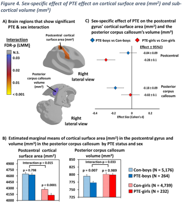 |
3 | Sex-specific effects of prenatal tobacco exposure on cognitive and brain morphometry measures in preadolescent children
Pedro Juan Rodriguez Rivera1, Amal Isaiah 1,2,3, Thomas Ernst1,4, Christine Cloak1, Huajun Liang1, and Linda Chang1,4,5
1Department of Diagnostic Radiology and Nuclear Medicine, University of Maryland School of Medicine, Baltimore, MD, United States, 2Department of Otorhinolaryngology, University of Maryland School of Medicine, Baltimore, MD, United States, 3Department of Pediatrics, University of Maryland School of Medicine, Baltimore, MD, United States, 4Department of Neurology, University of Maryland School of Medicine, Baltimore, MD, United States, 5Department of Neurology, Johns Hopkins University School of Medicine, Baltimore, MD, United States Prenatal tobacco exposure (PTE) can affect the offspring cognitive and brain development outcomes. Our study evaluated children who had PTE in the Adolescent Brain and Cognitive Development (ABCD) study and found poorer overall cognitive scores, especially working memory scores, lower cortical surface areas and subcortical volumes, with smaller surface areas in the posterior cingulate (PCC) and entorhinal cortices, the lingual and inferior parietal gyri, and abnormally smaller volumes in the thalamus and nucleus accumbens. Furthermore, we found that only PTE-girls had significantly smaller surface areas in the postcentral gyrus, while only PTE-boys had smaller volumes in the posterior corpus callosum. |
||
2736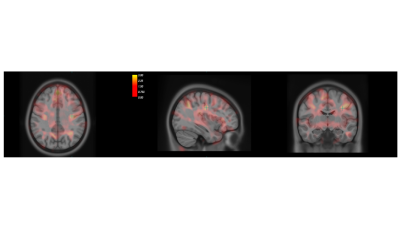 |
4 | Brain morphometry abnormalities in children with Childhood Apraxia of Speech.
Paolo Bosco1, Laura Biagi1, Simona Fiori1, Clara Bombonato1, Michela Tosetti1, and Anna Chilosi1
1IRCCS Stella Maris Foundation, Pisa, Italy
Childhood Apraxia of Speech (CAS) is a pediatric speech sound disorder in which precision and consistency of speech movements are impaired in absence of neuromuscular and structural deficit. The cause of CAS remains poorly understood and few neuroimaging studies still exist in children with CAS. In this case-control study we compared both at ROI and voxel level the cerebral gray matter of CAS and healthy controls subjects. The statistical analyses show that gray matter in CAS children is increased in areas of motor circuitries subserving speech and in areas involving sensorimotor learning.
|
||
2737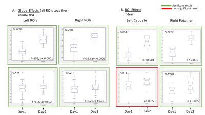 |
5 | ACUTE CAFFEINE ADMINISTRATION AS A CONFOUND FOR q-MRI and volumetric MRI STUDIES
Vishaal Sumra1,2,3 and Sofia Chavez1,2,4
1Institute of Medical Science, University of Toronto, Toronto, ON, Canada, 2Brain Health Imaging Centre, Centre for Addiction and Mental Health, Toronto, ON, Canada, 3Tanz Centre for Research in Neurodegenerative Diseases, University of Toronto, Toronto, ON, Canada, 4Department of Psychiatry, University of Toronto, Toronto, ON, Canada
Changes in cerebral blood flow (CBF) have the ability to confound structural and quantitative MRI studies. Caffeine is widely consumed and leads to strong decreases in CBF. Moderate caffeine users were scanned before and after caffeinated and decaffeinated coffee over two separate days. Percentage change in CBF maps, quantitative T1 maps, and estimates of grey matter volume (GMV) were assessed for the caffeinated coffee and decaffeinated coffee days. Robust decreases in CBF were observed, along with changes in T1 maps and estimates of GMV. Caffeine intake should be considered in studies relying on structural and quantitative MRI measures.
|
||
2738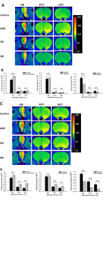 |
6 | Heroin addiction and relapse-induced axonal transport impairment in the brain and detected by in vivo MRI Video Not Available
Yueyuan Luo1, Jun Yang2, Chengde Liao2, and Zhongping Zhang3
1Department of Radiology., The Third Affiliated Hospital of Kunming Medical University, Yunnan Cancer Hospital, Kunming, China, 2Department of Radiology, The Third Affiliated Hospital of Kunming Medical University, Yunnan Cancer Hospital, Kunming, China, 3Philips Healthcare China, Guangzhou, China
Heroin plays role in heroin-induced cognitive dysfunction is unclear. We used manganese-enhanced magnetic resonance imaging for to evaluate the effect of heroin on axon transport in the body.
|
||
2739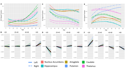 |
7 | Diffusion microstructure measurements across the brain and lifespan
Benjamin T Newman1 and T. Jason Druzgal1
1Department of Radiology & Medical Imaging, University of Virginia, Charlottesville, VA, United States
Understanding how the brain develops, matures, ages, and declines is one of the fundamental questions facing neuroscience1–3. Recent advances in diffusion microstructure analysis have allowed for detailed descriptions of neuronal change. However, it is essential that findings from these studies are appropriately contextualized to general age-related changes in the brain4,5. This study uses 3-tissue constrained spherical deconvolution (3T-CSD) to examine the relationship between brain diffusion microstructure and chronological age in a number of gross anatomical structures, subcortical gray matter, and cortex while additionally evaluating lateral differences in microstructural measurements. The results should serve as a benchmark for diffusion microstructure studies.
|
||
2740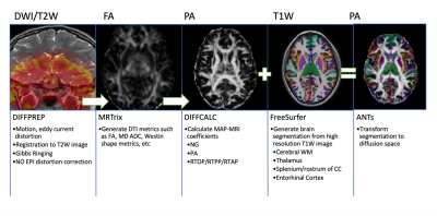 |
8 | Evaluating the feasibility of quantifying longitudinal microstructural changes in mild traumatic brain injury (mTBI) with MAP-MRI
Priyanka M Nadar1,2, Alexandru V Avram3,4, Luca Marinelli5, and Peter J Basser3
1Clinical Center, NIH, Bethesda, MD, United States, 2Drexel University College of Medicine, Philadelphia, PA, United States, 3NICHD, NIH, Bethesda, MD, United States, 4Henry M Jackson Foundation for the Advancement of Military Medicine, Bethesda, MD, United States, 5Global Research, General Electric, Niskayuna, NY, United States
Mean Apparent Propagator (MAP) MRI, an expansion of diffusion tensor imaging (DTI), explicitly measures the distribution of net 3D displacements of diffusing water molecules, providing better delineation of crossing white matter fiber tracts. This characteristic may be useful in characterizing unseen microstructural damage in patients with mild traumatic brain injuries whose clinical imaging scans are otherwise normal. An image processing pipeline for using mean apparent propagator (MAP) MRI to analyze diffusion weighted images in mTBI patients has been validated in healthy controls and shown to generate reliable data for patient brain scans.
|
||
2741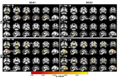 |
9 | Advanced multimodal MRI detects premanifest and early-stage alterations in SCA1 and SCA3 with high sensitivity
Jayashree Chandrasekaran1, Young Woo Park1, Emilien Petit2, Sophie Tezenas du Montcel2, Michal Povazan3, Guita Banan4, Romain Valabregue2, Philipp Ehses5, Jennifer Faber5, James M. Joers1, Pierrick Coupé6, Jose Vincente Manjón Herrera7, Chiadi U. Onyike3, Peter B. Barker3, Jeremy D. Schmahman8, Eva Maria Ratai8, S.H Subramony4, Thomas H. Mareci4, Khalaf O. Bushara9, Henry Paulson10,
Alexandra Durr2, Thomas Klockgether5, Tetsuo Ashizawa11, Christophe Lenglet1, and Gulin Oz1
1Center for Magnetic Resonance Research, University of Minnesota, Minneapolis, MN, United States, 2Sorbonne University, Paris, France, 3Johns Hopkins University, Baltimore, MD, United States, 4University of Florida, Gainesville, FL, United States, 5German Center for Neurodegenerative Diseases (DZNE), Bonn, Germany, 6University of Bordeaux, Bordeaux, France, 7Universidad Politécnica de Valencia, Valencia, Spain, 8Massachusetts General Hospital, Charlestown, MA, United States, 9University of Minnesota, Minneapolis, MN, United States, 10University of Michigan, Ann Arbor, MI, United States, 11The Houston Methodist Research Institute, Houston, TX, United States
Spinocerebellar ataxias are rare inherited neurodegenerative diseases that cause degeneration in the cerebellum and brainstem. The multi-site READISCA clinical trial readiness study aims to validate MR biomarkers at early stages of SCA1 and SCA3. SCA gene carriers (including individuals at pre-ataxic and ataxic stage) and matched controls (total N=107) were scanned at 3T to obtain structural and diffusion MRI and MR spectroscopy. Medulla, pons, and cerebellar peduncles were the earliest sites of involvement in both SCAs. Neurochemical and microstructural abnormalities were detected with very high sensitivity (AUC>0.9 in ROC analyses) prior to ataxia onset.
|
||
 Back to Meeting Home
 Back to the Program-at-a-Glance
The International Society for Magnetic Resonance in Medicine is accredited by the Accreditation Council for Continuing Medical Education to provide continuing medical education for physicians.
 Back to Meeting Home
 Back to the Program-at-a-Glance
 View the Presentation