Digital Poster
Imaging Nerves, Head & Neck
Joint Annual Meeting ISMRM-ESMRMB & ISMRT 31st Annual Meeting • 07-12 May 2022 • London, UK

| Computer # | ||||
|---|---|---|---|---|
1377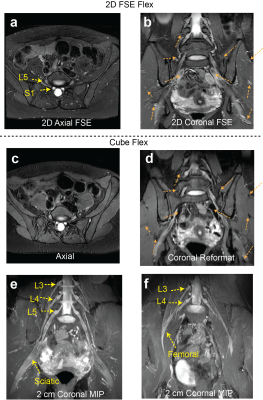 |
1 | Efficient Dixon-Based 3D Fast Spin-Echo MR Neurography of Lumbosacral Plexus
Misung Han1, Cynthia T Chin1, and Sharmila Majumdar1,2
1Radiology and Biomedical Imaging, University of California, San Francisco, San Francisco, CA, United States, 2Center for Digital Health Innovation, University of California, San Francisco, San Francisco, CA, United States
MR neurography (MRN) provides direct visualization of peripheral nerves and allows for detecting pathologies. Isotropic 3D MRN has been suggested as an effective method to evaluate overall nerve structures and subtle morphological changes; however, its clinical applications to the lumbosacral plexus have been limited. In this work, we optimized 3D FSE acquisition combined with a Dixon fat-water separation technique to provide uniform signal over the large volume and to suppress vessel signal. In vivo experiments with patients having a lower back pain at 3T demonstrated the clinical feasibility of using the proposed methods for evaluating nerve pathologies.
|
||
1378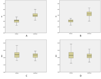 |
2 | Microstructural alteration of trigeminal nerve revealed by DTI and its correlation with vascular compression and pain Video Not Available
Tiantian Guo1, Chunqing Bu1, Chuanying Shi1, Daoqing Su2, Peng Wu3, and Chuanchen Zhang1
1Department of Radiology, Liaocheng People's Hospital, Liaocheng, China, 2Department of Neurosurgery, Liaocheng People's Hospital, Liaocheng, China, 3Philips Healthcare, Shanghai, China
Classical Trigeminal Neuralgia (CTN) is mainly caused by vascular compression of the trigeminal nerve. We analyzed the correlation between the fractional anisotropy (FA) / apparent diffusion coefficient (ADC) values of the bilateral trigeminal nerves and the degree of neurovascular compression (NVC) for CTN patients. Results shown the FA value is negatively correlated with the degree of NVC on the symptomatic side. FA values was lower if there was NVC on the asymptomatic side compared with no NVC. The correlation between FA and visual analogue scale (VAS) scores indicates that FA was a potential MR indicator for predicting patient clinical symptoms.
|
||
1379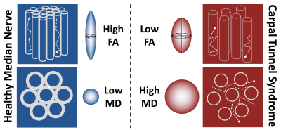 |
3 | Meta-Analysis of the Normal Diffusion Tensor Imaging Values of the Median Nerve And How They Change In Carpal Tunnel Syndrome Video Not Available
Djamila Rojoa 1, Firas Raheman 1, Joseph Rassam 1, and Ryckie George Wade2
1Leicester Royal Infirmary, Leicester, United Kingdom, 2University of Leeds, Leeds, United Kingdom
Carpal tunnel syndrome (CTS) is the most common compressive neuropathy worldwide. Compressed peripheral nerves exhibit distorted architecture, demyelination and perineurial fibrosis. Diffusion tensor imaging (DTI) generates proxy measures of nerve ‘health’ which are sensitive to myelination, axon diameter, fibre density and organisation. This meta-analysis included 32 studies of 2643 wrists, belonging to 1575 asymptomatic adults and 1068 patients with CTS. We summarise the normal FA (0·58 [95% CI 0·56-0·59]) and MD (1·138 x10-3 mm2/s [95% CI 1·101-1·174]) of the median nerve, then show that diffusion throughout the length of the median nerve is more isotropic in patients with CTS.
|
||
1380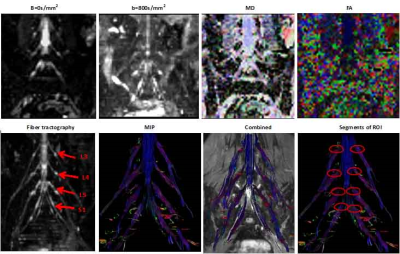 |
4 | Quantitative Evaluation of Normal Lumbosacral Plexus Nerve Using Diffusion Tensor Imaging with Multiband SENSE Video Not Available
Nan Zhang1, Caizhong Chen1, and Qingwei Song2
1Department of Radiology, Zhongshan Hospital affiliated to Fudan University, Shanghai, China, 2Department of Radiology, The First affiliated hospital of Dalian Medical University, Dalian, China
DTI can provide valuable structural information that may become an innovative tool in evaluating lumbosacral plexus nerve entrapment. Multiband SENSE technique could be used to accelerate the image acquisition .The present study aims to explore the feasibility of DTI with multiband SENSE on normal lumbosacral plexus nerve. The study showed that MB SENSE=2 was recommended for DTI on normal lumbosacral plexus nerve, which facilitated a 32% shorter image acquisition time than conventional SENSE accelerated diffusion tensor imaging.
|
||
1381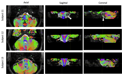 |
5 | Comparison of MR diffusion based tractography methods for reconstruction of the facial nerve in pre-operative vestibular schwannoma
Caitlin O'Brien1, Philip Touska2, Elizabeth Gabriel1, Haris Shuaib3, Karen Welsh4, and Steve Connor2,5
1Magnetic Resonance Physics, Guy’s & St. Thomas’ NHS Foundation Trust, London, United Kingdom, 2Department of Radiology, Guy’s & St. Thomas’ NHS Foundation Trust, London, United Kingdom, 3Clinical Scientific Computing, Guy’s & St. Thomas’ NHS Foundation Trust, London, United Kingdom, 4MRI, Guy’s and St. Thomas’ Hospitals & King’s College Hospital, London, United Kingdom, 5Neuroradiology, King’s College Hospital NHS Foundation Trust, London, United Kingdom
Four DWI tractography acquisitions are trialled across 13 pre-surgical patients with vestibular schwannomas: single shell EPI, ZOOMit, RESOLVE, & multi-shell RESOLVE, with an aim to reconstruct the path of the facial nerve. Data is analysed using MRtrix using multi-shell multi-tissue constrained spherical deconvolution (MSMT-CSD) and the probabilistic iFOD2 algorithm. The best reconstruction was achieved using a 30 direction multi-shell (b = 0, 300, 1000 s/mm2) RESOLVE acquisition scheme.
|
||
1382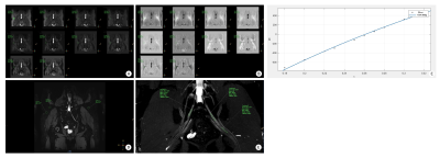 |
6 | 3D high-resolution Contrast-enhanced MR neurography of lumbosacral plexus with precise fat-suppression: A pilot image quality evaluation
Xiangchuang Kong1, Peng Sun2, QingPing Gu2, Tian Liao1, XiaoMing Liu1, and DingXi Liu1
1Radiology, Union Hospital, Tongji Medical College, Huazhong University of Science and Technology, Wuhan, China, 2Philips Healthcare, WuHan, China
The objective of this study was to investigate the values of precise fat-suppression for the robust visualization of 3D high-resolution contrast-enhanced MR neurography of lumbosacral plexus by inversion time (TI) scout technique. The precise TI of individuals could be calculated from several fast different TI experiments by measuring the fat signals recovery on the real and imaginary images. CNR (precise fat-suppression) (13.26±5.47) was about 425.4% higher than CNR (traditional TI) (2.39±0.55). Accurate fat suppression in MRN of lumbosacral plexus can help visual the branch of lumbosacral plexus nerves distinctly and improve the accuracy of diagnosis in diseases of lumbosacral plexus.
|
||
1383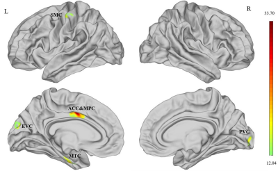 |
7 | Surface-Based Morphometry Analysis Shows Increased Cortical Sulcus Depth after Sound Therapy in Patients with Tinnitus Video Not Available
Xuan Wei1 and Jinxia Zhu2
1Department of Radiology, Beijing Friendship Hospital, Capital Medical University, Beijing, China, 2MR Collaboration, Siemens Healthcare Ltd., Beijing, China
In this study, we performed brain surface-based morphometry to evaluate changes in sulcal depth after sound therapy in patients with idiopathic tinnitus. Our results showed that sulcal depth was significantly reduced in the left medial temporal cortex (MTC) and right somatosensory and motor cortex (SMC) of patients with tinnitus compared to the healthy controls, but increased significantly at 24 weeks after sound therapy. Therefore, sulcal depth in the auditory sensory regions of the brain is a potential neuroimaging biomarker for evaluating treatment efficacy in tinnitus patients.
|
||
1384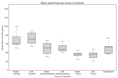 |
8 | Multi-delay pseudo-continuous arterial spin labeling in head and neck healthy tissues
Nienke D. Sijtsema1,2, Steven F. Petit1, Dirk H.J. Poot2, Gerda M. Verduijn1, Esther A.H. Warnert2, Mischa S. Hoogeman1,3, and Juan A. Hernandez-Tamames2
1Department of Radiotherapy, Erasmus MC Cancer Institute, Rotterdam, Netherlands, 2Department of Radiology and Nuclear Medicine, Erasmus MC, Rotterdam, Netherlands, 3Department of Medical Physics and Informatics, HollandPTC, Delft, Netherlands
Arterial spin labeling (ASL) has potential for response prediction of tumor and healthy tissue in the head and neck region after radiotherapy. Since little is known about blood flow (BF) values in this region, we measured BF and the repeatability of BF in several tissues simultaneously using multi-delay pseudo-continuous ASL. This enables investigation of the correlation between perfusion and severity of radiotherapy side-effects in a future study. Of the tissues we assessed, we found the parotids have the highest and the tonsils have the lowest perfusion.
|
||
1385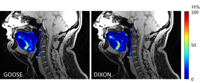 |
9 | Assessment of Fat Fractions of Tongues, Pharyngeal Walls, and Soft Palates by GOOSE and DIXON methods
Ruitian Song1, Scott N. Hwang2, Chris Goode1, Diana Storment1, Matthew Scoggins1, Zachary Abramson1, Claudia M Hillenbrand3, Belinda Mandrell4, Kevin Krull5, and Wilburn E. Reddick1
1Diagnostic Imaging, St Jude Children's Research Hospital, Memphis, TN, United States, 2Department of Radiology, Penn State Health Milton S Hershey Medical Center, Hershey, PA, United States, 3Research Imaging NSW, University of New South Wales, Sydney, Australia, 4Division Nursing Research, St Jude Children's Research Hospital, Memphis, TN, United States, 5Department of Epidemiology and Cancer Control, St Jude Children's Research Hospital, Memphis, TN, United States
Among 63 participants, the fat fractions (FFs) were estimated and compared in tongues, posterior pharyngeal walls (PPWs), and soft palates (SPs) using two-point DIXON and GOOSE (Globally Optimal Surface Estimation) methods. DIXON was seriously impaired by B0 inhomogeneity and underestimated FF relative GOOSE. There was a strong correlation between these two methods in the tongues and SPs, and a weak one in the PPWs. In our opinion, an IDEAL-based method is preferred for estimating fat content. Extra attention is needed when using the two-point DIXON method.
|
||
1386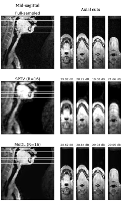 |
10 | Accelerated volumetric vocal tract MRI using model based deep learning
Wahidul Alam1 and Sajan Goud Lingala1,2
1Roy J. Carver Department of Biomedical Engineering, University of Iowa, Iowa city, IA, United States, 2Department of Radiology, University of Iowa, Iowa city, IA, United States
3D MRI is a powerful tool to safely visualize the various vocal-tract configurations during voice production [1-3]. Current accelerated 3D vocal-tract MRI schemes based on spatial total variation transform (SPTV) regularization are susceptible to non-trivial artifacts (e.g., blurring at air-tissue boundaries, patchy representation of small structures such as the epiglottis, glottis). In this work, we apply a model based deep learning reconstruction scheme that can significantly accelerate 3D vocal-tract imaging. We demonstrate it to produce images with high-spatial fidelity, natural-looking like contrast, and significantly robust to artifacts seen with current SPTV based schemes.
|
||
1387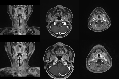 |
11 | Image quality of high-resolution three-dimensional neck MRI using CAIPIRINHA-VIBE and GRASP-VIBE: an intra-individual comparative study
Minkook Seo1, Jimin Yoon1, Yangsean Choi1, Jinhee Jang1, Na-Young Shin1, Kook-Jin Ahn1, and Bum-soo Kim1
1Radiology, Seoul St. Mary's Hospital, Seoul, Korea, Republic of
The image qualities of two high-resolution MRI sequences of the neck—CAIPIRINHA-VIBE and GRASP-VIBE—were compared. 173 patients clinically indicated for neck MRI were scanned using both sequences with isotropic (<1 mm) in-plane resolution. The image quality was qualitatively assessed by two radiologists. Quantitative assessments (i.e., non-uniformity, contrast-to-noise ratio and signal-to-noise ratio) were performed in all patients, a phantom and a healthy volunteer. GRASP-VIBE outperformed CAIPIRINHA-VIBE in all qualitative assessments except for the fat suppression degree. Quantitative assessments were significantly superior in GRASP-VIBE than in CAIPIRINHA-VIBE. Therefore, GRASP-VIBE may be a better alternative to CAIPIRINHA-VIBE for head and neck MRI.
|
||
 Back to Meeting Home
 Back to the Program-at-a-Glance
The International Society for Magnetic Resonance in Medicine is accredited by the Accreditation Council for Continuing Medical Education to provide continuing medical education for physicians.
 Back to Meeting Home
 Back to the Program-at-a-Glance
 View the Presentation