Digital Poster
White Matter Microstructure in Disease
Joint Annual Meeting ISMRM-ESMRMB & ISMRT 31st Annual Meeting • 07-12 May 2022 • London, UK

Digital Poster
White Matter Microstructure in Disease
| Computer # | ||||
|---|---|---|---|---|
2875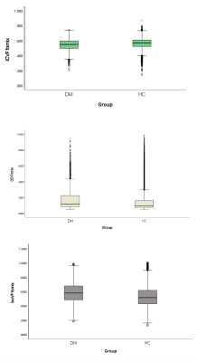 |
85 | Characterising Brain White Matter Alterations in Type 2 Diabetes: A UK Biobank Study of Neurite Orientation Dispersion and Density Imaging.
Abdulmajeed Alotaibi1,2,3, Amjad AlTokhis1,3, Anna Podlasek1,3, Chris Tench1,3, Sieun Lee1,3, Cris Constantinescu3,4,5, and Rob Dineen1,3,5
1Clinical Neurosciences, University of Nottingham, Nottingham, United Kingdom, 2Radiological Sciences, King Saud bin Abdulaziz University for Health Sciences, Riyadh, Saudi Arabia, 3Precision Imaging Beacon of Excellence, Nottingham, United Kingdom, 4Neurology, Cooper University Hospital, Camden, NJ, United States, 5NIHR Nottingham Biomedical Research Centre, Nottingham, United Kingdom
Type 2 diabetes is a metabolic disorder associated with subtle microstructural alteration of brain white matter. Diffusion tensor imaging (DTI) has been widely applied to evaluate white matter microstructural pathology in type 2 diabetes; however, DTI has limitations and lacks specificity. Using UK Biobank data, we applied neurite orientation dispersion and density imaging (NODDI) as an alternative advanced diffusion method to overcome DTI limitations. In this study, NODDI showed its potential role in giving a better biophysical characterisation of white matter neuroaxonal pathology in type 2 diabetes compared to DTI.
|
||
2876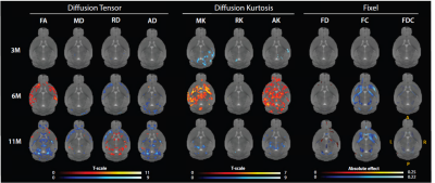 |
86 | Longitudinal Fixel-Based Analysis of diffusion MRI in the zQ175 Huntington’s disease mouse model
Nicholas Vidas-Guscic1,2, Joëlle van Rijswijk1,2, Johan Van Audekerke1,2, Ben Jeurissen2,3, Stephan Missault1,2, Dorian Pustina4, Haiying Tang4, Roger Cachope4, Longbin Liu4, Celia Dominguez4, Ignacio Munoz-Sanjuan4, Annemie Van der Linden1,2, and Marleen Verhoye1,2
1Bio-Imaging Lab, University of Antwerp, Antwerp, Belgium, 2µNEURO Research Centre of Excellence, University of Antwerp, Antwerp, Belgium, 3Vision-Lab, University of Antwerp, Antwerp, Belgium, 4CHDI Management/CHDI Foundation, Princeton, NJ, United States Huntington’s disease (HD) is a neurodegenerative disorder that affects motor and cognitive abilities. In this study, we present the outcome of a longitudinal multi-shell DWI investigation of the zQ175 HD mouse model using the diffusion tensor, diffusion kurtosis, and fixel-based analysis. This study reveals microstructural deficits at an early stage of the disease that mainly affect the diffusion tensor in corpus callosum and kurtosis in caudate putamen and grey matter, while fiber cross-section is reduced in major fiber bundles. At a late stage, many white matter fiber bundles show deficits that are indicative of differential myelination and potential axonal pathology. |
||
2877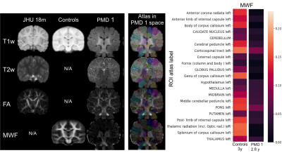 |
87 | Exploring Myelin Imaging Biomarkers in Pelizaeus-Merzbacher Disease
Caroline Koehler1, Paul Kuntke1, Prativa Sahoo2, Hannes Wahl1, Sean C Deoni3, Hagen H. Kitzler1, and Steffi Dreha-Kulaczewski2
1Institut for Diagnostic and Interventional Neuroradiology, University Hospital Carl Gustav Carus, Dresden, Germany, 2Pediatrics and Adolescent Medicine, University Medicine Göttingen, Goettingen, Germany, 3Advanced Baby Imaging Lab, Memorial Hospital of Rhode Island, Pawtucket, RI, United States
Advanced MRI techniques bear great potential to explore myelination in hypomyelinating leukodystrophies such as Pelizaeus-Merzbacher disease (PMD) in-vivo. Quantitative imaging biomarkers which reflect myelin, its spatial distribution, and dynamics during brain maturation are not readily available i.e. to monitor natural disease courses. In a pilot study including three PMD patients, multi-component relaxation which enables the evaluation of relative myelination by estimating the myelin water fraction (MWF) was obtained from 56 region of interest (ROI). Determination of ROIs was facilitated by coregistration of the patients data to a pediatric atlas and compared to age matched healthy controls.
|
||
2878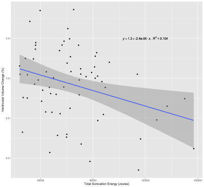 |
88 | Acute Global Structural and Diffusivity Changes Following Treatment with MRgFUS
Kain Kyle1,2, Yael Barnett3, Stephen Tisch3, Ben Jonker3, Arkiev D'Souza1, Jerome Maller4, Michael Barnett1,2, Joel Maamary3, and Chenyu Wang1,2
1University of Sydney, Sydney, Australia, 2Sydney Neuroimaging Analysis Centre, Sydney, Australia, 3St Vincent's Hospital Sydney, Sydney, Australia, 4GE Healthcare Australia, Melbourne, Australia MRgFUS is a non-invasive procedure for the treatment of tremor, involving the application of high-energy ultrasound waves to ablate tissue in the brain in regions implicated in tremor networks. There is a lack of data on the acute changes outside the ablated region following treatment with MRgFUS. This investigation sought to examine changes in brain volume and diffusion tensor metrics following treatment with MRgFUS. |
||
2879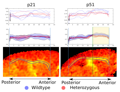 |
89 | Myelin Alterations during the Development of an Absence Seizure Mouse Model
Gustavo Chau Loo Kung1, Juliet Knowles2, Lijun Ni2, John Huguenard2, Michelle Monje2, and Jennifer McNab3
1Bioengineering Department, Stanford University, Stanford, CA, United States, 2Neurology Department, Stanford University, Stanford, CA, United States, 3Radiology Department, Stanford University, Stanford, CA, United States
Maladaptive myelination may be related to the increasing frequency of absence seizures. To explore this connection, we performed MRI microstructural measurements in ex vivo mouse brains from the Scn8amed+/- model of absence epilepsy in two cohorts of mice at two points of their development, both before and after seizures were well established. Our MRI g-ratio results strongly agree with our previous findings based on electron microscopy and show a clear alteration of myelination throughout the anterior portion of the corpus callosum.
|
||
2880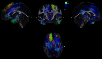 |
90 | Early MRI biomarkers in amyotrophic lateral sclerosis (ALS): DTI and functional connectivity.
Juan Carlos Quizhpilema1, Pablo Lecumberri2, Marta Vidorreta3, Tamara Laxe4, Mikel San Miguel5, Javier Díaz6, Inmaculada Pagola5, Marisol Gómez1, Ivonne Jericó5, and Teresa Cabada4
1Department of Statistics, Computer Science and Mathematics, UPNA, Pamplona, Spain, 2Movalsys, Pamplona, Spain, 3Siemens Healthineers, Madrid, Spain, 4Radiology department, HUN, Pamplona, Spain, 5Neurology department, HUN, Pamplona, Spain, 6Psychiatry department, HUN, Pamplona, Spain
Amyotrophic lateral sclerosis (ALS) is a neurodegenerative disease characterized by damage to motor neurons. The search for early biomarkers is relevant due to their rapid progression.This study aims to analyze structural and functional measures in a group of newly diagnosed ALS patients, compare them with a group of healthy controls, and evaluate changes longitudinally at a 6-month follow-up.Twenty patients and 15 controls were recruited. The MRI examination included a T1-weighted scan, DTI, rs-fMRI. ALS shows involvement in motor and non-motor areas as shown by DTI in cortical activity in the early phase and in time progression.
|
||
2881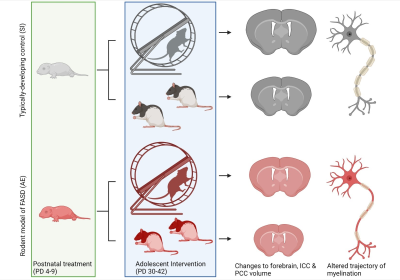 |
91 | Mind the Myelin: Investigating the therapeutic impact of exercise on white matter damage in a rodent model of Fetal Alcohol Spectrum Disorders Video Permission Withheld
Katrina Milbocker1, Eric Brengel1, Gillian LeBlanc1, and Anna Klintsova1
1Psychological & Brain Sciences, University of Delaware, NEWARK, DE, United States
Fetal Alcohol Spectrum Disorders (FASD) is an umbrella term used to identify individuals with a history of prenatal alcohol exposure which results in a spectrum of diagnostic disorders. 1 in 20 infants born in the U.S. has been diagnosed with an FASD, creating a major public health crisis. Deficits in corpus callosum myelination resulting from prenatal alcohol exposure have been correlated with impairments to perceptual learning and executive function in adolescents diagnosed with FASD. This study investigates the therapeutic potential of an exercise intervention to ameliorate alcohol-induced damage to corpus callosum myelination in a rodent model of FASD.
|
||
2882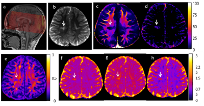 |
92 | Myelin Water Imaging and Diffusion Tensor Imaging in Pediatric Acute Lymphoblastic Leukemia Patients
Ruitian Song1, John O. Glass1, and Wilburn E. Reddick1
1Diagnostic Imaging, St Jude Children's Research Hospital, Memphis, TN, United States Myelin water imaging (MWI) and diffusion tensor imaging (DTI) was performed at two longitudinal time points, approximately two years apart, during chemotherapy in patients treated for acute lymphoblastic leukemia (ALL). DTI metrics correlated with MWF and LWF at time point 1, but the correlations were weaker at time point 2 (TP2). This may be caused by a possible decoupling of the relationship at TP2 as the brain is recovering from the impact of chemotherapy. |
||
2883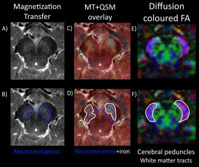 |
93 | Comprehensive brainstem imaging using MT, QSM and DTI for atypical parkinsonism diagnosis
Samy Abo Seada1, Anke W. van der Eerden1, Agnita Boon2, and Juan A. Hernandez-Tamames1,3
1Department of Radiology and Nuclear medicine, Erasmus MC, Rotterdam, Netherlands, 2Department of Neurology, Erasmus MC, Rotterdam, Netherlands, 3Department of Imaging Physics, TU Delft, Delft, Netherlands
Differentiating atypical parkinsonism (AP) from idiopathic Parkinson’s disease (PD) remains a challenge in clinical practice. Based on a recent review on differentiating atypical parkinsonisms using MRI, we developed a dedicated MRI protocol including magnetization transfer (MT), quantitative susceptibility mapping (QSM) and diffusion tensor imaging (DTI) for improving diagnosis. Using pilot data from a healthy volunteer, we show how the assessment of important structures (substantia nigra and locus coeruleus) can be improved using a comprehensive combination of MRI techniques.
|
||
2884 |
94 | Abnormal Cerebral Microstructures in Amyotrophic Lateral Sclerosis: a Diffusion Kurtosis Imaging Study Video Not Available
Jia Yan Shi1 and Hua Jun Chen2
1Fujian Medical University Union Hospital, Fuzhou, China, 2Radiology, Fujian Medical University Union Hospital, Fuzhou, China
Our study aim to investigate cerebral microstructural changes in Amyotrophic lateral sclerosis (ALS) using diffusion kurtosis imaging (DKI) for the first time. ALS patients showed mean kurtosis metrics (MK) reductions in several gray/white matter areas. The spatial distribution of the regions with reduced radial kurtosis(RK) was similar to those with decreased MK. No significant axial kurtosis(AK) difference was found between groups. The correlation analysis revealed significant associations between DKI metrics and clinical assessments. Additionally, several white matter regions showed between-group differences in conventional diffusion metrics; but the spatial extent was smaller than that with reduced DKI metrics.
|
||
 Back to Meeting Home
 Back to the Program-at-a-Glance
The International Society for Magnetic Resonance in Medicine is accredited by the Accreditation Council for Continuing Medical Education to provide continuing medical education for physicians.
 Back to Meeting Home
 Back to the Program-at-a-Glance
 View the Presentation