Digital Poster
Brain Tumors I
Joint Annual Meeting ISMRM-ESMRMB & ISMRT 31st Annual Meeting • 07-12 May 2022 • London, UK

| Computer # | ||||
|---|---|---|---|---|
2648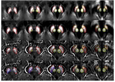 |
12 | Diagnosing Parkinson’s disease by Combining Neuromelanin and Iron Image Features Using Automatic SN Subregions Detection Approach Video Permission Withheld
Zhijia Jin1, Mojtaba Jokar2, Ying Wang2,3, Yan Li1, Zenghui Cheng1, Yu Liu1, Rongbiao Tang1, Xiaofeng Shi1, Youmin Zhang1, Jihua Min1, Fangtao Liu1, Naying He1, E. Mark Haacke1,2,3,4, and Fuhua Yan1
1Ruijin Hospital, Shanghai Jiao Tong University School of Medicine, Shanghai, China, 2Magnetic Resonance Innovations, Inc., Bingham Farms, MI, United States, 3Radiology, Wayne State University, Detroit, MI, United States, 4Biomedical Engineering, Wayne State University, Detroit, MI, United States
A total of 100 Parkinson’s disease (PD) patients and 100 age- and sex-matched healthy controls (HCs) were scanned using a single 3D gradient echo magnetization transfer sequence. We developed an automatic substantia nigra (SN) subregions segmentation approach to get neuromelanin (NM) and iron measurements in the SN. These measures along with their overlap region volume and the nigrosome-1 (N1) sign showed reliable results indicative of promising diagnostic biomarkers to differentiate PD patients from HCs.
|
||
2649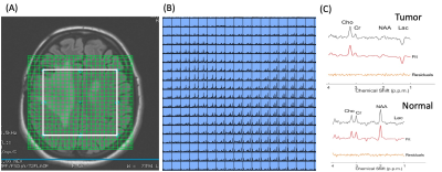 |
13 | Clinical Feasibility of Brain Tumor Multi-voxel MR Spectroscopic Imaging using Rapid Semi-LASER and EPSI Encoding at 3T
Sunitha B Thakur1, Olivia M Sutton2, Almir Bitencourt3, Ralph Noeske4, Tara Fahy1, Ricardo Otazo1, and Robert Young1
1Memorial Sloan Kettering Cancer Center, New York, NY, United States, 2Johns Hopkins Medicine, Baltimore, MD, United States, 3A.C.Camargo Cancer Center, Sao Paulo, SP, Brazil, 4GE HealthCare, Munich, Germany
The aim of this study was to evaluate the clinical feasibility of optimized multi-voxel MR spectroscopic imaging (MRSI) at 3T using rapid symmetric echo-planar spectroscopic imaging (EPSI) spatial encoding in patients with brain tumors. We performed the optimized MRSI at 3T using semi-localization by adiabatic selective refocusing pulses (semi-LASER), fast spatial encoding using symmetric EPSI, and robust water suppression with variable power and optimized relaxation delays. A radiologist with extensive experience in neuroimaging and spectroscopy assessed the overall spectral quality. This study is an attempt to integrate high-resolution MRSI in clinical practice for the assessment of brain cancer patients.
|
||
2650 |
14 | Amide proton transfer imaging for differentiation of glioblastoma from brain metastasis Video Permission Withheld
Utarat Kaewumporn1, Doonyaporn Wongsawaeng1, Siriwan Piyapittayanan1, Chanon Ngamsombat1, Panid Chaysiri1, Janiya Kittikornchaichan1, Kritdipha Ningunha2, Nanthasak Tisavipat2, Titima Itthimathin2, Ketkanok Rayaroji2, and Orasa Chawalparit1
1Radiology, Faculty of Medicine Siriraj hospital, Mahidol University, Bangkok, Thailand, 2Bangkok International Hospital, BDMS, Bangkok, Thailand Until now, the differentiation between glioblastoma and brain metastasis still has some limitations and inconclusive findings. |
||
2651 |
15 | Machine Learning-Based Radiomics Predicting the IDH1 Genotype of Diffuse Gliomas Video Permission Withheld
Qirui Zhao1, Zongfang Li1, Yi Lu1, Han Bao1, Zujun Hou2, Liuyang Chen3, Wei Xie1, Qing Wang1, Wei Zhao1, Tong-San Koh4, and Lisha Nie5
1Department of Radiology, The First Affiliated Hospital, Kunming Medical University, Kunming, China, 2Suzhou Institute of Biomedical Engineering and Technology, Chinese Academy of Sciences, Suzhou, China, 3Fisca Healthcare Ltd, Kunming, China, 4Department of Oncologic Imaging, National Cancer Center, Singapore, Singapore, 5GE Healthcare, MR Research, Beijing, China
The current study aims to evaluate the value of susceptibility weighted imaging (SWI) and contrast-enhanced T1-weighted imaging (CE-T1WI) radiomics features in predicting isocitrate dehydrogenase1 (IDH1) genotype of diffuse gliomas and build prediction models. It was concluded that SWI and CE-T1WI radiomics features can effectively predict the IDH1 genotype of diffuse gliomas, and CE-T1WI performed better. By combining SWI with CE-T1WI radiomics features, the prediction performance can be improved.
|
||
2652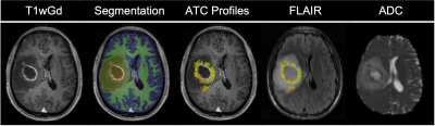 |
16 | A Novel GBM Infiltration Biomarker for Predicting Outcome: Clinical Study & Translational Preclinical Validation of ADC vs Tumor Cellularity
Antoine Vallatos1, Haitham Al-Mubarak2, Joanna Birch3, Adam Waldman1, William Holmes2, Anthony Chalmers3, and Gerard Thompson1
1Centre for Clinical Brain Sciences, University of Edinburgh, Edinburgh, United Kingdom, 2Glasgow Experimental MRI Centre, University of Glasgow, Glasgow, United Kingdom, 3Wolfson Wohl Translational Research Centre, University of Glasgow, Glasgow, United Kingdom
We report a novel clinical putative glioblastoma (GBM) biomarker of infiltration which predicts overall survival at presentation. Given the extreme challenges in obtaining comprehensive spatially matched MRI and tissue in humans to explore the biomarker biology, we assessed the relation between tumoral cellularity and ADC values in matched histology and MRI in a rodent infiltrating human xenograft GBM model. We demonstrate that the ADC slope measured along linear profiles perpendicular to the boundary of solid macroscopic tumor visible on ADC is a biomarker predictive of overall survival in a single centre human cohort. We demonstrate a strong correlation between ADC and tumor cellularity in the preclinical model, supporting the hypothesis that a radial vectorial profile analysis of tumor ADC maps could provide with a robust clinical biomarker of glioblastoma cell infiltration and therefore outcome.
|
||
2653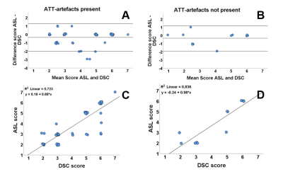 |
17 | Influence of Arterial Transit Time Delay in Arterial Spin Labeling on Differentiating Tumor Progression and Pseudo-Progression in Glioblastoma
Daniëlle van Dorth1, Janey Jiang2,3, Bárbara Schmitz-Abecassis1, Robert J. I. Croese4,5, Martin J. B. Taphoorn5, Marion Smits6, Johan A. F. Koekkoek5, Linda Dirven5, Jeroen de Bresser3, and Matthias J. P. van Osch1
1C. J. Gorter Center for High-Field MRI, Department of Radiology, Leiden University Medical Center, Leiden, Netherlands, 2Department of Radiology, HagaZiekenhuis, Den Haag, Netherlands, 3Department of Radiology, Leiden University Medical Center, Leiden, Netherlands, 4Department of Neurology, Haaglanden Medical Center, Den Haag, Netherlands, 5Department of Neurology, Leiden University Medical Center, Leiden, Netherlands, 6Department of Radiology and Nuclear Medicine, Erasmus MC, University Medical Center, Rotterdam, Netherlands
In the clinical follow-up of glioblastoma patients, presence of delayed arterial transit times (ATT) could affect the evaluation of ASL perfusion data. In this retrospective study the influence of the presence and severity of ATT-artifacts on perfusion assessment and differentiation between tumor progression and pseudo-progression were studied. The results show that the presence of ATT-artifacts lowers the agreement between radiological evaluation of DSC-MRI and ASL, although the severity of ATT-artifacts did not have significant influence. In conclusion, detection of ATT-artifacts is important as it could affect radiological evaluation of ASL-data. Future work aims to include additional quantitative perfusion measures.
|
||
2654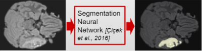 |
18 | Automated machine learning-based brain lesion segmentation on structural MRI acquired from traumatic brain injury patients
Ilkay Yildiz1, Rachael Garner1, Michael Douglas Morris2, Jesus Ruiz Tejeda2, Courtney Real2, Manuel Buitrago Blanco2, Paul Vespa2, and Dominique Duncan1
1Keck School of Medicine, University of Southern California, Los Angeles, CA, United States, 2David Geffen School of Medicine, University of California, Los Angeles, LOS ANGELES, CA, United States
Traumatic brain injury (TBI) can cause severe disorders, including post-traumatic epilepsy (PTE). Lesion segmentation is an MRI-based analysis to identify brain structures that correlate with PTE development post-TBI. Unfortunately, manual segmentation, considered the gold standard, is highly tedious and noisy. Thus, we propose the first automated machine-learning based lesion segmentation method for MRI of TBI patients enrolled in the Epilepsy Bioinformatics Study for Antiepileptogenic Therapy (EpiBioS4Rx). Experimental validation demonstrates considerable visual overlap of lesion predictions and ground-truths with 61% precision. Early and automated lesion segmentation via our approach can aid experts in MRI analysis and successful PTE identification following TBI.
|
||
2655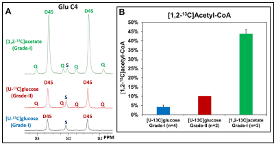 |
19 | Preferential utilization of acetate in human meningiomas
Omkar B Ijare1, David Baskin1, Suzanne Powell2, and Kumar Pichumani1
1Neurosurgery, Houston Methodist Hospital and Research Institute, Houston, TX, United States, 2Pathology and Genomic Medicine, Houston Methodist Hospital and Research Institute, Houston, TX, United States
Meningiomas are the most frequently reported central nervous system (CNS) tumors, accounting for 37% of all CNS tumors. Atypical meningiomas (WHO grade-II) show increased cellular proliferation and recurrence. Currently, no chemotherapy is part of the standard of care for meningiomas, and surgery followed by radiotherapy is the mainstay of treatment. Metabolism is the key to many biological processes including tumorigenesis. Unravelling metabolic phenotypes of various histological subtypes of meningiomas will identify new targets for the therapeutic intervention of these tumors. Here, we investigate the in vivo metabolism of [U-13C]glucose and [1,2-13C]acetate in meningioma patients using 13C NMR based isotopomer analysis.
|
||
2656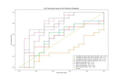 |
20 | IDH mutation prediction in glioma patients using 7T 3D-FID-MRSI
Sukrit Sharma1, Cornelius Cadrien1,2, Philipp Lazen 1, Julia Furtner3, Roxane Licandro4, Alexandra Lipkal5, Eva Heckova1, Lukas Hingerl1, Stanislav Motyka1, Stephan Gruber 1, Bernhard Strasser1, Barbara Kiesel2, Mario Mischkulnig2, Matthias Preusser6, Thomas Roetzer-Pejrimovsky7, Adelheid Wöhrer7, Michael Weber8, Christian Dorfer2, Karl Rössler2, Siegfried Trattnig5,
Wolfgang Bogner1, Georg Widhalm2, and Gilbert Hangel1,2
1High Field MR Centre, Department of Biomedical Imaging and Image-guided Therapy, Medical University of Vienna, Vienna, Austria, 2Department of Neurosurgery, Medical University of Vienna, Vienna, Austria, 3Division of Neuroradiology and Musculoskeletal Radiology, Department of Biomedical Imaging and Image-guided Therapy, Medical University of Vienna, Vienna, Austria, 4Department of Biomedical Imaging and Image-guided Therapy, Computational Imaging Research Lab (CIR), Medical University of Vienna, Vienna, Austria, 5Christian Doppler Laboratory for Clinical Molecular MR Imaging, Vienna, Austria, 6Division of Oncology, Department of Inner Medicine I, Medical University of Vienna, Vienna, Austria, 7Division of Neuropathology and Neurochemistry, Department of Neurology, Medical University of Vienna, Vienna, Austria, 8Division of Medical Imaging and Nuclear Medicine, Medical University of Vienna, Vienna, Austria
We used brain MRSI to explore the differentiation of gliomas regarding IDH-1 and Wildtype mutation. We found significant differencesin metabolites like Gln/NAA, Ins/NAA, and GPC+PCh/NAA using the Mann-Whitney-Wilcoxon test. More metabolites with signifi-cant differentiation were used for IDH classification using Random Forest, which also showed better classification results with additionalmetabolites than GPC+PCh/NAA alone. The ROC curve with more than GPC+PCh/NAA as feature yields an AUC value of 81 %.
|
||
2657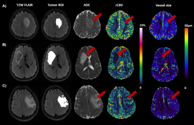 |
21 | Correlation of vessel size and cerebral blood volume measurements in glioma genetic subtypes
Fatemeh Arzanforoosh1, Sebastian R. Van Der Voort1,2, Fatih Incekara1,3, Arnaud J. P. Vincent 3, Martin Van den Bent2,3, Marion Smits1,2, and Esther A.H. Warnert1,2
1Department of Radiology & Nuclear Medicine, Erasmus MC, Rotterdam, Netherlands, 2Erasmus MC Cancer Institute, Erasmus MC, Rotterdam, Netherlands, 3Department of Neurology, Erasmus MC, Rotterdam, Netherlands
Deep insight about tumor microvasculature is important for diagnosis and prognosis of glioma patients. Relative Cerebral Blood Volume (rCBV) and vessel size are two parameters, derived from perfusion MRI, used for evaluation of tumor microvasculature in glioma. In this study, we investigated the clinical value of both rCBV and vessel size and their correlation for three subgroups of glioma based on the recent 2021 World Health Organization (WHO) classification scheme. The result showed that neither rCBV nor vessel size differed significantly between glioma subtypes, though correlation of these two parameters sheds light on the microvasculature characteristics of each subgroup.
|
||
2658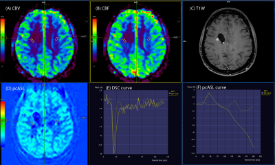 |
22 | Using combined DSC and pcASL perfusion imaging to assess treatment response of high-grade glioma.
Mariia Khurtsylava1 and Maksym Kovratko1
1Diagnostic department, Capital medical center universal clinic Oberig, Kyiv, Ukraine
To determine the efficacy and feasibility of using combined DSC and ASL perfusion imaging in a routine protocol to assess the response to treatment of high-grade gliomas in adults. Perfusion imaging is performed for perform differentiation of tumor recurrence from radiation necrosis, since it provides information about angiogenesis at the microscopic level. The data obtained show that the combined use of ASL and DSC perfusion in a routine protocol can be considered as possible additional methods in differentiating recurrent tumor from pseudoprogression.
|
||
2659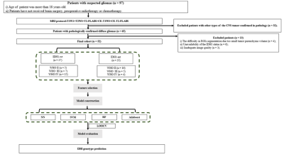 |
23 | Machine Learning-Based Contrast Enhanced T2-FLAIR Radiomics method for predicting IDH1 Genotype of Diffuse Gliomas
Han Bao1, Yi Lu1, Qirui Zhao1, Zujun Hou2, Liuyang Chen3, Wei Xie1, Qing Wang1, Wei Zhao1, Tong-San Koh4, Lisha Nie5, and Zongfang Li1
1Department of Radiology, The First Affiliated Hospital of Kunming Medical University, Kunming, China, 2Suzhou Institute of Biomedical Engineering and Technology,, Chinese Academy of Sciences, Suzhou, China, 3Fisca Healthcare Ltd, Kumming, China, 4Department of Oncologic Imaging, National Cancer Center,Duke-NUS Graduate Medical School, Singapore, Singapore, 5MR Research, GE Healthcare, Beijing, China
The current study aims to build isocitrate dehydrogenase 1 (IDH1) genotype prediction models based on selected radiomics features derived from contrast-enhanced T2 fluid attenuated inversion recovery (CE-T2-FLAIR) in predicting IDH1 genotype of diffuse gliomas. Radiomics features from CE-T2-FLAIR images go a step further to enrich the content of MRI-based radiomics. It was concluded that machine learning-based radiomics of CE-T2-FLAIR could efficiently predict the IDH1 genotype of diffuse gliomas.
|
||
2660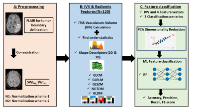 |
24 | ITSS vasculature volume and Radiomics Features from MR SWI: Performance in Glioma Grading
Rupsa Bhattacharjee1,2, Rakesh Kumar Gupta3, Rana Patir4, Sandeep Vaishya4, Suneeta Ahlawat5, and Anup Singh1,6
1Center for Biomedical Engineering, Indian Institute of Technology (IIT), Delhi, New Delhi, India, 2Department of Radiology & Biomedical Engineering, University of California, San Francisco (UCSF), San Francisco, CA, United States, 3Department of Radiology and Imaging, Fortis Memorial Research Institute, Gururam, India, 4Department of Neurosurgery, Fortis Memorial Research Institute, Gurugram, India, 5SRL Diagnostics, Fortis Memorial Research Institute, Gurugram, India, 6Department of Biomedical Engineering, All India Institute of Medical Sciences, New Delhi, India Majority of the glioma studies rely on using radiomic-features extracted only from conventional MRI and noticeably limited in SWI. Main objective of the current study is to evaluate the role of SWI in glioma grading aided by machine-learning (ML) methods, in terms of ITSS-vasculature-volume(IVV), radiomic features and by combining both. We conclude SWI to be the one of the useful sequences in the glioma-analysis as it contributes to calculate the IVV, which can singlehandedly improve the glioma grading. When IVV is combined with radiomic-analysis used in combination with PCA feature reduction and RF ML classifier, the performance is marginally improved. |
||
2661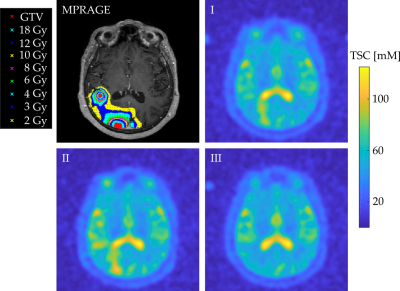 |
25 | Tissue Sodium Concentration Quantifies Cell Response after Radiation Therapy with Stereotactic Radiosurgery
Anne Adlung1, Sherif A Mohamed2,3, Michaela AU Hoesl1, Frank A Giordano4,5, Eva Neumaier Probst2, Lothar R Schad1, and Arne M Ruder4,6
1Computer Assisted Clinical Medicine, Medical Faculty Mannheim, Heidelberg University, Mannheim, Germany, 2Department of Neuroradiology, Medical Faculty Mannheim, Heidelberg University, Mannheim, Germany, 3Department of Diagnostic and Interventional Radiology, University Hospital Heidelberg, Heidelberg, Germany, 4Department of Radiation Oncology, Medical Faculty Mannheim, Heidelberg University, Mannheim, Germany, 5Department of Radiation Oncology, University Hospital Bonn, Bonn, Germany, 6Department of Radiation Oncology, University Hospital Heidelberg, Heidelberg, Germany
We investigated TSC within Gross Tumor Volume (GTV), and surrounding tissue and healthy tissue (HR). 12 patients with a total of 14 GTV was included. 23Na MRI were acquired at three different time-points (2days pre-, 5days post-, 40days post-SRS). TSC was measured within GTV, HR and isodose-areas between 2 and 18Gy at all time-points. Results show changes in TSC within GTV shortly after SRS and subsequent TSC recovery. TSC was higher within higher isodose-areas showing a correlation of radiation exposure and TSC. Findings suggest that TSC might be able to quantify cell response to radiation of tumorous and healthy tissue.
|
||
2662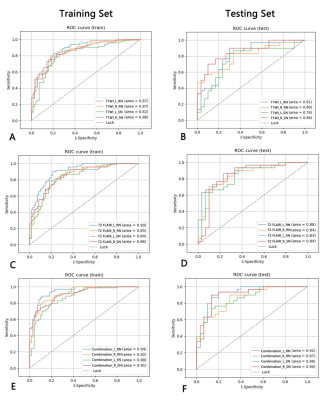 |
26 | Utility of T1WI and T2 FLAIR for Diagnosis of Parkinson’s Disease Using Radiomics
Di Wang1, Baohui Lou2, Sicong Wang3, Pu-Yeh Wu3, Chunmei Li2, and Min Chen2
1Beijing Hospital, National Center of Gerontology; Institute of Geriatric Medicine, Chinese Academy of Medical Sciences, P.R. China, Beijing, China, 2Beijing Hospital, Beijing, China, 3GE Healthcare, Beijing, China, Beijing, China
Radiomic features of bilateral RN and SN based on T1WI and T2 FLAIR from 100 PD patients and 100 healthy controls showed good performance in differentiation of PD, the radiomic model of combine T1WI and T2 FLAIR had best performance than individual T1WI and T2 FLAIR. Radiomics may provide MR imaging biomarkers for PD, and the routine MR sequence may help to diagnose PD using radiomics.
|
||
 Back to Meeting Home
 Back to the Program-at-a-Glance
The International Society for Magnetic Resonance in Medicine is accredited by the Accreditation Council for Continuing Medical Education to provide continuing medical education for physicians.
 Back to Meeting Home
 Back to the Program-at-a-Glance
 View the Presentation