Digital Poster
Image Reconstruction & Signal Models: Low Rank, Sparse, Rapid & Artifact-Free I
Joint Annual Meeting ISMRM-ESMRMB & ISMRT 31st Annual Meeting • 07-12 May 2022 • London, UK

Digital Poster
Image Reconstruction & Signal Models: Low Rank, Sparse, Rapid & Artifact-Free I
| Computer # | ||||
|---|---|---|---|---|
1603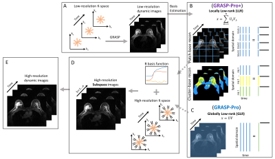 |
58 | GRASP-Pro+: GRASP reconstruction with locally low-rank subspace constraint for DCE-MRI
Eddy Solomon1, Jonghyun Bae1,2, Elcin Zan2, Linda Moy2, Yulin Ge2, Li Feng3, and Gene Sungheon Kim1
1Department of Radiology, MRI Research Institute, Weill Cornell Medicine, New York, NY, United States, 2Department of Radiology, New York University, New York, NY, United States, 3BioMedical Engineering and Imaging Institute, Icahn School of Medicine at Mount Sinai, New York, NY, United States
While the globally low-rank (GLR) model has been demonstrated to be effective in representing global contrast change, it is expected to be less effective for spatially localized signal dynamics. In this work, we propose an improved reconstruction framework, which extends GRASP-Pro using a locally low-rank (LLR) model to represent spatially localized dynamics based on clusters or patches information. This approach has been tested in multiple DCE applications including both cancer and healthy subjects. In addition, we propose an anatomical cluster-based reconstruction approach for brain DCE-MRI.
|
||
1604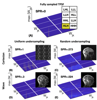 |
59 | Rapid CS-Wave MPRAGE acquisition with automated parameter selection
Gabriel Varela-Mattatall1,2,3, Tae Hyung Kim3, Jaejin Cho3, Wei-Ching Lo4, Borjan A. Gagoski5,6, Ravi S. Menon1,2, and Berkin Bilgic3,7
1Centre for Functional and Metabolic Mapping (CFMM) | Robarts Research Institute | Western University, London, ON, Canada, 2Department of Medical Biophysics | Schulich School of Medicine and Dentistry | Western University, London, ON, Canada, 3Department of Radiology | Athinoula A. Martinos Center for Biomedical imaging | Massachusetts General Hospital & Harvard Medical School, Charlestown, MA, United States, 4Siemens Medical Solutions USA, Inc., Charlestown, MA, United States, 5Department of Radiology | Harvard Medical School, Boston, MA, United States, 6Fetal-Neonatal Neuroimaging & Developmental Science Center | Boston Children's Hospital, Boston, MA, United States, 7Harvard-MIT Division of Health Sciences and Technology, Massachusetts Institute of Technology, Cambridge, MA, United States
Wave encoding mitigates g-factor noise amplification in highly accelerated parallel imaging but achieving ultra-high acceleration factors is precluded by the intrinsic “√R” SNR penalty. To overcome this limitation, we propose a compressed sensing-based reconstruction with automatic selection of the regularization weighting. Moreover, we show that CS-Wave is flexible enough to perform well with uniform undersampling. We compare reconstruction performance of CS-Wave against the state-of-art Wave-LORAKS which requires parameter tuning, and evaluate different undersampling patterns at R=12-fold acceleration. Results indicate higher reconstruction quality and showcase the feasibility of ultra-fast Wave-MPRAGE acquisitions.
|
||
1605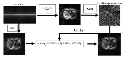 |
60 | EigenGRASP: Subject-Specific Temporal-Learning Radial Sampling Image Reconstruction in Dynamic Contrast-Enhanced MRI
Ramin Jafari1, Masoud Zarepisheh1, Richard Kinh Gian Do2, and Ricardo Otazo1
1Memorial Sloan Kettering Cancer Center, New York, NY, United States, 2Memorial Sloan Kettering Cancer Center, New york, NY, United States
GRASP is an image reconstruction algorithm for free-breathing dynamic contrast-enhanced MRI which uses universal L1-type regularization to suppress undersampling artifacts. We propose to replace it with a subject-specific data-driven L2-type regularization which can improve image quality and decrease reconstruction time.
|
||
1606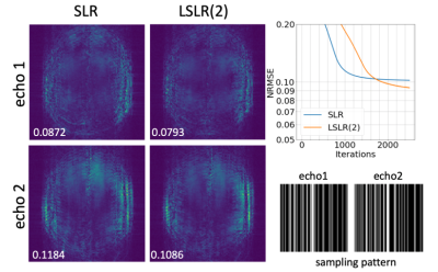 |
61 | A Locally Structured Low-Rank Tensor Method Using Submatrix Constraints for Joint Multi-echo Image Reconstruction
Xi Chen1, Wenchuan Wu1, and Mark Chiew1
1Wellcome Centre for Integrative Neuroimaging, FMRIB, Nuffield Department of Clinical Neurosciences, University of Oxford, Oxford, United Kingdom Recently, we proposed an improved structured low-rank (SLR) reconstruction - locally structured low-rank (LSLR) method, which forces low-rank constraints on submatrices of the Hankel structured matrix. This simple modification leads to a robust improvement over conventional SLR reconstruction, which has been validated on the low-rank tensor reconstruction for the multi-echo GRE data. |
||
1607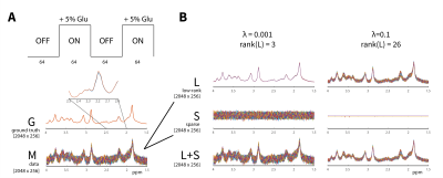 |
62 | Applying L+S decomposition to improve sensitivity to dynamic changes in functional MR spectroscopy
Adam Berrington1 and I. Betina Ip2
1Sir Peter Mansfield Imaging Centre, University of Nottingham, Nottingham, United Kingdom, 2Wellcome Centre for Integrative Neuroimaging, University of Oxford, Oxford, United Kingdom
Functional MRS (fMRS) is a powerful technique to measure metabolite responses over time. However, noise and spectral contamination limit the ability to study individual metabolite time-courses. In this work, we propose to model fMRS spectra as a superposition of low-rank (L) and sparse components (S). L+S decomposition resulted in separation of temporally-correlated signal from noise in simulation. In vivo, L+S spectra had higher SNR compared to original data (P=0.007) and the mean glutamate time-course, using L+S spectra, was more strongly correlated to stimulus. L+S decomposition is a promising data-driven method to enhance sensitivity to dynamic changes in fMRS.
|
||
1608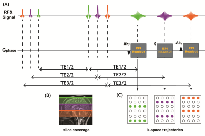 |
63 | Joint reconstruction of multi-TE diffusion MRI acquired using TDM-EPI with complementary k-space sampling
Yang Ji1,2, Congyu Liao3, William Scott Hoge4, Berkin Bilgic5, Yogesh Rathi1,4, and Lipeng Ning1
1Department of Psychiatry, Brigham and Women’s Hospital, Harvard Medical School, Boston, MA, United States, 2Wellcome Centre for Integrative Neuroimaging, FMRIB Division, Nuffield Department of Clinical Neurosciences, University of Oxford, Oxford, United Kingdom, 3Department of Radiology, Stanford University, Stanford, CA, United States, 4Department of Radiology, Brigham and Women’s Hospital, Harvard Medical School, Boston, MA, United States, 5Athinoula A. Martinos Center for Biomedical Imaging, Massachusetts General Hospital, Harvard Medical School, Boston, MA, United States
Combined diffusion-relaxometry has demonstrated promising capability to noninvasively probe tissue microstructure by joint modeling of relaxation coefficients and diffusivity. Our recent work has introduced a sequence based on the time-division multiplexing technique to accelerate the acquisition of relaxation-diffusion MRI. In this work, we further developed the TDM-EPI sequence by integrating ky-shifted k-space sampling strategies for data acquired at different TEs. Moreover, we implemented and compared several reconstruction methods to integrate complementary k-space samples to joint estimate images at different TEs. The results showed that the joint reconstruction approach can improve image quality and reduce artifact compared with conventional reconstruction methods.
|
||
1609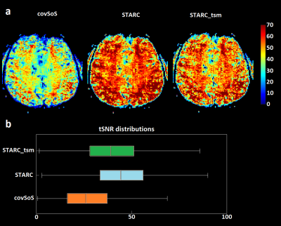 |
64 | Pitfalls of data-driven tSNR optimized coil combination for fMRI
Redouane Jamil1, Franck Mauconduit1, Caroline Le Ster1, Philipp Ehses2, Benedikt A Poser3, Alexandre Vignaud1, and Nicolas Boulant1
1CEA, CNRS, BAOBAB, NeuroSpin, Gif-sur-Yvette, France, France, 2German Center for Neurodegenerative Diseases (DZNE), Bonn, Germany, 3Department of Cognitive Neuroscience, Maastricht Brain Imaging Centre, Faculty of Psychology and Neuroscience, Maastricht University, Maastricht, Netherlands
For MRI with a multi-receiver RF coil, one image per coil element and per time frame is obtained. The final image is typically calculated from the root sum of squares (rSoS) combination across channels. While this combination approach is quasi-optimal for SNR, it is not necessarily optimal for temporal SNR (tSNR) of the time-series. We present two analytical and voxel-wise coil combination expressions reaching optimality in tSNR and t-score for the mean (TSM) respectively. Their BOLD sensitivity is compared to the gold standard covariance root sum of squares. Both improved tSNR and TSM but yielded weaker t-scores than covSoS.
|
||
1610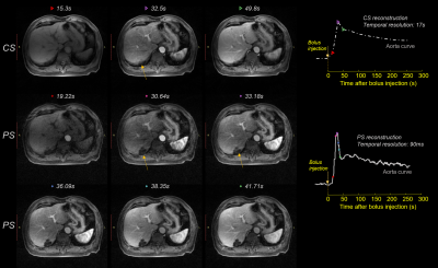 |
65 | Fast 3D DCE MRI using partially separable model: autopilot Multitasking
Jingyuan Lyu1, Qi Liu1, Yu Ding1, Jian Xu1, Xiaomao Gong2, Weiguo Zhang1, Shengxiang Rao3,4,5, Wentao Wang3,4,5, and Mengsu Zeng3,4,5
1UIH America, Inc., Houston, TX, United States, 2United Imaging Healthcare, Shanghai, China, 3Department of Radiology, Zhongshan Hospital, Fudan University, Shanghai, China, 4Department of Medical Imaging, Shanghai Medical College, Fudan University, Shanghai, China, 5Shanghai Institute of Medical Imaging, Shanghai, China
Inspired by Multitasking, we have developed a fast reconstruction method for 3D stack-of-star (SoS) acquisitions to achieve high temporal resolution (0.1s) DCE MRI without additional navigator data. The proposed method is fully compatible with existing sequence using 3D SoS trajectories, but with less computation complexity.
|
||
 Back to Meeting Home
 Back to the Program-at-a-Glance
The International Society for Magnetic Resonance in Medicine is accredited by the Accreditation Council for Continuing Medical Education to provide continuing medical education for physicians.
 Back to Meeting Home
 Back to the Program-at-a-Glance
 View the Presentation