Digital Poster
Advances in Parkinson's Disease Imaging II
Joint Annual Meeting ISMRM-ESMRMB & ISMRT 31st Annual Meeting • 07-12 May 2022 • London, UK

Digital Poster
Advances in Parkinson's Disease Imaging II
| Computer # | ||||
|---|---|---|---|---|
2372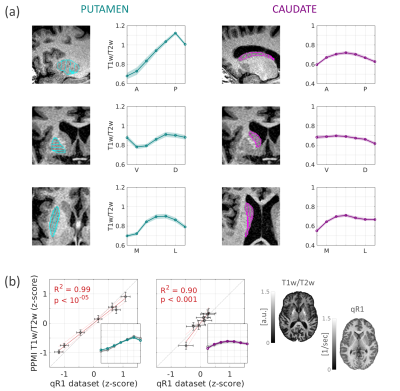 |
1 | Parkinson’s disease local atrophies measured in the striatum in vivo explain patients’ asymmetric motor symptoms and dopamine loss
Elior Drori1, Shai Berman1, and Aviv Mezer1
1The Edmond and Lily Safra Center for Brain Sciences, The Hebrew University of Jerusalem, Jerusalem, Israel
The striatum is a heterogeneous brain structure with microstructural gradients along its main axes. Changes in its organization are associated with Parkinson’s disease (PD). Yet the spatial variability in the human striatum is not well characterized and is mostly limited to animal and postmortem studies. We developed a non-invasive MRI method for measurement of microstructural gradients along axes of the striatum in individuals in vivo. Using clinical data, we found a T1w/T2 gradient along the anterior-posterior axis of the putamen, which is decreased in PD. This decrease explains the patients’ asymmetries in dopamine loss and motor symptoms.
|
||
2373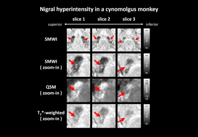 |
2 | Observing hyperintensity region within substantia nigra in cynomolgus monkey
Chungseok Oh1, Chang-Yeop Jeon2, Jincheol Seo2, Hyeong-Geol Shin1, Sooyeon Ji1, Seongbeom Park3, Soohwa Song3, Dong Hoon Shin3, Youngjeon Lee2, and Jongho Lee1
1Department of Electrical and Computer Engineering, Seoul National University, Seoul, Korea, Republic of, 2National Primate Research Center, Korea Research Institute of Bioscience and Biotechnology (KRIBB), Cheongju, Korea, Republic of, 3Heuron Co.,Ltd, Incheon, Korea, Republic of
In this study, a hyperintensity region within substantia nigra is explored in cynomolgus monkeys over a wide range of ages. This region is similar to that of humans, which is often called as a swallow tail sign in healthy subjects. This nigral hyperintensity appears consistently in SMWI, QSM, and T2*-weighted images, particularly in aged animals.
|
||
2374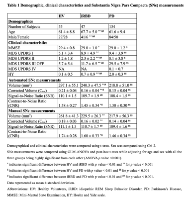 |
3 | Nigral Neuromelanin MRI Changes in Isolated Rapid Eye Movement Sleep Behavior Disorder Using Deep Learning
Rahul Gaurav1,2, Romain Valabrègue2, Nadya Pyatigorskaya1,2,3, Graziella Mangone4, Smaranda Leu-Semenescu5, Jean-Christophe Corvol4,6, Marie Vidailhet1,6, Isabelle Arnulf1,5, and Stephane Lehericy1,2,3
1Movement Investigations and Therapeutics Team (MOV’IT), Paris Brain Institute (ICM), Paris, France, 2CENIR, Paris Brain Institute (ICM), Paris, France, 3Department of Neuroradiology, Pitié-Salpêtrière Hospital, AP-HP, Paris, France, 4INSERM, Clinical Investigation Center for Neurosciences (CIC), Paris Brain Institute (ICM), Paris, France, 5Sleep Disorders Unit, Pitié-Salpêtrière Hospital, AP-HP, Paris, France, 6Department of Neurology, Pitié-Salpêtrière Hospital, AP-HP, Paris, France
Isolated REM sleep behavior disorder (iRBD) is considered a prodromal stage of parkinsonism. Neurodegenerative changes in the substantia nigra pars compacta (SNc) in parkinsonism can be detected using neuromelanin-sensitive MRI. In this cross-sectional, observational, case-control study, we investigated changes in neuromelanin in participants with iRBD compared to Parkinson’s disease and healthy volunteers in the SNc segmented automatically using convolutional neural network-based U-net architecture. The iRBD participants had reduced neuromelanin content at an intermediate level between the values in HV and PD patients.
|
||
2375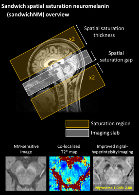 |
4 | Multiecho sandwich spatial saturation neuromelanin imaging: improved nigral hyperintensity and need for T2* correction in NM-sensitive images
Sooyeon Ji1, Eun-Jung Choi1, Beomseok Sohn2, Kyoungwon Baik2, Eung Yeop Kim3, In Seong Kim4, Jin Wook Choi5, Seongbeom Park6, Soohwa Song6, Dong Hoon Shin6, Phil Hyu Lee7, and Jongho Lee1
1Seoul National University, Seoul, Korea, Republic of, 2Severance Hospital, Seoul, Korea, Republic of, 3Department Radiology, Samsung Medical Center, Seoul, Korea, Republic of, 4Siemens Healthineers Ltd., Seoul, Korea, Republic of, 5Department of Radiology, Ajou University School of Medicine, Ajou University Medical Center, Suwon, Korea, Republic of, 6Heuron Co. Ltd., Incheon, Korea, Republic of, 7Department of Neurology, Severance Hospital, Seoul, Korea, Republic of
Multi-echo sandwich spatial saturation neuromelanin (sandwichNMmulti-echo) imaging is proposed to simultaneously acquire a high-quality neuromelanin-sensitive image, a co-localized T2* map, and an improved susceptibility map weighted image for nigral hyperintensity. When this method is applied for PD patients and healthy controls, the results suggest that the reduced neuromelanin contrast in PD is partially explained by shorter T2* in substantia nigra of the PD patients. Initial results at 7T further support the finding, suggesting a need for T2* correction in assessing the NM-sensitive images.
|
||
2376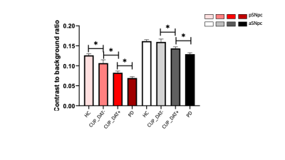 |
5 | Clinically uncertain parkinsonism shows nigral depigmentation in the presence and absence of dopaminergic deficit: a multimodal imaging study
YUE XING1,2,3, Saadnah Naidu1,2,3, Marta Gennaro4,5, Andreas Antonios-Roussakis6, Nicholas P. Lao-Kaim4, Halim Abdul-Sapuan1,2,3, Jonathan Evans7, Gillian Sare7, Antonio Martin-Bastida4,8, Schwarz T. Stefan1,2,9, Paola Piccini4,6, and Dorothee P Auer1,2,3
1Mental Health and Clinical Neuroscience Unit, University of Nottingham, Nottingham, United Kingdom, 2Sir Peter Mansfield Imaging Centre, University of Nottingham, Nottingham, United Kingdom, 3NIHR Nottingham Biomedical Research Centre, University of Nottingham, Nottingham, United Kingdom, 4Division of Neurology, Imperial College London, London, United Kingdom, 5Nuclear Medicine department, Royal Brompton & Harefield hospitals, London, United Kingdom, 6NHLI Department, Imperial College London, London, United Kingdom, 7Nottingham University Hospitals, Nottingham, United Kingdom, 8Department of Neurology and Neurosciences, Clínica Universidad de Navarra, Pamplona-Madrid, Spain, 9Department of Radiology, Cardiff and Vale University Health Board, Cardiff, United Kingdom
Brain dopamine transporter (DAT) imaging with 123I-FP-CIT SPECT has been used for detecting striatal deficits in clinically uncertain parkinsonism (CUP). Neuromelanin(NM)-MRI assesses nigral depigmentation with high accuracy in confirmed PD; but its diagnostic value in CUP is unclear. Here, we compare nigral NM in CUP with (DAT+) or without (DAT-) striatal dopaminergic deficits, to established PD and healthy controls. CUP with DAT+ had more nigral NM deficits (vs. controls and DAT-CUP) but were less pronounced than in established PD. Interestingly, DAT- cases showed ventrolateral nigral deficits. Our results suggest that nigral depigmentation possibly starts before striatal dopaminergic decline.
|
||
2377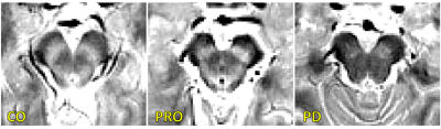 |
6 | Examination of nigral volumetric and microstructural changes in prodromal and overt Parkinson’s disease
Jason Langley1, Kristy S. Hwang2,3, Daniel E. Huddleston4, and Xiaoping Hu1,5
1Center for Advanced Neuroimaging, University of California Riverside, Riverside, CA, United States, 2Department of Neurosciences, University of California San Diego, San Diego, CA, United States, 3Parkinson & Other Movement Disorders Center, University of California San Diego, San Diego, CA, United States, 4Department of Neurology, Emory University, Atlanta, GA, United States, 5Department of Bioengineering, University of California Riverside, Riverside, CA, United States
We examine nigral volume and tissue microstructure in prodromal and symptomatic Parkinson’s disease. Decreases in nigral volume were observed in the symptomatic Parkinson's disease group relative to the control group (p<10-3) and prodromal Parkinson's disease (p<10-3) group. A reduction in nigral volume was seen in the prodromal Parkinson's disease group relative to the control group (p=0.025). An increase in nigral free water was also found in the symptomatic Parkinson’s disease group relative to the control group (p=0.014).
|
||
2378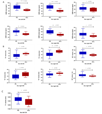 |
7 | Quantitative Analysis of Deep Nuclei in Patients with Parkinson’s disease (PD) using STrategically Acquired Gradient Echo (STAGE) Imaging Video Not Available
Zhaoxi Liu1, Yiwei Zhang2, Han Wang1, Dan Xu1, Bo Wu3, E. Mark Haackeb4, Hui You1, and Feng Feng1
1Peking Union Medical College Hospital, Bejing, China, 2Peking university first hospital, Bejing, China, 3SpinTech, Inc., Bingham Farms, MI, United States, 4Wayne State University, Detroit, MI, United States
Patients with Parkinson’s disease (PD) showed increased iron deposition in the substantia nigra, red nucleus, dentate nucleus and globus pallidus compared with healthy controls. Besides, the T1 value, T2* value and R2* value of deep nucleus also altered in PD patients. Which suggested the STrategically Acquired Gradient Echo (STAGE) imaging could provide a good performance in quantitative analysis in multi-modal parameters.
|
||
2379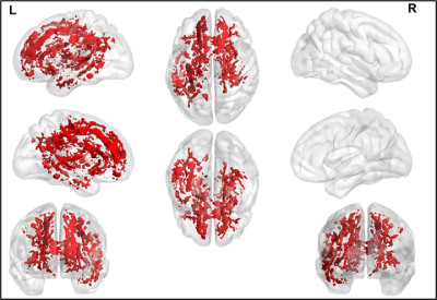 |
8 | Free-Water Corrected Diffusion MRI Measures Reveal White Matter Disorganization in Parkinson’s Disease Patients with Memory Impairment.
Virendra R Mishra1, Karthik R Sreenivasan1, Jessica Caldwell2, Aaron Ritter3, and Zoltan K Mari3
1Imaging, Cleveland Clinic Lou Ruvo Center for Brain Health, Las Vegas, NV, United States, 2Neuropsychology, Cleveland Clinic Lou Ruvo Center for Brain Health, Las Vegas, NV, United States, 3Neurology, Cleveland Clinic Lou Ruvo Center for Brain Health, Las Vegas, NV, United States
Diffusion MRI (dMRI) could be used to understand cognitive impairment in Parkinson’s disease (PD). However, single-tensor (ST)-derived fractional anisotropy (FA) measures are biased due to the presence of crossing-fibers and contamination from cerebrospinal fluid (CSF). CSF contamination can be corrected by fitting a bi-tensor model to estimate free water (FW) contamination. Hence, in this study, we compared FW and FW-corrected ST dMRI-derived measures between PD patients (with and without cognitive impairment) and healthy controls (HC) estimated with multishell dMRI data. Our analysis suggests that FW-corrected dMRI analyses are more powerful in understanding WM disorganization in PD patients with cognitive impairment.
|
||
2380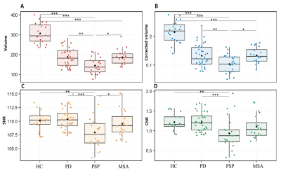 |
9 | Regional patterns of nigral degeneration in the substantia nigra in atypical Parkinsonism using neuromelanin-sensitive MRI
LYDIA CHOUGAR1,2, EMINA ARSOVIC1, RAHUL GAURAV3, EMMA BIONDETTI4, NADYA PYATIGORSKAYA1, and STEPHANE LEHERICY1
1PITIE SALPETRIERE, PARIS, France, 2PARIS BRAIN INSTITUTE, PARIS, France, 3Paris Brain Institute, PARIS, France, 4Università degli Studi "G. d'Annunzio" Chieti, CHIETI, Italy
We investigated the regional selectivity of neurodegenerative changes in the SNc in patients with Parkinson’s disease (PD) and atypical parkinsonism using neuromelanin-sensitive MRI. We confirmed that neuromelanin volume and signal were reduced in parkinsonian disorders. The spatial pattern of changes differed between progressive supranuclear palsy (PSP) and synucleinopathies. Compared to healthy controls (HC), subjects with PD and multisystem atrophy (MSA), PSP subjects had greater changes in the associative region. Signal-to-noise in the sensorimotor territory was preserved in PSP, but reduced in PD patients and there was a trend in MSA patients. There was no significant difference between MSA and PD.
|
||
2381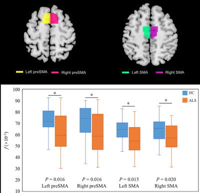 |
10 | Sensorimotor-Related Area Abnormalities in Amyotrophic Lateral Sclerosis: An Intravoxel Incoherent Motion Magnetic Resonance Imaging Study Video Not Available
Jia Yan Shi1 and Hua Jun Chen2
1Fujian Medical University Union Hospital, Fuzhou, China, 2Radiology, Fujian Medical University Union Hospital, Fuzhou, China We conducted the first assessment of the sensorimotor-related areas(SMA) alterations in ALS using IVIM MRI and atlas-based analysis, which may provide useful information for localization of specific cortex and white matter(WM) tract damage in ALS. In grey matter areas (GM), ALS patients showed a reduction in f in the bilateral preSMA and SMA, while in WM areas, ALS patients exhibited an increase in D in a WM tract projecting to the right ventral premotor cortex(PMv). Correlation analysis revealed that the average D in this right PMv tract was negatively correlated to ALS disease severity. |
||
2382 |
11 | Resting-State Functional Connectivity of the Central Autonomic Network (CAN) in Parkinson’s Disease
Pohchoo Seow1, Yao-Chia Shih2, Septian Hartono3, Li Rong Yin1, Aeden Zi Cheng Kuek1, Samuel Yong Ern Ng3, Eng King Tan3, Louis Tan3, and Ling Ling Chan1
1Diagnostic Radiology, Singapore General Hospital, Singapore, Singapore, 2Graduate Institute of Medicine, Yuan Ze University, Taipei, Taiwan, 3Neurology, National Neuroscience Institute, Singapore, Singapore
Vast evidence for network-level functional dysfunctions have been reported in Parkinson’s disease (PD). However, brain abnormalities that underlie common autonomic symptoms in PD have not been fully investigated. Resting-state functional integration of the Central Autonomic Network (CAN) of 79 PD patients and 43 healthy controls (HC) were evaluated using seed-based analysis. Significantly decreased functional connectivity between the right anterior insular seed and left lateral occipital cortex, right lingual gyrus, and left occipital pole were found in HC compared to PD. Functional connectivity analysis of the CAN facilitates further understanding of the potential mechanisms underlying altered autonomic regulation in PD.
|
||
2383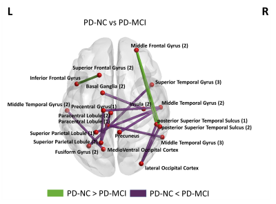 |
12 | Disrupted brain functional network connectivity in Parkinson’s disease patients with Mild Cognitive Impairment.
Karthik R Sreenivasan1, Xiaowei Zhuang1, Jessica Caldwell1, Aaron Ritter1, Dietmar Cordes1, Zoltan Mari1, and Virendra Mishra1
1Cleveland Clinic Lou Ruvo Center for Brain Health, Las Vegas, NV, United States
The underlying cause of cognitive deficits in PD (PD-MCI) is not well understood and there are no biomarkers to diagnose PD-MCI. Although previous studies have found disrupted functional connectivity several factors make it difficult to generalize findings from fMRI studies. In the current study, we investigate functional network abnormalities in PD-MCI and PD patients who are cognitively normal (PD-NC). Our results were in line with earlier studies that showed altered connectivity was observed involving the frontal and striatal regions in the PD-MCI group, and we also found altered connectivity between regions in the temporal and the parietal cortex.
|
||
2384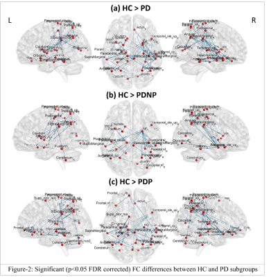 |
13 | Altered functional connectivity in Parkinson’s disease with psychosis Video Permission Withheld
Apoorva Safai1, Shweta Prasad2,3, Jitender Saini4, Abhishek Lenka2, Pramod Pal2, and Madhura Ingalhalikar1
1Symbiosis Centre for Medical Image Analysis, Symbiosis International University, Pune, India, 2Department of Neurology, National Institute of Mental Health & Neurosciences, Bangalore, India, 3Department of Clinical Neuroscience, National Institute of Mental Health & Neurosciences, Bangalore, India, 4Department of Neuroimaging & Interventional Radiology, National Institute of Mental Health & Neurosciences, Bangalore, India
Visual hallucinations (VH) is a commonly occurring psychosis symptom in Parkinson’s disease (PD). However, neuropathology of VH in PD is not clearly known, thereby limiting the efficacy of therapeutic strategies. To mitigate this gap, we evaluated functional connectivity (FC) changes and network organisation, related to VH in PD. PD patients exhibited widespread reduction in interhemispheric FC and local network topology of temporal, frontal, parietal, striatal, limbic, cerebellar, occipital and sensory motor regions. Patients with psychosis displayed larger FC reductions, particularly in cerebellar regions and their connections with frontal and occipital regions. Topological changes in temporal regions seen across PD patients
|
||
 Back to Meeting Home
 Back to the Program-at-a-Glance
The International Society for Magnetic Resonance in Medicine is accredited by the Accreditation Council for Continuing Medical Education to provide continuing medical education for physicians.
 Back to Meeting Home
 Back to the Program-at-a-Glance
 View the Presentation