Digital Poster
Advances in Epilepsy & Neurology Imaging
Joint Annual Meeting ISMRM-ESMRMB & ISMRT 31st Annual Meeting • 07-12 May 2022 • London, UK

Digital Poster
Advances in Epilepsy & Neurology Imaging
| Computer # | ||||
|---|---|---|---|---|
1923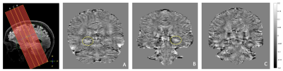 |
18 | 7T metabolic MRI in focal epilepsy
Sarah M Jacobs1, Zahra Shams1, Evita C Wiegers1, Jannie P Wijnen1, Dennis W Klomp1, Edwin Versteeg1, Jeroen C.W. Siero1,2, Angelika Mühlebner3,4, Wim Van Hecke3, Pieter van Eijsden5, Pierre A Robe5, Maeike Zijlmans5,6, and Anja G van der Kolk1,7
1Department of Radiology and Nuclear Medicine, University Medical Center Utrecht, Utrecht, Netherlands, 2Spinoza Centre for Neuroimaging Amsterdam, Amsterdam, Netherlands, 3Department of Pathology, University Medical Center Utrecht, Utrecht, Netherlands, 4Department of Pathology, Amsterdam University Medical Center, location AMC, Amsterdam, Netherlands, 5UMC Utrecht Brain Center, Department of Neurology and Neurosurgery, University Medical Center Utrecht, Utrecht, Netherlands, 6Stichting Epilepsie Instellingen Nederland (SEIN), Heemstede, Netherlands, 7Department of Radiology, Radboud University Medical Center, Nijmegen, Netherlands In this clinical study, we combined different (metabolic) MRI sequences at 7T to characterize focal epileptogenic lesions, and uncover potential metabolic markers that could help identifying the culprit lesion in MRI-negative epilepsy patients. Using QSM, we observed increased iron deposition in the affected hippocampus of two HS patients that was not found in the contralateral hippocampus, neither in a suspected HS patient with no abnormal tissue, nor in matched healthy volunteers. No increased iron deposition was found in patients with FCD or matched healthy volunteers. No significantly different metabolite ratios between patients and healthy volunteers were found using SV 1H-MRS. |
||
1924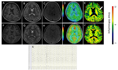 |
19 | Diffusion-weighted MRI-based Virtual Elastography is feasibility for the evaluate of Rolandic epilepsy Video Not Available
Lu Gao1, Xianjun Li1, Yuying Feng2, Chao Jin2, Hua Zhang3, Huanfa Li3, Haipeng Hu4, Xiaocheng Wei5, and Jian Yang 2
1Departemnt of diagnostic Radiology, The First Affiliated Hospital of Xi'an Jiaotong University, Xi'an, China, 2The First Affiliated Hospital of Xi'an Jiaotong University, Xi'an, China, 3Departemnt of Neurosurgery, The First Affiliated Hospital of Xi'an Jiaotong University, Xi'an, China, 4Departemnt of Pediatrics, The First Affiliated Hospital of Xi'an Jiaotong University, Xi'an, China, 5GE Healthcare, MR Research China, Beijing, Beijing, China
Conventional MRI is difficult to discover the subtle changes caused by epilepsy. Virtual magnetic resonance elastography (vMRE) is based on diffusion MRI, which is sensitive for variety of physiologic and pathologic conditions. This study use vMRE to evaluated left Rolandic epilepsy. We found that left precentral gyrus, left postcentral gryus and left superior temporal gyrus is more stiffer than controls. The longer the seizure duration the stiffer of the brain tissues. vMRE has the potential to noninvasive evaluate epilepsy.
|
||
1925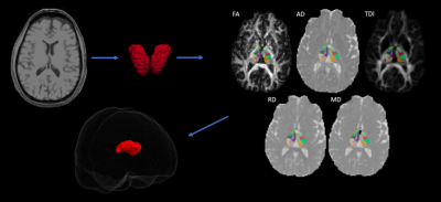 |
20 | Microstructural Reorganizations in Thalamic Nuclei Across Characteristics of Temporal Lobe Epilepsy
KiChang Kang1, Anish V. Sathe1, Isaiah Ailes1, India Shelley1, Mashaal Syed1, Christopher Miller1, Feroze Mohamed1, Ashwini Sharan1, and Mahdi Alizadeh1
1Thomas Jefferson University, Philadelphia, PA, United States
Temporal lobe epilepsy (TLE) is the most common form of epilepsy which can become medically refractory and referred for surgery. The thalamus has been shown to demonstrate both structural and functional changes in TLE. However, little is known about microstructural alterations within individual thalamic nuclei. In this study, we used diffusion tensor imaging (DTI) to determine if subjects with unilateral TLE demonstrate hemispheric and intra-hemispheric differences in thalamic nuclei across clinical characteristics. We report statistically significant differences in nuclei in subjects with tonic-clonic seizures and mesial temporal sclerosis in both hemispheric and intra-hemispheric comparisons.
|
||
1926 |
21 | Segmentation and Quantification of Venous structures and Perivascular Spaces in the Thalamus in Epilepsy Patients at 7T
Mackenzie Langan1,2, Gaurav Verma1, Derek Smith1, Bradley Delman3, Madeline Fields4, Rebecca Feldman*1,5, and Priti Balchandani*1
1Biomedical Engineering and Imaging Institute, Icahn School of Medicine at Mount Sinai, New York, NY, United States, 2Icahn School of Medicine, Graduate School of Biomedical Sciences, New York, NY, United States, 3Diagnostic, Molecular and Interventional Radiology, Icahn School of Medicine at Mount Sinai, New York, NY, United States, 4Department of Neurology, Mount Sinai Hospital, New York, NY, United States, 5Department of Computer Science, Math, Physics, and Statistics, University of British Columbia, Vancouver, BC, Canada
Here we outline a preliminary analysis using a novel method which leverages UHF neuroimaging to measure detectable differences in vasculature within the thalamus that may not be detectable at lower field strengths. We provide a tool for detection and quanitifcation of vessels, perivascular spaces, and subsequent overlaps within the thalamus which may be relevant to uncover possible underlying neuroinflammatory processes in focal epilepsy patients. In our analysis, we found a significant difference in the number of thalamic vessels in patients compared to controls, providing a possible marker to measure abnormal or disordered vessel growth possibly associated with increased seizure activity.
|
||
1927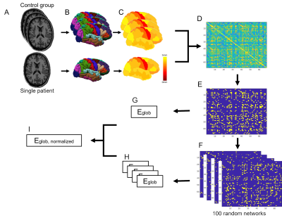 |
22 | Network efficiency of structural covariance networks relate to cognitive performance in children with childhood absence epilepsy
Merel JA Eussen1,2, Jacobus FA Jansen2,3, Twan Voncken4, Mariette HJA Debeij-van Hall5, Jos GM Hendriksen4,5, Jeroen R Vermeulen4, Sylvia Klinkenberg4, Walter H Backes2,3, and Gerhard Drenthen2,3
1Department of Biomedical Technology, Eindhoven University of Technology, Eindhoven, Netherlands, 2Department of Radiology & Nuclear Medicine, Maastricht University Medical Center, Maastricht, Netherlands, 3School for Mental Health & Neuroscience, Maastricht University, Maastricht, Netherlands, 4Department of Neurology, Maastricht University Medical Center, Maastricht, Netherlands, 5Department of Behavioral Sciences, Kempenhaeghe, Heeze, Netherlands
Cognitive deficits have been reported in children with childhood absence epilepsy (CAE). Regional alterations in morphology in children with CAE are likely related to the changes in the underlying network structure. Structural covariance networks (SCNs) based on interregional correlations of cortical thicknesses can describe these changes. To relate cognitive performance to network efficiency, individual SCNs are derived from anatomical MR images of the control group and one patient. The global efficiency calculated from the resulting SCNs showed a negative relation with cognitive performance for children with CAE.
|
||
1928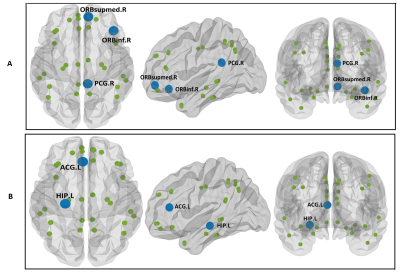 |
23 | Structural Connectivity in Default Mode Network May Lateralize Temporal Lobe Epilepsy: A Graph-Theory Approach
Fatemeh Salimi 1, Saeed Masoudnia 2, Alireza Fallahi 2,3, Narges Hoseini Tabatabaei4, Mohammadreza Ay1,2, and Mohammad-Reza Nazem-Zadeh1,2
1Medical Physics and Biomedical Engineering, Tehran University of Medical Sciences, Tehran, Iran (Islamic Republic of), 2Research Center for Molecular and Cellular Imaging, Tehran University of Medical Sciences, Tehran, Iran (Islamic Republic of), 3Biomedical Engineering, Hamedan University of Technology, Hamedan, Iran (Islamic Republic of), 4Medical School, Tehran University of Medical Sciences, Tehran, Iran (Islamic Republic of)
Difficulties in lateralizing the epileptogenic side of the brain challenge the temporal lobe epilepsy (TLE) surgery. We examined structural connectivity in default mode network (DMN) based on graph network analysis for specifying the epileptogenic side of the brain in TLE patients. The results showed significantly different connectivity in DMN nodes among the TLE patients compared to the control cohort. DMN regions with abnormal structural connectivity in TLE subjects corresponded with the epileptogenic brain sides.
|
||
1929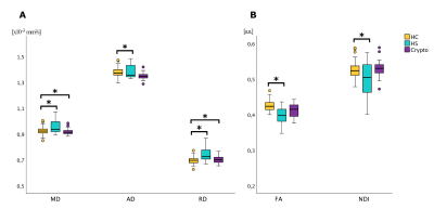 |
24 | White matter microstructure characterisation in hippocampal sclerosis and cryptogenic temporal lobe epilepsy
Nicolò Rolandi1, Fulvia Palesi1, Francesco Padelli2, Isabella Giachetti2, Domenico Aquino2, Paul Summers3, Elena Tartara4, Giancarlo Germani3, Valeria Mariani5, Egidio D'Angelo1,6, Laura Tassi5, Claudia Angela Michela Gandini Wheeler-Kingshott1,6,7, and Paolo Vitali3,8
1Department of Brain and Behavioral Science, University of Pavia, Pavia, Italy, 2Neuroradiology, I.R.C.C.S. Istituto Neurologico Carlo Besta, Milano, Italy, 3Neuroradiology, IRCCS Mondino Foundation, Pavia, Italy, 4Epilepsy Centre, IRCCS Mondino Foundation, Pavia, Italy, 5“C. Munari” Epilepsy Surgery Centre, ASST Niguarda, Milano, Italy, 6Brain Connectivity Center Research Unit, IRCCS Mondino Foundation, Pavia, Italy, 7NMR Research Unit, Queen Square MS Centre, Department of Neuroinflammation, UCL Queen Square Institute of Neurology, UCL Queen Square Institute of Neurology, London, United Kingdom, 8Radiology, IRCCS Policlinico San Donato, San Donato Milanese, Italy
Temporal lobe epilepsy is characterised by heterogeneous aetiology. In this work we focused on hippocampal sclerosis and cryptogenic groups. In order to characterize microstructure white matter alterations underlying the pathology, we performed a regional analysis of the temporal lobe white matter. Our results showed less disruptive microstructural changes in cryptogenic patients. Alterations in Axial Diffusivity and Neurite Density Index in hippocampal sclerosis potentially indicate significant axonal degeneration. Further investigations are needed to ascertain the value of Orientation Dispersion Index as a possible biomarker to predict surgery outcome.
|
||
1930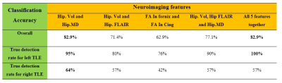 |
25 | Application of Machine Learning in Comparison between Multimodal Neuroimaging Markers of Laterality in Temporal Lobe Epilepsy
Alireza Fallahi 1,2, Mohammad Pooyan3, Jafar Mehvari-Habibabadi 4, Narges Hoseini Tabatabaei5, Mohammadreza Ay1,6, and Mohammad-Reza Nazem-Zadeh1,6
1Research Center for Molecular and Cellular Imaging, Tehran University of Medical Sciences, Tehran, Iran (Islamic Republic of), 2Biomedical Engineering, Hamedan University of Technology, Hamedan, Iran (Islamic Republic of), 3Biomedical Engineering, Shahed University, Tehran, Iran (Islamic Republic of), 4Isfahan Neuroscience Research Center, Isfahan University of Medical Sciences, Isfahan, Iran (Islamic Republic of), 5Medical School, Tehran University of Medical Sciences, Tehran, Iran (Islamic Republic of), 6Medical Physics and Biomedical Engineering, Tehran University of Medical Sciences, Tehran, Iran (Islamic Republic of)
Five neuroimaging markers including T1 volume, FLAIR signal intensity, and mean diffusivity in hippocampus, and fractional anisotropy in both posteroinferior cingulum and crus of fornix were used for lateralization of temporal lobe epilepsy (TLE). Support vector machine (SVM) was used as a classifier and for measuring the importance of neuroimaging attributes. The classification results demonstrated that the hippocampal volumetric and mean diffusivity showed the highest correct classification rate and the largest area under the curve (AUC) for the receiver operating characteristic (ROC), thus considered as the most important attributes of TLE laterality among all markers investigated in this study.
|
||
1931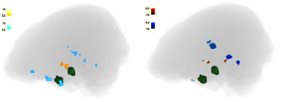 |
26 | fMRI Can Detect Functional Connections of Seizure Spread Regions to Onset Zones in Patients with Mesial Temporal Sclerosis
Anish Vinay Sathe1, Michael Kogan2, KiChang Kang1, Jingya Miao1, Mashaal Syed1, Isaiah Ailes1, Caio Matias1, Feroze Mohamed1, Ashwini Sharan1, and Mahdi Alizadeh1
1Thomas Jefferson University, Philadelphia, PA, United States, 2The University of New Mexico, Albuquerque, NM, United States
Epilepsy is a disease involving seizure initiation and often spread. sEEG has shown that areas of seizure spread are correlated with onset regions. We find that seed-to-voxel analysis via amplitude synchronization of fMRIs of epilepsy patients can also detect significant correlations between seizure onset and spread zones. These results validate fMRI analysis as a promising noninvasive method to detect other correlated brain regions that may be involved in seizure propagation.
|
||
1932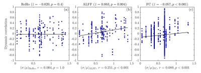 |
27 | Static and Dynamic Resting-State fMRI Features to Localize Brain Areas Generating Epileptic Spikes
Siu Lung Tang1, Kristina Sabaroedin2, Will Wilson2, Daniel Pittman2, Paolo Federico2, and Pierre LeVan2
1Department of Pediatrics, University of Calgary, Calgary, AB, Canada, 2Department of Radiology, University of Calgary, Calgary, AB, Canada
The potential of using features derived from resting-state fMRI time series to localize epileptic brain areas was studied. Static and dynamic correlations between local spike rates measured with intracranial EEG and regional homogeneity (ReHo), amplitude of low-frequency fluctuations (ALFF), and functional connectivity (FC), respectively, were analyzed based on data collected from 13 subjects with refractory epilepsy. While static measures of ReHo and ALFF were not statistically correlated with spike rates measured during long-term monitoring, static correlation with FC was apparent. However, in dynamic analysis, temporal variations in instantaneous spike rates were associated with synchronous fluctuations of both ALFF and FC.
|
||
1933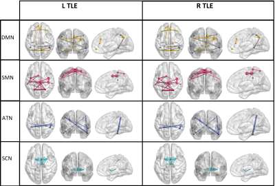 |
28 | A Graph Theoretical Approach for Lateralization of Temporal Lobe Epilepsy Using Nodal Functional Connectivity
Alireza Fallahi 1, Mohammad Pooyan2, Jafar Mehvari-Habibabadi 3, Narges Hoseini Tabatabaei4, Mohammadreza Ay1,5, and Mohammad-Reza Nazem-Zadeh1,5
1Research Center for Molecular and Cellular Imaging, Tehran University of Medical Sciences, Tehran, Iran (Islamic Republic of), 2Biomedical Engineering, Shahed University, Tehran, Iran (Islamic Republic of), 3Isfahan Neuroscience Research Center, Isfahan University of Medical Sciences, Isfahan, Iran (Islamic Republic of), 4Medical School, Tehran University of Medical Sciences, Tehran, Iran (Islamic Republic of), 5Medical Physics and Biomedical Engineering, Tehran University of Medical Sciences, Tehran, Iran (Islamic Republic of)
In this study, six graph theoretical measures were identified as nodal level epileptogenicity in TLE patients using functional connectivity analysis. The aim of this study is to define brain nodes that have significant difference between left and right TLE patients using resting state functional connectivity analysis. Clustering coefficient, degree centrality, betweenness centrality, node neighbor’s degree, closeness centrality, and page rank were calculated as graph theoretical characteristics and multi-dimensional scaling (MDS) was used as a statistical method. Results of the applied method suggested significant nodes for prediction of laterality in TLE patients.
|
||
1934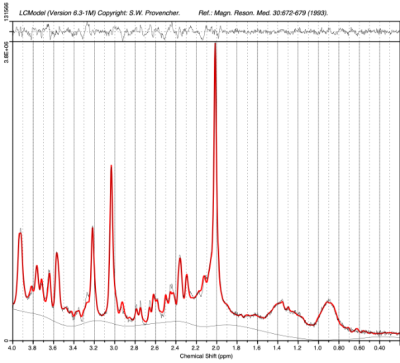 |
29 | Magnetic Resonance Spectroscopy in Children and Adolescents with Functional Neurological Disorder
Vishwa Shukla1, Molly Faith Charney1, Sheryl Foster2,3, Wufan Zhao1, Han Sam Jiang1, Kasia Kozlowska4,5,6, and Alexander P Lin1
1Radiology, Brigham and Women's Hospital, Boston, MA, United States, 2Sydney School of Health Sciences, The University of Sydney, Sydney, Australia, 3Radiology, Westmead Hospital, Westmead, Australia, 4The Children’s Hospital at Westmead Clinical School, Sydney, Australia, 5Psychiatry, Child & Adolescent Health, University of Sydney Medical School, Sydney, Australia, 6Westmead Institute for Medical Research, Westmead, Australia
Functional Neurological Disorder (FND) is characterized by a broad array of neurological symptoms that are thought to reflect aberrant activity in neural networks. This study aims to compare the neurochemical levels in the posterior default mode network (pDMN) in children and adolescents with FND and healthy controls using magnetic resonance spectroscopy (MRS). FND patients exhibited lower levels of tNAA/tCr in the pDMN compared to controls, indicating potential neuronal alterations or modified neuronal-glial signaling.
|
||
1935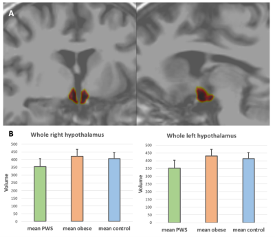 |
30 | Imaging the hypothalamus in Prader-Willi Syndrome
Stephanie S. G. Brown1, Katherine Manning1, and Anthony Holland1
1Department of Psychiatry, University of Cambridge, Cambridge, United Kingdom
Prader-Willi Syndrome (PWS) is a neurodevelopmental disorder characterised by intellectual disability, emotional dysregulation and importantly, marked overeating behaviour. The hypothalamus controls food intake and satiety by endocrine control, and evidence from animal studies has shown that lesions of the hypothalamic nuclei result in hyperphagia. However, studies examining the hypothalamus in humans in vivo are scarce. We show here significantly lower volumes of the hypothalamus and hypothalamic nuclei in PWS compared to healthy controls and general population obese individuals. Our findings are strongly suggestive that impaired appetite control and problematic eating behaviours are a consequence of abnormal hypothalamic structure in PWS.
|
||
 Back to Meeting Home
 Back to the Program-at-a-Glance
The International Society for Magnetic Resonance in Medicine is accredited by the Accreditation Council for Continuing Medical Education to provide continuing medical education for physicians.
 Back to Meeting Home
 Back to the Program-at-a-Glance
 View the Presentation