Digital Poster
New Look of Neurofluids Physiology I
Joint Annual Meeting ISMRM-ESMRMB & ISMRT 31st Annual Meeting • 07-12 May 2022 • London, UK

Digital Poster
New Look of Neurofluids Physiology I
| Computer # | ||||
|---|---|---|---|---|
1205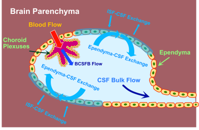 |
1 | Cerebrospinal fluid-tissue exchange revealed by phase alternate labeling with null recovery MRI
Anna M Li1 and Jiadi Xu1,2
1F.M. Kirby Research Center for Functional Brain Imaging, Kennedy Krieger Research Institute, Baltimore, MD, United States, 2Russell H. Morgan Department of Radiology and Radiological Science, The Johns Hopkins University School of Medicine, Baltimore, MD, United States
Our understanding of the CSF exchange with surrounding tissues and its correlation with brain function is limited. We presented the first non-invasive MRI methods to quantify this process with two novel T1 and ADC Phase Alternate LAbeling with Null recovery (PALAN) schemes. The CSF signal was nulled, while the partial recovery of other tissues was labeled by alternating the phase of the pulse by using the significantly different T1 and ADC values between CSF and the other tissues. Rapid and major CSF water exchange occurs on the surface of the ventricles.
|
||
1206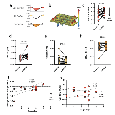 |
2 | Caffeine acutely reduces aqueductal CSF flow in humans
Tekla Maria Kylkilahti1,2, Max Wictor1,2, Johannes Töger3, Karin Markenroth Bloch 4,5, and Iben Lundgaard1,2
11Department of Experimental Medical Science, Lund University, Lund, Sweden, 2Wallenberg Centre for Molecular Medicine, Lund University, Lund, Sweden, 3Department of Clinical Sciences, Diagnostic Radiology, Lund University, Skåne University Hospital, Lund, Sweden, 4Department of Medical Radiation Physics, Lund University, Lund, Sweden, 5Lund University Bioimaging Center, Lund University, Lund, Sweden
In this ultra-high field 7T MRI study, we find that caffeine acutely reduces CSF net flow in humans. Interestingly, regular caffeine consumption seems to be protective against this reduction. We used non-invasive 2D PC 7T MRI in healthy volunteers to measure CSF flow in the cerebral aqueduct. Ventricular CSF flow is upstream of the glymphatic system and may thus reflect or affect the function of CSF-mediated clearance pathways that have been suggested to be important for waste product removal in the brain. Better understanding of CSF dynamics may help us find new targets for treating proteinopathies such as Alzheimer’s disease.
|
||
1207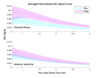 |
3 | Assessment of Choroid Plexus Perfusion and the Blood-CSF Barrier with Multi Post Label Delay Arterial Spin Labeling MRI and Vasodilation
Yufei David Zhu1, Moss Zhao2, Bin Shen2, Greg Zaharchuk2, and Audrey Peiwen Fan1,3
1Biomedical Engineering, University of California, Davis, Davis, CA, United States, 2Radiology, Stanford University, Palo Alto, CA, United States, 3Neurology, University of California, Davis, Davis, CA, United States
The choroid plexus contributes to the production of cerebrospinal fluid (CSF), maintains electrolyte and metabolic balance, and plays important immunological roles. Here, we sought to determine whether multi-post label delay (PLD) arterial spin labeling (ASL) MRI could robustly measure cerebral blood flow (CBF) in the choroid plexus and its response to a vasodilatory challenge. We saw choroid plexus perfusion increase by 30 ml/100g/min (p=0.0003) with acetazolamide. We also calculated the ratio of the choroid plexus to anterior ventricle ASL signal (rCV) as a clinically useful biomarker of blood-CSF passage and observed an average increase of 0.529 in rCV post vasodilation.
|
||
1208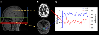 |
4 | Simultaneous measurement of dynamic CBF and CSF signal changes with pseudo-continuous arterial spin labeling
Jae-Geun Im1, Jun-Hee Kim1, and Sung-Hong Park1
1Department of Bio and Brain Engineering, Korea Advanced Institute of Science and Technology, Daejeon, Korea, Republic of
To measure CBF and CSF signals simultaneously, we proposed a new 2D EPI-based pCASL with overlapping portion of the labeling plane and imaging region. Our results showed that signal difference between label and control scans in the overlapping portion were higher in CSF than in other tissues. In addition, the pCASL-based CSF signals were negatively correlated with CBF, which was more significant than correlations of CBF with other tissues or baseline raw CSF signals. Based on the results, pCASL can be used for simultaneous measurement of dynamic CBF and CSF signal changes with our technique, which warrants further investigation.
|
||
1209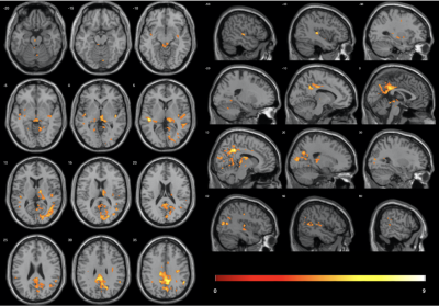 |
5 | Using diffusion kurtosis imaging to measure glymphatic system related changes during human sleep
Balázs Örzsik1, Mara Cercignani2, and Neil Harrison2
1BSMS, University of Sussex, Brighton, United Kingdom, 2Cardiff University, Cardiff, United Kingdom
Glymphatic clearance has been associated with an increase in interstitial volume fraction during sleep. Higher order diffusion weighted MRI images were acquired of awake and sleeping participants to investigate glymphatic system related changes. Sleep was associated with a significant decrease in kurtosis measures. Clusters with significant decrease in radial kurtosis were located in default mode network areas, occipital cortex, thalamus, midbrain and temporal lobe. Decrease in kurtosis is in line with the glymphatic system hypothesis, as diffusion becomes more Gaussian when it is less restricted, which expected with the increase in interstitial space volume.
|
||
1210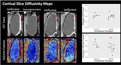 |
6 | Detection of enhanced cerebrospinal fluid transport during dexmedetomidine anesthesia in the rat using diffusion, T2 and morphometric MRI
Elizabeth B Hutchinson1, Anakaren Romero-Lozano1, Jean-Pierre Galons1, Christine Howison1, Alexandru Avram2, Nathan Williamson2, Michal Komlosh2, and Peter J Basser2
1Biomedical Engineering, University of Arizona, Tucson, AZ, United States, 2NICHD/NIH, Bethesda, MD, United States
The enhancement of CSF transport in a robust experimental model was measurable using in-vivo quantitative MRI.TBM and voxelwise T2 mapping analysis were both able to changes in regions that are known to be associated with CSF transport based on contrast-enhancement studies. These two methods do not require contrast and if validated could provide a non-contrast outcome metric for health of the CSF transport system. Diffusion MRI outcomes suggest that it may be possible to sensitize the diffusion MRI paradigm to CSF transport and this is an area for future research.
|
||
1211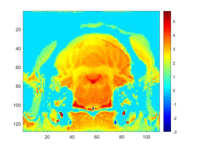 |
7 | In vivo CSF Flow During Central Sensitization Using Fourier Velocity Encoded MRI at 21.1 T
Dayna L. Richter1,2, Samuel W. Holder1,2, and Samuel C. Grant1,2
1National High Magnetic Field Laboratory, Florida State University, Tallahassee, FL, United States, 2Chemical & Biomedical Engineering, FAMU-FSU College of Engineering, Tallahassee, FL, United States
Cerebrospinal fluid (CSF) flow was evaluated temporally in a preclinical migraine model at 21.1 T. As elevated sodium levels are implicated in the progression of migraine, evaluating how sodium is transported in the ventricular system and brain should provide insight on bulk sodium accumulation around, among and within anatomical regions implicated in migraine. A series of Fourier Velocity Encoding MRI was acquired over 2 h post-nitroglycerin injection to evaluate CSF flow during the progression of central sensitization.
|
||
1212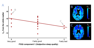 |
8 | Sleep and waste clearance: The association of sleep quality with a 7T IVIM imaging derived proxy of interstitial fluid
Merel M. van der Thiel1,2, Gerhard S. Drenthen1,2, Paulien H.M. Voorter1,2, Thorsten Feiweier3, Inez H.G.B. Ramakers2,4, Walter H. Backes1,2,5, and Jacobus F.A. Jansen1,2,6
1Department of Radiology & Nuclear Medicine, Maastricht University Medical Center, Maastricht, Netherlands, 2School for Mental Health & Neuroscience, Maastricht University, Maastricht, Netherlands, 3Siemens Healthcare, Erlangen, Germany, 4Department of Psychiatry & Neuropsychology, Maastricht University, Maastricht, Netherlands, 5School for Cardiovascular Disease, Maastricht University, Maastricht, Netherlands, 6Department of Electrical Engineering, Eindhoven University of Technology, Eindhoven, Netherlands
Cerebral clearance is most active during sleep, therefore reduced sleep quality might induce impaired clearance function. Interstitial fluid (ISF) washes waste products from in-between cells through the parenchyma and its volume is found to be regulated by the sleep-wake cycle. Assessment of the ISF-fraction through IVIM can be a potential, non-invasive method to determine sleep-related variations in ISF, without contamination of parenchymal or microvascular diffusion. The current exploratory study investigates the potential of the IVIM-derived ISF-fraction to assess ISF-volume changes in relation to sleep by examining whether these changes are driven by actual hours of sleep or self-reported sleep quality.
|
||
1213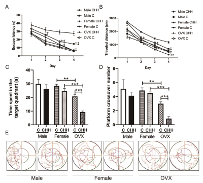 |
9 | Ovariectomized Female Rats Suffer Hippocampal Neuroinflammation and the Blood-Brain Barrier Disruption Easier than Male and Intact Female Rats following Prolonged Exposure of a Hypobaric and Hypoxic Environment at High Altitude Video Not Available
Dongyong Zhu1, Bo He1, Yixuan Wan1, Mengdi Zhang1, and Fabao Gao1
1Department of Radiology, West China Hospital of Sichuan University, Chengdu, China
Ovariectomized Female Rats Suffer Hippocampal Neuroinflammation and the Blood-Brain Barrier Disruption Easier than Male and Intact Female Rats following Prolonged Exposure of a Hypobaric and Hypoxic Environment at High Altitude
|
||
1214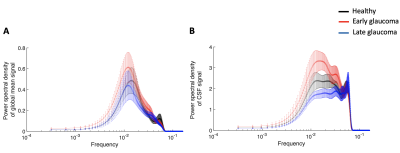 |
10 | Changes in cerebrospinal fluid dynamics and its coupling with global brain activity in early glaucoma patients
Ji Won Bang1, Eva Yarsky1, Gadi Wollstein1, Joel S Schuman1, and Kevin C Chan1,2
1Department of Ophthalmology, New York University Grossman School of Medicine, New York, NY, United States, 2Department of Radiology, New York University Grossman School of Medicine, New York, NY, United States
Neurodegenerative diseases are thought to involve changes in the cerebrospinal fluid (CSF). Since glaucoma is a neurodegenerative disease of the visual system, here, we examined the CSF dynamics in glaucoma patients. Specifically, using resting-state fMRI, we observed that early glaucoma patients present greater power in the low frequency of the CSF flow and greater coupling between CSF flow and global brain activity compared to late glaucoma patients. Further, the coupling between CSF flow and global brain activity is associated with the volumes of the optic nerve and optic chiasm. Overall, our results suggest that glaucoma involves changes in CSF flow.
|
||
 Back to Meeting Home
 Back to the Program-at-a-Glance
The International Society for Magnetic Resonance in Medicine is accredited by the Accreditation Council for Continuing Medical Education to provide continuing medical education for physicians.
 Back to Meeting Home
 Back to the Program-at-a-Glance
 View the Presentation