Digital Poster
New Look of Neurofluids Physiology II
Joint Annual Meeting ISMRM-ESMRMB & ISMRT 31st Annual Meeting • 07-12 May 2022 • London, UK

Digital Poster
New Look of Neurofluids Physiology II
| Computer # | ||||
|---|---|---|---|---|
1295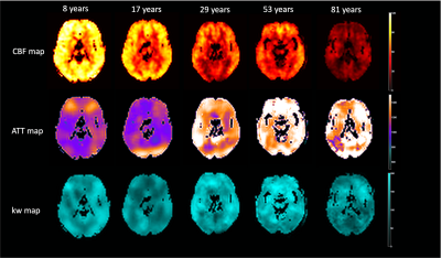 |
1 | Age Dependent Changes of Water Exchange Rate in Blood Brain Barrier (BBB) Assessed by Diffusion-Prepared Arterial Spin Labeling
Qinyang Shou1, Xingfeng Shao1, Kimberly Felix2, Brandon Ojogho1, Xuejuan Jiang2,3, Megan M Herting2, and Danny JJ Wang1
1Laboratory of Functional MRI Technology (LOFT), Stevens Neuroimaging and Informatics Institute, University of Southern California, Los Angeles, CA, United States, 2Department of Preventive Medicine, Keck School of Medicine, University of Southern California, Los Angeles, CA, United States, 3Department of Ophthalmology, Keck School of Medicine, University of Southern California, Los Angeles, CA, United States
The purpose of this study was to investigate the relationship between water exchange rate (kw) of blood brain barrier and age. Diffusion Prepared pCASL technique was used to measure kw in four datasets with a wide range of age (8 to 81 years). Global and regional kw values as well as cerebral blood flow and arterial transit time were analyzed. The results show an overall trend of decreasing kw with age, in parietal and parahippocampal regions there are U-shaped and inverted U-shaped changes of kw with age.
|
||
1296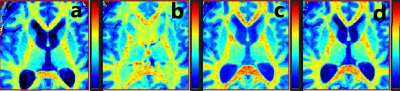 |
2 | Assessment of gadolinium contrast in the brain using a compressed sense 3D Look Locker sequence after intrathecal contrast administration
Tryggve Holck Storås1, Svein Are Vatnehol2,3, Geir Ringstad4, Kyrre Holck Emblem5, and Per Kristian Eide6,7
1Radiology and Nuclear Medicine, Oslo University Hospital, Oslo, Norway, 2The Intervention Centre, Oslo University Hospital, Oslo, Norway, 3Faculty of Health and Social Sciences, University of South-Eastern Norway, Drammen, Norway, 4Department of Radiology, Oslo University Hospital, Oslo, Norway, 5Department of Radiology and Nuclear Medicine, Oslo University Hospital, Oslo, Norway, 6Institute of Clinical Medicine, Faculty of Medicine, University of Oslo, Oslo, Norway, 7Department of Neurosurgery, Oslo University Hospital – Rikshospitale, Oslo, Norway
Quantitative assessment of T1 enhancement after intrathecal administration of Gd-contrast may remove present challenges due to image scaling varying between acquisitions. The compressed SENSE technique allow 3D-Look-Locker-TFE acquisitions with isotropic 1 mm resolution and full brain coverage to be run in clinically feasible times. A clinical example is demonstrated and a small phantom study exploring further improvements based on trade-offs between undersampling and reduced saturation effects is presented.
|
||
1297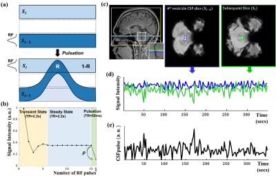 |
3 | Measurement of CSF pulsation from EPI-based human fMRI
Jun-Hee Kim1, Jae-Geun Im1, and Sung-Hong Park1
1Department of Bio and Brain Engineering, Korea Advanced Institute of Science and Technology, Daejeon, Korea, Republic of
To study CSF dynamics and brain functional activity simultaneously with quantitative aspect, we proposed a new method for quantifying CSF pulsatility information based on an interslice CSF pulsation model in 4th ventricle of EPI-based fMRI (CSFpulse). The proposed CSFpulse successfully detected the higher CSF flow during the resting state than the typical task states, both in quantitative and time course aspects. Also, CSFpulse was significantly correlated with stroke volume measured using phase contrast MRI during functional states. Based on these results, CSFpulse can be used for investigating functional changes in BOLD and CSF pulsation simultaneously based on conventional EPI-based fMRI.
|
||
1298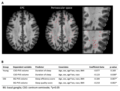 |
4 | Effects of Sleep on perivascular space in healthy population
Nien-Chu Shih1, Karen Lincoln2, Farshid Sepehrband1, and Jeiran Choupan1
1USC Stevens Neuroimaging and Informatics Institute, Keck School of Medicine, University of Southern California, Los Angeles, CA, United States, 2Suzanne Dworak-Peck School of Social Work, University of Southern California, Los Angeles, CA, United States
The perivascular space (PVS) has been reported to clear Amyloid-β (Aβ) and metabolic wastes during sleep. However, the relationship between sleep and PVS in a healthy cohort is still unclear. We investigate the association between PVS and sleep across different age groups. We found that the effect of sleep on PVS volume varies in the young and older population.
|
||
1299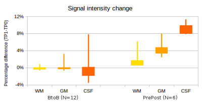 |
5 | Early post gadolinium T2-W FLAIR signal intensity change in normal brain tissues and CSF: significance for clinical neuroimaging and neurofluids
Nivedita Agarwal1,2,3, Denis Peruzzo1, John Dewitt Port4, Roxana Octavia Carare5, and Graeme Bydder6
1Neuroradiology, I.R.C.C.S. Eugenio Medea, Bosisio Parini, Italy, 2Radiology, Azienda Provinciale per i Servizi Sanitari, Rovereto, Italy, 3Neurosciences, University of Southampton, Southampton, United Kingdom, 4Neuroradiology, Mayo Clinic, Rochester, MN, United States, 5University of Southampton, Southampton, United Kingdom, 6University of San Diego, California, CA, United States
Apart from the study of meningeal diseases, cortical metastases and multiple sclerosis, T2-W FLAIR is used for analyzing the CSF drainage of fluids. However, little is known regarding gadolinium induced signal intensity (SI) changes in the normal gray (GM) and white matter (WM). The SI ratios GM/WM and CSF/WM increase by 2.5% and 7% respectively whereas the SI in WM does not change. These findings are important for the interpretation of clinical findings and will provide better understanding of the movement of neurofluids in the human brain at early time points (e.g.4-5 minutes after GBCA injection).
|
||
1300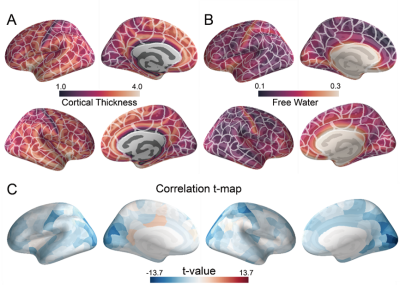 |
6 | Identifying the role of free water in human cortical macroscopic organization Video Not Available
Lei Wei1, MIng Ding1, yuwen zhang1, and he wang1,2
1Institute of Science and Technology for Brain-Inspired Intel, ShangHai City, China, 2Human Phenome Institute, Fudan University, Shanghai, China, ShangHai City, China
Free water imaging is a novel tool to study human brain. Recently, the free water has been used in aging disease researach. However, the role of free water in human cortex macroscopic is still unknown. This work shows the relationship between the cortical free water and the cortical thickness, and investigate the underlying molecular and genetic mechanism of the association. Neurons and astrocytes are strongly associated with the free water and the macroscopic organization.
|
||
1301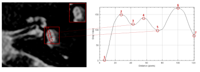 |
7 | Optimization of an MRI protocol for non-contrast saccule and utricle visualization at 1.5T
Alba Iruela Sanchez1, Valentin H. Prevost2, Alicia Palomar Garcia1, Wolter de Graaf3, and Bruno Triaire2
1Canon Medical Systems Spain and Portugal, Barcelona, Spain, 2Canon Medical Systems Corporation, Tochigi, Japan, 3Canon Medical Systems Europe, Zoetermeer, Netherlands
This study evaluates the feasibility of different approaches for the visualization of endolymphatic content in inner ear structures on 1.5T MR systems without the need of contrast injection. The results show that using an optimized high-resolution 3D T2w sequence combined with DLR-based denoising tools it is possible to visualize the utricle and saccule structures on healthy volunteers. The next step will be testing this technique on patients with endolymphatic hydrops.
|
||
 Back to Meeting Home
 Back to the Program-at-a-Glance
The International Society for Magnetic Resonance in Medicine is accredited by the Accreditation Council for Continuing Medical Education to provide continuing medical education for physicians.
 Back to Meeting Home
 Back to the Program-at-a-Glance
 View the Presentation