Digital Poster
Multiple Sclerosis I
Joint Annual Meeting ISMRM-ESMRMB & ISMRT 31st Annual Meeting • 07-12 May 2022 • London, UK

| Computer # | ||||
|---|---|---|---|---|
0776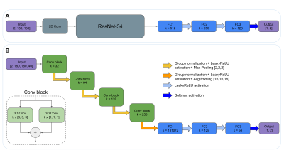 |
1 | Classification of MS patients into disability levels using deep learning approaches based solely on routinely-acquired MRI
Llucia Coll1, Pere Carbonell-Mirabent1, Álvaro Cobo-Calvo1, Georgina Arrambide1, Ángela Vidal-Jordana1, Manuel Comabella1, Joaquín Castilló1, Breogán Rodríguez-Acevedo1, Ana Zabalza1, Ingrid Galán1, Luciana Midaglia1, Carlos Nos1, Annalaura Salerno2, Cristina Auger2, Manel Alberich2, Jordi Río1, Jaume Sastre-Garriga1, Arnau Oliver3, Xavier Montalban1, Àlex Rovira2,
Mar Tintoré1, Deborah Pareto2, Xavier Lladó3, and Carmen Tur1
1Multiple Sclerosis Centre of Catalonia (Cemcat), Department of Neurology/Neuroimmunology, Hospital Universitari Vall d’Hebron, Universitat Autònoma de Barcelona, Barcelona, Spain, 2Section of Neuroradiology, Department of Radiology (IDI), Vall d'Hebron University Hospital, Spain. Universitat Autònoma de Barcelona, Barcelona, Spain, 3Research institute of Computer Vision and Robotics, University of Girona, Girona, Spain The ability of existing MRI biomarkers to predict MS patients’ prognosis is limited and inaccurate to be used at the individual level. We aimed to assess the ability of Convolutional Neural Networks (CNNs) to classify relapse-onset MS patients into non-disabled and markedly disabled using only MRI images. T1w and T2w-FLAIR images of 538 MS patients were used to train and test two CNN approaches which were compared also with (conventional) logistic regression models. The results showed that the CNN models performed better, having the intrinsic potential to improve after the inclusion of regional priors and other valuable clinical data. |
||
0777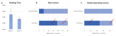 |
2 | Improved Detection, Description and Efficiency of Multiple Sclerosis Lesions’ Assessment using a Dedicated Software
Christian Federau1, Guangming Zhu2, Nicolin Hainc3, Silvio Paganucci1, Lukas Kipp2, and Max Wintermark2
1AI Medical, Zollikon, Switzerland, 2Stanford University, Stanford, CA, United States, 3University Zürich, Zürich, Switzerland
Yearly multiple sclerosis MRI follow-ups require the tedious and time-consuming manual comparing and counting of multiple demyelinating lesions, which can reach hundreds in some cases. We evaluated a semi-automatic software for the efficient assessment of such images, and found that a significant increase in the number of reported new multiple sclerosis lesions can be achieved in two-and-a-half minutes reading on average per case. In contrast, current standard reports missed 80% of the new lesions. In addition, The Jazz software permits the efficient reporting of slowly evolving lesions, an entity growing in clinical relevance and typically overlooked in current reporting.
|
||
0778 |
3 | “Better together”: a combination of machine learning and radiomics to predict long-term disability in Multiple Sclerosis
Sirio Cocozza1, Renato Cuocolo1, Giuseppe Pontillo1, Lorenzo Ugga1, Maria Petracca1, Teresa Costabile1, Roberta Lanzillo1, Vincenzo Brescia Morra1, Mario Quarantelli2, and Arturo Brunetti1
1University of Naples Federico II, Naples, Italy, 2National Research Council, Naples, Italy
The identification of early biomarkers to predict the disability accumulation is crucial in Multiple Sclerosis (MS). We performed a combined radiomics and Machine Learning (ML) study to predict long-term clinical changes in MS. Radiomics data were extracted from data of 177 patients with a 10-years clinical follow-up available. The model based on the recursive elimination of the features combined with the Extra Trees classifier was able to obtain a maximum precision for each endpoint of 0.71 and 0.69 for cognitive and motor disability, respectively.This combined radiomics-ML approach seems to be a feasible tool for long-term clinical prediction in MS.
|
||
0779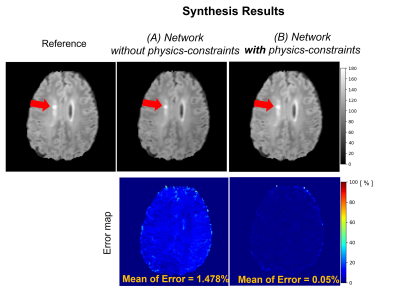 |
4 | Physics-Constrained Neural Network for Synthesis of MR Parameter Maps and Clinical Contrast
Gawon Lee1, Ji Wan Son1, Ken SaKaie2, Kunio Nakamura3, Yufan Zheng3, Daniel Ontaneda4, Bruce Trapp5, Mark Lowe2, Dong Hye Ye6, and Se-hong Oh7
1Division of Biomedical engineering, Hankuk University of Foreign Studies, Yongin-si, Gyeonggi-do, Korea, Republic of, 2Imaging institute, Cleveland Clinic, Cleveland, OH, United States, 3Biomedical Engineering, Lerner Research Institue, Cleveland Clinic, Cleveland, OH, United States, 4Mellen Center for Multiple Sclerosis, Cleveland Clinic, Cleveland, OH, United States, 5Department of Neurosciences, Cleveland Clinic, Cleveland, OH, United States, 6Department of Electrical and Computer Engineering, Marquette University, Milwaukee, WI, United States, 7Biomedical engineering, Hankuk University of Foreign Studies, Yongin-si, Gyeonggi-do, Korea, Republic of
When a clinical MR scan is acquired, there might be missing tissue contrasts due to the corruption by patient’s motion during long scan time. In this study, we propose a method to synthesize the missing T2-weighted or FLAIR contrasts from a T1-weighted image using physics-constrained neural network. We incorporate the Bloch equations that generate MR contrast images from tissue parameter maps based on MR physical models into a synthesis neural network and show the improved performance compared with the existing U-Net directly synthesizing from a T1-weighted image.
|
||
0780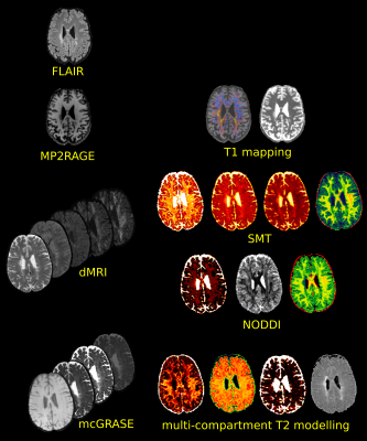 |
5 | Multimodal lesion characterization in multiple sclerosis: a pilot study
Simona Schiavi1, Gian Franco Piredda2,3,4, Caterina Lapucci5, Francesco Tazza1, Domenico Zacà6, Luca Roccatagliata7,8, Tom Hilbert2,3,4, Tobias Kober2,3,4, Matilde Inglese1,9, and Mauro Costagli1
1Department of Neuroscience, Rehabilitation, Ophthalmology, Genetics, Maternal and Child Health (DINOGMI), University of Genoa, Genoa, Italy, 2Advanced Clinical Imaging Technology, Siemens Healthcare AG, Lausanne, Switzerland, 3Department of Radiology, Lausanne University Hospital and University of Lausanne, Lausanne, Switzerland, 4LTS5, Ecole Polytechnique Fédérale de Lausanne (EPFL), Lausanne, Switzerland, 5HNSR, IRRCS Ospedale Policlinico San Martino, Genoa, Italy, 6Siemens Healthcare s.r.l, Milan, Italy, Milan, Italy, 7Department of Neuroradiology, IRCCS Ospedale Policlinico San Martino, Genoa, Italy, 8Dipartimento di Scienze della Salute (DISSAL), University of Genoa, Genoa, Italy, 9IRCCS Ospedale Policlinico San Martino, Genoa, Italy Using a comprehensive multimodal quantitative MRI protocol, we study the relationships between different microstructural metrics inside lesional tissue and investigate the heterogeneous pathological processes underlying alterations visible as hyperintense plaques in FLAIR images. Our preliminary results suggest that, although all the metrics can detect differences between lesions and normal appearing white matter, not all are directly associated with clinical status. Moreover, many metrics are intercorrelated and should not be considered as independent information when analyzing clinical outcomes. Understanding the clinical value of these parameters can advance the understanding of complex microstructural processes in neurological diseases and informs MRI protocol designs. |
||
0781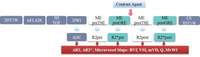 |
6 | Microvascular Morphology Alteration Using Relaxation Rate Change with Gd-based MRI Contrast Agent in the Patient with Alzheimer’s Disease
Chang-Woo Ryu1, Xiao-Yi Guo2, HyeokJung Kwon2, Soonchan Park1, Hak Young Rhee3, A-Rang Cho4, and Geon-Ho Jahng1
1Radiology, Kyung Hee University Hospital at Gangdong, Seoul, Korea, Republic of, 2Medicine, Kyung Hee University, Seoul, Korea, Republic of, 3Neurology, Kyung Hee University Hospital at Gangdong, Seoul, Korea, Republic of, 4Psychiatry, Kyung Hee University Hospital at Gangdong, Seoul, Korea, Republic of
To assess the cerebral microvascular alteration in the demented patients relative to a non-demented population using in vivo microvascular index maps by using Gd-based contrast agent at a 3T MRI system, we included 11 non-demented participants and 11 AD patients. Compared with the non-demented group, BVf and MvWI were significantly increased in the demented group. Both mVD and VSI were only significantly decreased in the demented group at the white matter hyperintensity (WMHI) area. BVf and MvWI were significantly positively correlated with age and VSI was significantly positively correlated with MMSE.
|
||
0782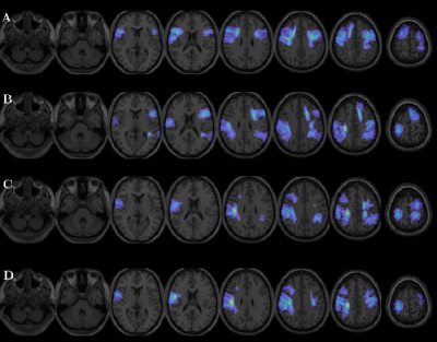 |
7 | Identification of Subcortical White Matter Biomarkers in Multiple Sclerosis Patients according to AVLT performance using Random Forest
Cristian Montalba1,2,3, Pamela Franco1,3,4, Tomás Labbé5, Marcelo Andia1,2, Miguel Guevara6, Jean-François Mangin7, Juan Pablo Cruz2, Ethel Ciampi8,9, Claudia Cárcamo5,8, Pamela Guevara6, and Sergio Uribe1,2,3
1Biomedical Imaging Center, Pontificia Universidad Católica de Chile, Santiago, Chile, 2Radiology Department, School of Medicine, Pontificia Universidad Católica de Chile, Santiago, Chile, 3Millennium Nucleus for Cardiovascular Magnetic Resonance, Santiago, Chile, 4Electrical Engineering Department, School of Engineering, Pontificia Universidad Católica de Chile, Santiago, Chile, 5Interdisciplinary Center of Neurosciences, Pontificia Universidad Católica de Chile, Santiago, Chile, 6Faculty of Engineering, Universidad de Concepción, Concepción, Chile, 7UNATI, Neurospin, CEA, Université Paris-Saclay, Gif-sur-Yvette, France, 8Neurology Department, School of Medicine, Pontificia Universidad Católica de Chile, Santiago, Chile, 9Neurology Service, Hospital Dr. Sótero del Río, Santiago, Chile
Radiological biomarkers of cognitive impairment in Multiple Sclerosis (MS) is still scarce. This study aimed to identify subcortical white matter biomarkers of cognitive impairment related to verbal episodic memory in MS patients and healthy controls, by using a random forest approach.
|
||
0783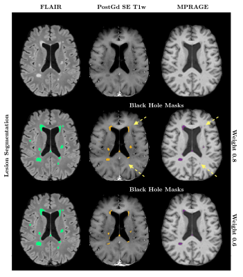 |
8 | Identification of ’black hole’ T1 hypointense lesions using different intensity thresholds on gradient-echo T1w MRI
Vanessa Wiggermann1, Mie Arnau Martinez1, Jasmin Merhout1, Jeppe Romme Christensen2, Morten Blinkenberg2, Finn Sellebjerg2,3, Henrik Lundell1,4, and Hartwig Roman Siebner1,5
1Danish Research Centre for Magnetic Resonance, Centre for Functional and Diagnostic Imaging and Research, Copenhagen University Hospital - Amager and Hvidovre, Hvidovre, Denmark, 2Neurology, Danish Multiple Sclerosis Center, Rigshospitalet, Glostrup, Denmark, 3Clinical Medicine, University of Copenhagen, Copenhagen, Denmark, 4Health Technology, Technical University of Denmark, Lyngby, Denmark, 5Neurology, Copenhagen University Hospital Bispebjerg, Copenhagen, Denmark
Black holes (BHs), T1 hypointense lesions, are known predictors of multiple sclerosis disease severity, however, their identification is strongly intensity dependent. As traditional spin-echo T1 scans are being replaced by 3D gradient-echo images, we need to understand how hypointense lesions on these images compare. We assessed whether an intensity threshold for MPRAGE images exists that would allow to match spin-echo black hole definitions. Simple thresholding was only able to reproduce black hole segmentations to an intermediate degree.
|
||
0784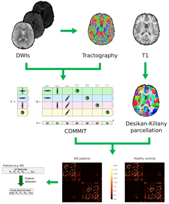 |
9 | Improving the accuracy of multiple sclerosis classification via robust feature selection based on quantitative tractography
Maria Petracca1,2, Alberto Azzari3, Antonella Mensi3, Nicole Graziano2, Alessandro Daducci3, Manuele Bicego3, Matilde Inglese2,4,5,6, and Simona Schiavi3,4
1Department of Human Neurosciences, Sapienza University of Rome, Rome, Italy, 2Department of Neurology, Icahn School of Medicine at Mount Sinai, New York, NY, United States, 3Department of Computer Science, University of Verona, Verona, Italy, 4Department of Neuroscience, Rehabilitation, Ophthalmology, Genetics, Maternal and Child Health (DINOGMI), University of Genoa, Genoa, Italy, 5Department of Radiology, Icahn School of Medicine at Mount Sinai, New York, NY, United States, 6IRCCS Ospedale Policlinico San Martino, Genoa, Italy
Improving image-based classification accuracy in multiple sclerosis while characterizing biological relevant features remains an open challenge. To this aim we applied a robust feature selection (FS) procedure based on a leave-one-out cross-validation scheme on structural connectivity features derived from connectomes computed with convex optimization modelling for microstructure informed tractography. We computed classification accuracy for different classifiers (NN, KNN, SVM-LIN, SVM-RBF, RF) before and after the application of the FS procedure. The highest overall accuracy (91%) was obtained using the FS procedure on the whole connectome. The biological meaningfulness of the selected features is supported by their correlations with clinical scores.
|
||
0785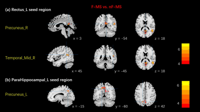 |
10 | Multiple sclerosis related fatigue: Altered resting-state functional connectivity of the Default Mode Network. Video Not Available
Yujing Li1, Jun Wang1, Tingli Yang1, Pengfei Zhang1, Kai Ai2, Xiaoli He1, Haiying Zhao1, Yuan Ding1, Rui Wang1, Min Li1, Xinying Ren1, Diaohan Xiong1, and Jing Zhang1
1Department of Magnetic Resonance, Lanzhou University Second Hospital, Lanzhou, China, 2Philips Healthcare, Xi’an, China
The objective of this study was to elucidate the no-motor component of fatigue mechanism in multiple sclerosis (MS) patients by studying the resting state functional connectivity (RS-FC) of default mode network (DMN). 12 with fatigue (F) and 10 without fatigue (nF) MS underwent resting sate magnetic resonance imaging (MRI). We selected 11 priori regions within the DMN as the regions of interests (ROIs) and ROI to ROI and ROI to global FC were calculated. Compared to nF-MS patients, F-MS patients showed abnormal increased functional connectivity in brain regions associated with cognitive function within the DMN.
|
||
0786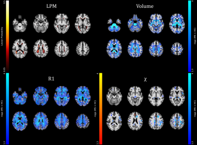 |
11 | Clinical correlates of brain iron and myelin changes in Multiple Sclerosis: a multi-parameter quantitative MRI study
Giuseppe Pontillo1, Maria Petracca1, Serena Monti2, Mario Quarantelli2, Roberta Lanzillo1, Teresa Costabile3, Antonio Carotenuto1, Fabio Tortora1, Andrea Elefante1, Vincenzo Brescia Morra1, Arturo Brunetti1, Giuseppe Palma2, and Sirio Cocozza1
1University of Naples "Federico II", Naples, Italy, 2Institute of Biostructure and Bioimaging, National Research Council, Naples, Italy, 3Università degli Studi della Campania "Luigi Vanvitelli", Naples, Italy
In this cross-sectional study, voxel-based morphometry and voxel-based quantification analyses of longitudinal relaxation rate (R1) and quantitative susceptibility (QSM) maps, reflecting myelin and iron, respectively, were conducted on 117 patients with MS (pwMS) and 53 healthy controls. Between-group differences and correlations with clinical variables (global disability, cognitive and motor performance) were assessed. We found widespread atrophy and R1 decrease in pwMS, with smaller clusters of reduced susceptibility in the thalami and cerebral WM. While subcortical atrophy and WM iron depletion contributed to global and motor disability, deep GM atrophy and R1 decrease in lesional areas correlated with cognitive performance.
|
||
0787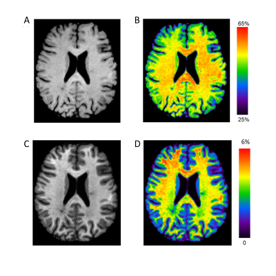 |
12 | Magnetisation transfer saturation and MTR in multiple sclerosis: sensitivity to longitudinal change and clinical disability
Elizabeth York1, Rozanna Meijboom1, Michael J. Thrippleton1, Agniete Kampaite1, Maria Valdes Hernandez1, Peter Connick2, Siddharthan Chandran1,2, David P.J. Hunt1, and Adam D. Waldman1
1Centre for Clinical Brain Sciences, University of Edinburgh, Edinburgh, United Kingdom, 2Anne Rowling Regenerative Neurology Clinic, University of Edinburgh, Edinburgh, United Kingdom
Myelin-sensitive magnetisation transfer saturation (MTsat) corrects magnetisation transfer ratio (MTR) signal variance, accounting for T1 recovery and B1 inhomogeneities, but its validity as a longitudinal imaging marker in multiple sclerosis (MS) is not yet clear. Here, we examine longitudinal change in cerebral MTsat and MTR in a cohort of people with newly diagnosed MS (n=62). MTsat, but not MTR, decreased in normal-appearing white matter over one year and was associated with clinical disability progression. In white matter lesions, both MTsat and MTR increased longitudinally. We conclude that MTsat is more sensitive to subtle myelin damage over time than MTR.
|
||
0788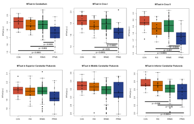 |
13 | MTsat is more sensitive to cerebellar myelin abnormality in multiple sclerosis compared to MTR
Lisa Eunyoung Lee1,2, Irene Vavasour3, Enedino Hernandez-Torres4, Anthony Traboulsee5, Roger Tam3,5,6, Shannon Kolind3,5,7, Tom Schweizer2,8, and Jiwon Oh1,2
1Medicine (Neurology), University of Toronto, Toronto, ON, Canada, 2Medicine, Keenan Research Centre for Biomedical Research, Toronto, ON, Canada, 3Radiology, University of British Columbia, Vancouver, BC, Canada, 4Copenhagen University Hospital, Copenhagen, Denmark, 5Medicine (Neurology), University of British Columbia, Vancouver, BC, Canada, 6Biomedical Engineering, University of British Columbia, Vancouver, BC, Canada, 7Physics and Astronomy, University of British Columbia, Vancouver, BC, Canada, 8Medicine (Neurosurgery), University of Toronto, Toronto, ON, Canada
Magnetization transfer saturation (MTsat) is suggested to be superior to conventional MT ratio (MTR) in quantifying myelin as it reduces bias from T1 effect and B1 inhomogeneity and is more sensitive to tissue damage. This study aimed to compare the ability of MTsat and MTR to detect cerebellar myelin differences across multiple sclerosis (MS) subtypes and to evaluate whether MTsat or MTR demonstrates stronger correlations with clinical disability. We found that MTsat demonstrated greater group differences and more strongly correlated with clinical disability in cerebellum than MTR suggesting that MTsat may be more sensitive to cerebellar myelin abnormalities than MTR.
|
||
0789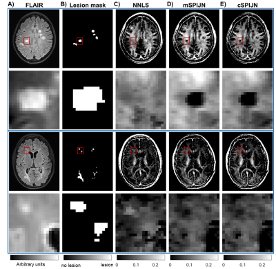 |
14 | Comparing the SPIJN algorithm for myelin water fraction mapping with conventional NNLS evaluation in healthy and multiple sclerosis brains
Ronja C. Berg1, Thomas Amthor2, Irene Vavasour3, Mariya Doneva2, and Christine Preibisch1
1Department of Neuroradiology, School of Medicine, Technical University of Munich, Munich, Germany, 2Philips Research Europe, Hamburg, Germany, 3Department of Radiology, University of British Columbia, Vancouver, BC, Canada
Myelin water fraction (MWF) mapping provides information on myelin concentration, which can be affected by neurological diseases. Most commonly, a non-negative least squares (NNLS) algorithm is used to obtain MWF. A faster alternative is the Sparsity Promoting Iterative Joint NNLS (SPIJN) algorithm. Here, we compared both methods in several brain regions from healthy participants and normal-appearing and lesion tissue from MS patients (EDSS 0-1.5). We found that NNLS-based lesion-average MWF was rather comparable to white matter while SPIJN-based MWF was lower. Thus, SPIJN could be more sensitive to demyelination in lesions but comparisons to gold standard techniques are clearly needed.
|
||
0790 |
15 | Multimodal MRI Investigation of Basal Forebrain Damage in Patients with Mild Cognitive Impairment
Qian Chen1, Jilei Zhang2, and Bing Zhang1,3
1Department of Radiology, Drum Tower Hospital, Clinical College of Nanjing Medical University, Nanjing, China, 2Philips Healthcare, Shanghai, China, 3Department of Radiology, Drum Tower Hospital, Medical School of Nanjing University, Nanjing, China
Cholinergic degeneration in the basal forebrain (BF) plays a crucial role in Alzheimer’s disease. In the present study, reduced BF volumes and damaged BF-associated tracts were observed in patients with mild cognitive impairment (MCI) compared to those with normal aging. Correlation and mediation analyses highlighted that volume reductions in the BF mainly accounted for deficits in global cognition, memory, and visuospatial ability, while disruption in BF-cortical tracts mainly contributed to executive and processing speed dysfunction. Collectively, this study may provide a better understanding of BF damage underlying cognitive impairment and provide guidance for intervention targets in MCI patients.
|
||
 Back to Meeting Home
 Back to the Program-at-a-Glance
The International Society for Magnetic Resonance in Medicine is accredited by the Accreditation Council for Continuing Medical Education to provide continuing medical education for physicians.
 Back to Meeting Home
 Back to the Program-at-a-Glance
 View the Presentation