Digital Poster
Multiple Sclerosis II
Joint Annual Meeting ISMRM-ESMRMB & ISMRT 31st Annual Meeting • 07-12 May 2022 • London, UK

| Computer # | ||||
|---|---|---|---|---|
0881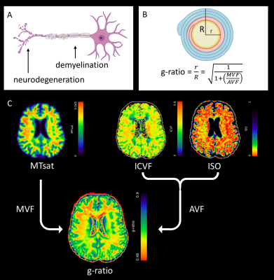 |
1 | MR-derived g-ratio is sensitive to longitudinal change and associated with disability progression in early multiple sclerosis
Elizabeth York1, Rozanna Meijboom1, Mark Bastin1, Agniete Kampaite1, Maria Valdes Hernandez1, Michael J. Thrippleton1, Peter Connick2, Siddharthan Chandran1,2, David P.J. Hunt1, and Adam D. Waldman1
1Centre for Clinical Brain Sciences, University of Edinburgh, Edinburgh, United Kingdom, 2Anne Rowling Regenerative Neurology Clinic, University of Edinburgh, Edinburgh, United Kingdom
There is a pressing need for longitudinal in vivo biomarkers sensitive to heterogeneous pathology in relapsing-remitting multiple sclerosis (RRMS). The MR-g-ratio may be derived from myelin-sensitive magnetisation transfer saturation (MTsat) and multishell diffusion-weighted MRI but its relevance as a longitudinal biomarker and correlate of disability progression in RRMS is previously unexplored. Fifty-nine patients with recently diagnosed RRMS contributed g-ratio data at baseline and one year. G-ratio showed a significant decrease in normal-appearing white matter (NAWM) but not T2 FLAIR white matter lesions over one year. Both g-ratio in NAWM and lesions were, however, associated with clinical disability progression.
|
||
0882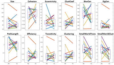 |
2 | Network analysis of multiple sclerosis using pair-wise matched controls and cohesive parcellation of rsfMRI
Ajay Nemani1, Katherine Koenig2, Xuemei Huang1, and Mark J Lowe2
1Imaging Institute, Cleveland Clinic, Cleveland, OH, United States, 2Cleveland Clinic, Cleveland, OH, United States
Multiple sclerosis (MS) is characterized by degenerative changes in white matter, resulting in motor and cognitive deficits. The corresponding damage to functional brain networks is not well understood. We studied several graph theoretic network features of multiple sclerosis patients imaged with rsfMRI. Using a data-driven parcellation designed for optimal node-based representation as well as sex and age-matched controls, we find no significant differences. This result was robust to individual and group parcellations, as well as paired and group comparisons. This differs from previous studies based on standard parcellations and pooled group comparisons. Methodologic reasons for the different observations are discussed.
|
||
0883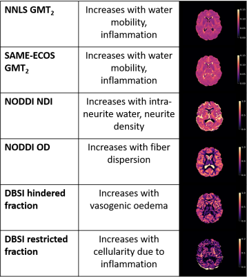 |
3 | Correlations of inflammation metrics from multi echo T2 relaxation and multi-shell diffusion imaging in multiple sclerosis
Tigris S. Joseph1,2, Hanwen Liu2,3,4, Shannon H. Kolind1,2,4,5, Guojun Zhao4, Peng Sun6, Robert Carruthers4, Alice Schabas4, Ana-Luiza Sayao4, Virginia Devonshire4, Roger Tam5,7, G. R. Wayne Moore2,4,8, David K. B. Li4,5, Sheng-Kwei Song9, Anthony Traboulsee4, Irene M. Vavasour2,5, and Cornelia Laule1,2,5,8
1Physics and Astronomy, University of British Columbia, Vancouver, BC, Canada, 2International Collaboration on Repair Discoveries, University of British Columbia, Vancouver, BC, Canada, 3Montreal Neurological Institute - Hospital, McGill University, Montreal, QC, Canada, 4Medicine, University of British Columbia, Vancouver, BC, Canada, 5Radiology, University of British Columbia, Vancouver, BC, Canada, 6Imaging Physics, MD Anderson Cancer Center, Houston, TX, United States, 7School of Biomedical Engineering, University of British Columbia, Vancouver, BC, Canada, 8Pathology & Laboratory Medicine, University of British Columbia, Vancouver, BC, Canada, 9Radiology, Washington University, St. Louis, MO, United States
Inflammation is a key component in multiple sclerosis (MS) pathology. Quantitative MRI metrics reflecting inflammation are important for understanding disease processes in vivo. Metrics from multi-echo T2 relaxation, Diffusion Basis Spectrum Imaging (DBSI) and Neurite Orientation Dispersion and Density Imaging (NODDI) were compared in participants with MS and healthy controls. NODDI neurite density index and DBSI restricted fraction were correlated and may be sensitive to similar pathology. GMT2 correlated negatively with cellularity, but positively with oedema, suggesting oedema may be a key mechanism driving GMT2 increases. Our findings support a complementary T2 and diffusion approach to probe inflammation in MS.
|
||
0884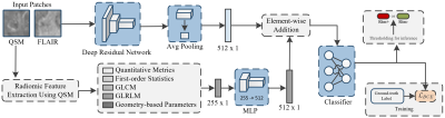 |
4 | QSMRim-Net: Fusing Radiomic and Convolutional Features for Identification of Chronic Active MS Lesions on Quantitative Susceptibility Maps
Hang Zhang1, Jinwei Zhang2, Melanie Marcille3, Pascal Spincemaille4, Thanh D. Nguyen3, Susan A. Gauthier3, Yi Wang3, and Elizabeth M. Sweeney5
1Electrical & Computer Engineering, Cornell University, New York, NY, United States, 2Biomedical Engineering, Cornell University, New York, NY, United States, 3Department of Radiology, Cornell University, New York, NY, United States, 4Cornell University, New York, NY, United States, 5Department of Population Health Sciences, Cornell University, New York, NY, United States
Chronic active multiple sclerosis (MS) lesions are characterized on Quantitative susceptibility mapping (QSM) by a paramagnetic rim (rim+) at the edge of the lesion. We present QSMRim-Net, a deep neural network that fuses lesion-level radiomic and convolutional image features together for automated identification of rim+ lesions on MRI. On the lesion-level, using five-fold cross validation, the proposed QSMRim-Net detected rim+ lesions with an area under the receiver operating characteristic curve of 0.965 and an area under the precision recall curve of 0.655. QSMRim-Net out-performed other state-of-the-art methods on both metrics.
|
||
0885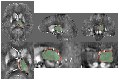 |
5 | Effect of limited segmentation performance on regional qMRI parameter estimations using FSL FIRST in anatomical regions with poor T1 contrast
Fahad Salman1, Niels Bergsland1,2, Michael Dwyer1,3, Bianca Weinstock-Guttman4, Robert Zivadinov1,3, and Ferdinand Schweser1,3
1Buffalo Neuroimaging Analysis Center, Department of Neurology, Jacobs School of Medicine and Biomedical Sciences at the University at Buffalo, Buffalo, NY, United States, 2IRCCS, Fondazione Don Carlo Gnocchi ONLUS, Milan, Italy, 3Center for Biomedical Imaging, Clinical and Translational Science Institute, The State University of New York, Buffalo, NY, United States, 4Jacobs Multiple Sclerosis Center, Department of Neurology, Jacobs School of Medicine and Biomedical Sciences, University at Buffalo, The State University of New York, Buffalo, NY, United States
Most T1w imaging sequences produce only weak deep gray matter (DGM) contrast compared to iron-sensitive techniques such as QSM. In particular, using multi-modal information in both template generation and brain normalization steps has the potential to result in improved segmentation performance. In this study, we compared FIRST to a custom multi-contrast T1w-QSM atlas segmentation approach for estimating thalamic volumes and susceptibility. Our study suggested that an advanced atlas-based approach may result in better regional segmentations of regions with weak T1w contrast and result in more accurate parameter estimates.
|
||
0886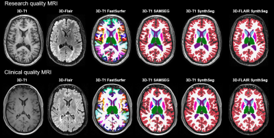 |
6 | Brain tissue segmentation on 3D-FLAIR weighted images in multiple sclerosis
Samantha Noteboom1, Martijn D. Steenwijk1, David R. van der Nederpelt2, Eva M.M. Strijbis3, Bastiaan Moraal2, Frederik Barkhof2,4, Jeroen J.G. Geurts1, Matthan W. A. Caan5, Hugo Vrenken2, and Menno M Schoonheim1
1Anatomy and Neurosciences, MS Center Amsterdam, Amsterdam Neuroscience, Amsterdam UMC, Vrije Universiteit Amsterdam, Amsterdam, Netherlands, 2Radiology and Nuclear Medicine, MS Center Amsterdam, Amsterdam Neuroscience, Amsterdam UMC, Vrije Universiteit Amsterdam, Amsterdam, Netherlands, 3Neurology, MS Center Amsterdam, Amsterdam Neuroscience, Amsterdam UMC, Vrije Universiteit Amsterdam, Amsterdam, Netherlands, 4Neurology and Healthcare Engineering, UCL London, Amsterdam, United Kingdom, 5Biomedical Engineering & Physics, Amsterdam UMC, location AMC, Amsterdam, Netherlands Conventional brain segmentation approaches typically require high resolution 3D-T1 weighted images, which are often unavailable in clinical multiple sclerosis (MS) protocols. Recently, SynthSeg was released which allows 3D-FLAIR to be used for segmentations. This study compared segmentation on 3D-FLAIR with SynthSeg to segmentation in the same patients on 3D-T1 (SynthSeg, FastSurfer and SAMSEG). Brain segmentation was performed on 100 patients with 3D-T1 and 3D-FLAIR images from research and clinical datasets. ICC results showed good comparability for brain tissue, ventricle and grey matter assessments in research data. In clinical data only good comparability was found for ventricle segmentation. |
||
0887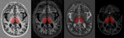 |
7 | Quantitative MRI measures of thalamic microstructural integrity in patients with multiple sclerosis
Alessandro Cagol1, Reza Rahmanzadeh1, Muhamed Barakovic1, Po-Jui Lu1, Matthias Weigel2, Lester Melie-Garcia1, Antoine Lutti3, Than D. Nguyen4, Yi Wang4, Jens Kuhle5, Ludwig Kappos1, and Cristina Granziera1
1Translational Imaging in Neurology (ThINK) Basel, Department of Biomedical Engineering, Translational Imaging in Neurology (ThINK) Basel, Department of Biomedical Engineering, Faculty of Medicine, University Hospital Basel and University of Basel, Basel, Switzerland. Neurologic Clinic and Policlinic, MS Center and Research Center for Clinical Neuroimmunology and Neuroscience Basel (RC2NB), University Hospital Basel and University of Basel, Basel, Switzerland, Basel, Switzerland, 2Translational Imaging in Neurology (ThINK) Basel, Department of Biomedical Engineering, Faculty of Medicine, University Hospital Basel and University of Basel, Basel, Switzerland. Neurologic Clinic and Policlinic, MS Center and Research Center for Clinical Neuroimmunology and Neuroscience Basel (RC2NB), University Hospital Basel and University of Basel, Basel, Switzerland. Division of Radiological Physics, Department of Radiology, University Hospital Basel, Basel, Switzerland, Basel, Switzerland, 3Laboratory for Research in Neuroimaging, Department of Clinical Neuroscience, Lausanne University Hospital and University of Lausanne, Switzerland, Lausanne, Switzerland, 4Department of Radiology, Weill Cornell Medical College, New York, NY, USA, New York, NY, United States, 5Neurologic Clinic and Policlinic, MS Center and Research Center for Clinical Neuroimmunology and Neuroscience Basel (RC2NB), University Hospital Basel and University of Basel, Basel, Switzerland, Basel, Switzerland Thalamus represents a pivotal structure to study MS-associated neurodegeneration. In this study we investigated the alterations in thalamic microstructure of MS patients by using magnetization transfer saturation (MTsat), T1-relaxometry, and myelin water fraction (MWF). Compared to healthy controls (HCs), MS patients presented significant modifications in the thalamic quantitative MRI metrics, suggesting ongoing microstructural and myelin loss. The thalamic quantitative MRI metrics explored showed variable degrees of association with MS lesion burden, brain atrophic changes, as well as with clinical and cognitive disability. |
||
0888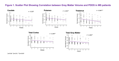 |
8 | Gray Matter Correlates of Patient-Reported Indices of Disability in Multiple Sclerosis
Wafaa Sweidan1, Jenny Chen1, Benjamin Ades-Aron1, Tamar Bacon2, Matthew Lee1, Santiago Coelho1, Dmitry S Novikov1, Timothy Shepherd1, Ilya Kister2, and Els Fieremans1
1Radiology, NYU, New York, NY, United States, 2Neurology, NYU, New York, NY, United States
In a cohort of 172 MS subjects, we identified thalamic atrophy as a significant gray matter correlate of patient-reported indices of motor disability.
|
||
0889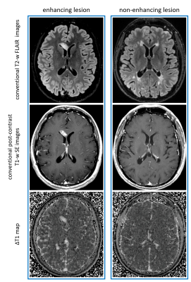 |
9 | Magnetic Resonance Fingerprinting enables measuring T1-shortening in multiple sclerosis lesions after contrast media administration
Graziella Donatelli1,2, Paolo Cecchi1,2, Gianmichele Migaleddu1, Matteo Cencini3, Claudio D'Amelio4, Guido Buonincontri3, Luca Peretti2,3,4, Livia Pasquali4, Michela Tosetti2,3, Mirco Cosottini4, and Mauro Costagli3,5
1Azienda Ospedaliero Universitaria Pisana, Pisa, Italy, 2IMAGO 7 Research Foundation, Pisa, Italy, 3IRCCS Stella Maris, Pisa, Italy, 4University of Pisa, Pisa, Italy, 5Department of Neuroscience, Rehabilitation, Ophtalmology, Genetics, Maternal and Child Sciences, University of Genoa, Genoa, Italy
Quantitative maps of T1-shortening (ΔT1) related to intra-venous contrast media administration are obtained in patients with Multiple Sclerosis by using 3D Quantitative Transient-state Imaging. Statistically significant ΔT1 is observed not only in enhancing lesions, but also in some non-enhancing lesions, suggesting a potential role of this method as a quantitative imaging biomarker to detect subtle blood-brain barrier damage.
|
||
0890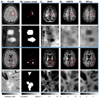 |
10 | Comparing myelin-sensitive markers MWF, ihMTR, and MTsat in healthy and normal-appearing brain tissue and multiple sclerosis lesions
Ronja C. Berg1, Viola Pongratz2, Markus Lauerer2, Thomas Amthor3, Guillaume Gilbert4, Aurore Menegaux1, Claus Zimmer1, Christian Sorg1, Mariya Doneva3, Irene Vavasour5, Mark Mühlau2, and Christine Preibisch1
1Department of Neuroradiology, School of Medicine, Technical University of Munich, Munich, Germany, 2Department of Neurology, School of Medicine, Technical University of Munich, Munich, Germany, 3Philips Research Europe, Hamburg, Germany, 4MR Clinical Science, Philips Healthcare, Mississauga, ON, Canada, 5Department of Radiology, University of British Columbia, Vancouver, BC, Canada
Measurement of myelin concentration could provide valuable information on the integrity of brain tissue. Several myelin-sensitive magnetic resonance imaging methods have been developed. Here, we compared myelin water fraction (MWF), inhomogeneous magnetization transfer ratio (ihMTR) and magnetization transfer saturation (MTsat) in healthy volunteers and patients with multiple sclerosis (MS). We found highest correlation between MWF and ihMTR but all three measures showed clearly reduced values in MS lesions compared to healthy or normal-appearing white matter. However, the measures varied in differentiating between various WM regions and between peri-lesional tissue and more distant normal-appearing WM.
|
||
0891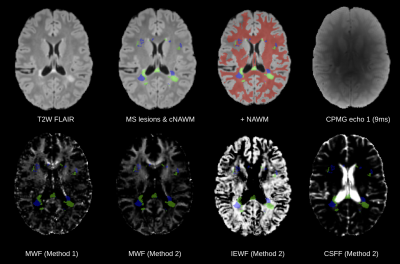 |
11 | Multi-Component T2 Characterization of Lesions in Pediatric Onset Multiple Sclerosis
Serge Didenko Vasylechko1,2, Fedel Machado-Rivas1,2, Camilo Jaimes1,2, Mark P. Gorman2,3, Leslie Benson2,3, Simon K. Warfield1,2, Sila Kurugol1,2, and Onur Afacan1,2
1Radiology, Boston Children's Hospital, Boston, MA, United States, 2Harvard Medical School, Boston, MA, United States, 3Neurology, Boston Children's Hospital, Boston, MA, United States
This study evaluated myelin water fraction (MWF) estimates in chronic MS lesions against contralateral normal appearing white matter (NAWM) of pediatric onset MS patients (POMS). Accelerated 2D multi-slice CPMG acquisitions provided near full brain coverage. Two methods were used to estimate the myelin fractions, as well as intra-/extra- axonal (IE) associated water and CSF water. Significant differences were found between the group measures. Further comparison of NAWM in POMS showed significant differences with whole-white matter in a control group. Our results support that a multi-component T2 analysis can provide quantitative insights into lesional and non-lesional disease burden in POMS.
|
||
0892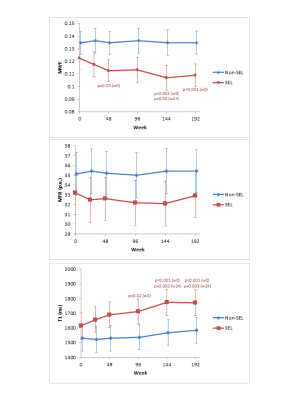 |
12 | Differences in myelin measures between slowly evolving and non-slowly evolving lesions
Irene Margaret Vavasour1, Colm Elliott2, Doug Arnold2,3, Laura Gaetano4, David Clayton5, Stefano Magon4, Ulrike Bonati4, Wei Wei4, Anthony Traboulsee1, and Shannon Kolind1
1University of British Columbia, Vancouver, BC, Canada, 2NeuroRX Research, Montreal, QC, Canada, 3McGill University, Montreal, QC, Canada, 4F Hoffmann-La Roche Ltd, Basel, Switzerland, 5Genentech Inc, San Francisco, CA, United States
Multiple sclerosis (MS) slowly evolving lesions (SELs) are defined on MRI as contiguous regions of pre-existing lesions with constant and concentric local expansion on conventional T1-weighted and T2-weighted images. Myelin-related changes using myelin water fraction (MWF) and magnetization transfer ratio (MTR) in SELs and non-SELs were measured over 192 weeks in patients with relapsing MS. SELs showed reduced myelin measures at baseline compared to non-SELs. Furthermore, over the 4 years, only SELs, and not non-SELs, showed a significant mean decrease in MWF and increase in T1. Both results are indicative of progressive demyelination in this subgroup of chronic lesions.
|
||
0893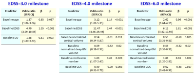 |
13 | Spinal Cord Lesions and Brain Grey Matter Atrophy Predict 5-Year Disease Worsening in Multiple Sclerosis: A Multicentre Study Video Permission Withheld
Paola Valsasina1, Maria A. Rocca1,2,3, Alessandro Meani1, Loredana Storelli1, Claudio Gobbi4,5, Chiara Zecca4,5, Frederik Barkhof6,7, Menno M Schoonheim8, Eva Strijbis7, Hugo Vrenken6,7, Antonio Gallo9, Alvino Bisecco9, Olga Ciccarelli10, Marios Yiannakas10, Alex Rovira11, Jaume Sastre-Garriga12, Jacqueline Palace13, Lucy Matthews13, Achim Gass14, Philipp Eisele14,
Carsten Lukas15, Barbara Bellenberg15, Monica Margoni1, Paolo Preziosa1,2, and Massimo Filippi1,2,3,16,17
1Neuroimaging Research Unit, Division of Neuroscience, IRCCS San Raffaele Scientific Institute, Milan, Italy, 2Neurology Unit, IRCCS San Raffaele Scientific Institute, Milan, Italy, 3Vita-Salute San Raffaele University, Milan, Italy, 4Neurology Clinic, MS Center/ Headache Center, Neurocenter of Southern Switzerland, Lugano, Switzerland, 5Faculty of Biomedical Sciences, Università della Svizzera Italiana, Lugano, Switzerland, 6Radiology and Nuclear Medicine, MS Center Amsterdam, Amsterdam UMC, location Vumc, Amsterdam, Netherlands, 7Department of Neurology, MS Center Amsterdam, Amsterdam UMC, location Vumc, Amsterdam, Netherlands, 8Department of Anatomy and Neurosciences, MS Center Amsterdam, Amsterdam UMC, location Vumc, Amsterdam, Netherlands, 9Department of Advanced Medical and Surgical Sciences, and 3T MRI-Center, University of Campania “Luigi Vanvitelli”, Naples, Italy, 10NMR Research Unit, Queen Square MS Centre, Department of Neuroinflammation, UCL Institute of Neurology, London, United Kingdom, 11Section of Neuroradiology, Department of Radiology, Hospital Universitari Vall d'Hebron, Barcelona, Spain, 12Department of Neurology/Neuroimmunology, Multiple Sclerosis Centre of Catalonia, Hospital Universitari Vall d'Hebron, Barcelona, Spain, 13Nuffield Department of Clinical Neurosciences, University of Oxford, Oxford, United Kingdom, 14Department of Neurology, Universitätsmedizin Mannheim, University of Heidelberg, Mannheim, Germany, 15Institute of Neuroradiology, St. Josef Hospital, Ruhr-University Bochum, Bochum, Germany, 16Neurorehabilitation Unit, IRCCS San Raffaele Scientific Institute, Milan, Italy, 17Neurophysiology Service, IRCCS San Raffaele Scientific Institute, Milan, Italy
Aim of this study was to evaluate the independent role of brain and cervical cord damage in predicting 5-year clinical disability worsening in a multicentre cohort of multiple sclerosis patients. Results showed that cortical atrophy and spinal cord damage, together with baseline disability and progressive disease phenotype, independently predicted 5-year disability worsening (C-index=0.81). Older age, higher clinical disability and cord lesion number independently predicted conversion to a progressive disease phenotype (C-index=0.91). Focal spinal cord lesions and cortical atrophy had also a role in predicting clinically-relevant disability milestones at 5 years.
|
||
0894 |
14 | Diffusion Tensor Imaging Helps to Differentiate Neuromyelitis Optica Spectrum Disorders and Multiple Sclerosis-Related Optic Neuritis Video Not Available
Yan Xie1, Yan Zhang1, Weiyin Vivian Liu2, and Wenzhen Zhu1
1Tongji Hospital, Tongji Medical College, Huazhong University of Science and Technology, Wuhan, China, 2GE Healthcare, MR Research China, Beijing, China
The purpose of this study was to assess the differences in the patterns of NMOSD and MS-related optic nerve damage by DTI. In this study, the intraorbital optic nerve was divided anatomically in three equal parts. We found that NMOSD-related optic neuritis exhibited extensive optic nerve damage, particularly in the posterior segment of the optic nerve, whereas MS-related optic neuritis tended to be more anterior-middle optic nerve damage. In addition, the combination of FA with conventional MRI showed better differential diagnostic efficacy for NMOSD and MS-related optic neuritis.
|
||
 Back to Meeting Home
 Back to the Program-at-a-Glance
The International Society for Magnetic Resonance in Medicine is accredited by the Accreditation Council for Continuing Medical Education to provide continuing medical education for physicians.
 Back to Meeting Home
 Back to the Program-at-a-Glance
 View the Presentation